
The azygos vein is a vein running up the right side of the thoracic vertebral column draining itself towards the superior vena cava. It connects the systems of superior vena cava and inferior vena cava and can provide an alternative path for blood to the right atrium when either of the venae cavae is blocked.
The small saphenous vein is a relatively large superficial vein of the posterior leg.

In human anatomy, the superior mesenteric vein (SMV) is a blood vessel that drains blood from the small intestine. Behind the neck of the pancreas, the superior mesenteric vein combines with the splenic vein to form the portal vein that carries blood to the liver. The superior mesenteric vein lies to the right of the similarly named artery, the superior mesenteric artery, which originates from the abdominal aorta.

In human anatomy, the hepatic veins are the veins that drain venous blood from the liver into the inferior vena cava. There are usually three large upper hepatic veins draining from the left, middle, and right parts of the liver, as well as a number (6-20) of lower hepatic veins. All hepatic veins are valveless.

The coronary sinus is the largest vein of the heart. It drains over half of the deoxygenated blood from the heart muscle into the right atrium. It begins on the backside of the heart, in between the left atrium, and left ventricle; it begins at the junction of the great cardiac vein, and oblique vein of the left atrium. It receives multiple tributaries. It passes across the backside of the heart along a groove between left atrium and left ventricle, then drains into the right atrium at the orifice of the coronary sinus.

The straight sinus, also known as tentorial sinus or the sinus rectus, is an area within the skull beneath the brain. It receives blood from the inferior sagittal sinus and the great cerebral vein, and drains into the confluence of sinuses.
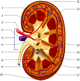
The renal circulation supplies the blood to the kidneys via the renal arteries, left and right, which branch directly from the abdominal aorta. Despite their relatively small size, the kidneys receive approximately 20% of the cardiac output.
In many Animalia, including humans, an ileocolic structure or problem is something that concerns the region of the gastrointestinal tract from the ileum to the colon. In Animalia that have ceca, the ileocecal region is a subset of the ileocolic region, and the entire range can also be described as ileocecocolic, whereas in some Animalia, the ileocolic region contains no cecum, as the ileum joins the colon directly.

The cerebellar veins are veins which drain the cerebellum. They consist of the superior cerebellar veins and the inferior cerebellar veins. The superior cerebellar veins drain to the straight sinus and the internal cerebral veins. The inferior cerebellar veins drain to the transverse sinus, the superior petrosal sinus, and the occipital sinus.
The middle colic vein drains the transverse colon. It is a tributary of the superior mesenteric vein, and follows the path of its corresponding artery, the middle colic artery. As the superior mesenteric vein drains to the hepatic portal vein, the middle colic vein is considered part of the hepatic portal system. This vein also carries nutrients absorbed from the large intestine to the liver.

The basivertebral veins are large, tortuous veins of the trabecular bone of vertebral bodies that drain into the internal and external vertebral venous plexuses.

The anterior cardiac veins are a variable number of small veins which drain blood from the anterior portion of the right ventricle into the right atrium.
The cystic veins drain venous blood from the gallbladder and the cystic duct. The cystic veins either drain into various branches and tributaries of the hepatic portal vein.

The vaginal venous plexus is a group of veins draining blood from the vagina. It lies around the sides of the vagina. Its blood eventually drains into the internal iliac veins.

The right gastric vein drains blood from the lesser curvature of the stomach into the hepatic portal vein. It is part of the portal circulation.

The central retinal vein is a vein that drains the retina of the eye. It travels backwards through the centre of the optic nerve accompanied by the central retinal artery before exiting the optic nerve together with the central retinal artery to drain into either the superior ophthalmic vein or the cavernous sinus.

The anterior auricular veins are veins which drain the anterior aspect of the external ear. The veins drains to the superficial temporal vein.
The appendicular vein is the vein which drains blood from the vermiform appendix. It is located in the mesoappendix and accompanies the appendicular artery. The appendicular vein drains into the ileocolic vein.
The uterine vein is a vein of the uterus. It is found in the cardinal ligament. It drains into the internal iliac vein. It follows a similar course to the uterine artery. It helps to drain blood from the uterus, and removes waste from blood in the placenta during pregnancy.

The superior mesenteric lymph nodes may be divided into three principal groups:
