Portal vein

Last updated
Portal vein
Gray591-Portal vein.png
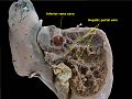
The portal vein (in light blue) and its tributaries. It is formed by the superior mesenteric vein, inferior mesenteric vein, and splenic vein. Lienal vein is an old term for splenic vein.
Details
System Hepatic portal system
Drains from Gastrointestinal tract, spleen, pancreas
Source Splenic vein, superior mesenteric vein, inferior mesenteric vein, pancreatic vein
Drains to Liver sinusoid
Identifiers
Latin vena portae hepatis
MeSH D011169
TA98 A12.3.12.001
TA2 5092
FMA 50735
Anatomical terminology

The portal vein or hepatic portal vein (HPV) is a blood vessel that carries blood from the gastrointestinal tract, gallbladder, pancreas and spleen to the liver. This blood contains nutrients and toxins extracted from digested contents. Approximately 75% of total liver blood flow is through the portal vein, with the remainder coming from the hepatic artery proper. The blood leaves the liver to the heart in the hepatic veins.

Contents

The portal vein is not a true vein, because it conducts blood to capillary beds in the liver and not directly to the heart. It is a major component of the hepatic portal system, one of three portal venous systems in the human body; the others being the hypophyseal and renal portal systems.

The portal vein is usually formed by the confluence of the superior mesenteric, splenic veins, inferior mesenteric, left, right gastric veins and the pancreatic vein.

Conditions involving the portal vein cause considerable illness and death. An important example of such a condition is elevated blood pressure in the portal vein. This condition, called portal hypertension, is a major complication of cirrhosis. In abdominal obesity fats, inflammatory cytokines and other toxic substances are transported by the portal vein from visceral fat into the liver, leading to hepatic insulin resistance and non-alcoholic fatty liver disease. [1] [2]

Structure

Measuring approximately 8 cm (3 inches) long in adults, [3] the portal vein is located in the right upper quadrant of the abdomen, originating behind the neck of the pancreas. [4]

In most individuals, the portal vein is formed by the union of the superior mesenteric vein and the splenic vein. [5] For this reason, the portal vein is occasionally called the splenic-mesenteric confluence. [4] Occasionally, the portal vein also directly communicates with the inferior mesenteric vein, although this is highly variable. Other tributaries of the portal vein include the cystic and the left and right gastric veins. [6] and also pararumbilical vein and prepyloric vein.

Tributaries of the hepatic portal vein [6]

Immediately before reaching the liver, the portal vein divides into right and left. It ramifies further, forming smaller venous branches and ultimately portal venules. Each portal venule courses alongside a hepatic arteriole and the two vessels form the vascular components of the portal triad. These vessels ultimately empty into the hepatic sinusoids to supply blood to the liver. [6]

Portacaval anastomoses

The portal venous system has several anastomoses with the systemic venous system. In cases of portal hypertension these anastomoses may become engorged, dilated, or varicosed and subsequently rupture.

Accessory hepatic portal veins

Accessory hepatic portal veins are those veins that drain directly into the liver without joining the hepatic portal vein. These include the paraumbilical veins as well as veins of the lesser omentum, falciform ligament, and those draining the gallbladder wall. [4]

Function

The portal vein and hepatic arteries form the liver's dual blood supply. Approximately 75% of hepatic blood flow is derived from the portal vein, while the remainder is from the hepatic arteries. [4]

Unlike most veins, the portal vein does not drain into the heart. Rather, it is part of a portal venous system that delivers venous blood into another capillary system, the hepatic sinusoids of the liver. In carrying venous blood from the gastrointestinal tract to the liver, the portal vein accomplishes two tasks: it supplies the liver with metabolic substrates and it ensures that substances ingested are first processed by the liver before reaching the systemic circulation. This accomplishes two things. First, possible toxins that may be ingested can be detoxified by the hepatocytes before they are released into the systemic circulation. Second, the liver is the first organ to absorb nutrients just taken in by the intestines. After draining into the liver sinusoids, blood from the liver is drained by the hepatic vein.

Clinical significance

Portal hypertension

Increased blood pressure in the portal vein, called portal hypertension, is a major complication of liver disease, most commonly cirrhosis. [7] A dilated portal vein (diameter of greater than 13 or 15 mm) is a sign of portal hypertension, with a sensitivity estimated at 12.5% or 40%. [8] On Doppler ultrasonography, the main portal vein (MPV) peak systolic velocity normally ranges between 20 cm/s and 40 cm/s. [9] A slow velocity of <16 cm/s in addition to dilatation in the MPV are diagnostic of portal hypertension. [9]

Clinical signs of portal hypertension include those of chronic liver disease: ascites, esophageal varices, spider nevi, caput medusae, and palmar erythema. [10]

Pulsatility

Doppler ultrasonography of the portal vein over 5 seconds, showing peaks of maximal velocity, as well as points of minimal velocity. Doppler ultrasonography of the portal vein in cirrhosis.jpg
Doppler ultrasonography of the portal vein over 5 seconds, showing peaks of maximal velocity, as well as points of minimal velocity.

Portal vein pulsatility can be measured by Doppler ultrasonography. An increased pulsatility may be caused by cirrhosis, as well as increased right atrial pressure (which in turn may be caused by right heart failure or tricuspid regurgitation). [9] Portal vein pulsatility can be quantified by pulsatility indices (PI), where an index above a certain cutoff indicates pathology:

Pulsatility indices (PI)
IndexCalculationCutoff
Average-based(Max - Min) / Average [9] 0.5 [9]
Max-based(Max - Min) / Max [11] 0.5 [11] [12] - 0.54 [12]

Infection

Pylephlebitis is infection of the portal vein, usually arising from an infectious intra-abdominal process such as diverticulitis. [13] [14]

Portal venous gas

Hepatic portal venous gas is a rare finding on radiological exams. Gas is shown to enter the portal venous system. It is most commonly caused by intestinal ischemia but has also been associated with colon cancer. [15]

Additional images

Related Research Articles

<span class="mw-page-title-main">Ascites</span> Abnormal build-up of fluid in the abdomen

Ascites is the abnormal build-up of fluid in the abdomen. Technically, it is more than 25 ml of fluid in the peritoneal cavity, although volumes greater than one liter may occur. Symptoms may include increased abdominal size, increased weight, abdominal discomfort, and shortness of breath. Complications can include spontaneous bacterial peritonitis.

<span class="mw-page-title-main">Vascular surgery</span> Medical specialty, operative procedures for the treatment of vascular disorders

Vascular surgery is a surgical subspecialty in which vascular diseases involving the arteries, veins, or lymphatic vessels, are managed by medical therapy, minimally-invasive catheter procedures and surgical reconstruction. The specialty evolved from general and cardiovascular surgery where it refined the management of just the vessels, no longer treating the heart or other organs. Modern vascular surgery includes open surgery techniques, endovascular techniques and medical management of vascular diseases - unlike the parent specialities. The vascular surgeon is trained in the diagnosis and management of diseases affecting all parts of the vascular system excluding the coronaries and intracranial vasculature. Vascular surgeons also are called to assist other physicians to carry out surgery near vessels, or to salvage vascular injuries that include hemorrhage control, dissection, occlusion or simply for safe exposure of vascular structures.

<span class="mw-page-title-main">Portal hypertension</span> Abnormally increased portal venous pressure

Portal hypertension is defined as increased portal venous pressure, with a hepatic venous pressure gradient greater than 5 mmHg. Normal portal pressure is 1–4 mmHg; clinically insignificant portal hypertension is present at portal pressures 5–9 mmHg; clinically significant portal hypertension is present at portal pressures greater than 10 mmHg. The portal vein and its branches supply most of the blood and nutrients from the intestine to the liver.

<span class="mw-page-title-main">Caput medusae</span> Appearance of distended and engorged superficial epigastric veins

Caput medusae is the appearance of distended and engorged superficial epigastric veins, which are seen radiating from the umbilicus across the abdomen. The name caput medusae originates from the apparent similarity to Medusa's head, which had venomous snakes in place of hair. It is also a sign of portal hypertension. When the portal vein, that transfers the blood from the gastrointestinal tract to the liver, is blocked, the blood volume increases in the peripheral blood vessels making them appear engorged. It is caused by dilation of the paraumbilical veins, which carry oxygenated blood from mother to fetus in utero and normally close within one week of birth, becoming re-canalised due to portal hypertension caused by liver failure.The appearance is due to cutanous portosystemic collateral formation between distended and engorged paraumbilical veins that radiate from the umbilicus across the abdomen to join systemic veins.

<span class="mw-page-title-main">Celiac artery</span> First major branch of the abdominal aorta

The celiacartery, also known as the celiac trunk or truncus coeliacus, is the first major branch of the abdominal aorta. It is about 1.25 cm in length. Branching from the aorta at thoracic vertebra 12 (T12) in humans, it is one of three anterior/ midline branches of the abdominal aorta.

<span class="mw-page-title-main">Superior mesenteric artery</span> Artery which supplies blood to the intestines and pancreas

In human anatomy, the superior mesenteric artery (SMA) is an artery which arises from the anterior surface of the abdominal aorta, just inferior to the origin of the celiac trunk, and supplies blood to the intestine from the lower part of the duodenum through two-thirds of the transverse colon, as well as the pancreas.

<span class="mw-page-title-main">Superior mesenteric vein</span> Vein which drains blood from the small intestine

In human anatomy, the superior mesenteric vein (SMV) is a blood vessel that drains blood from the small intestine. Behind the neck of the pancreas, the superior mesenteric vein combines with the splenic vein to form the portal vein that carries blood to the liver. The superior mesenteric vein lies to the right of the similarly named artery, the superior mesenteric artery, which originates from the abdominal aorta.

<span class="mw-page-title-main">Splenic vein</span> Vein that drains blood from the spleen, stomach and pancreas

In human anatomy, the splenic vein is a blood vessel that drains blood from the spleen, the stomach fundus and part of the pancreas. It is part of the hepatic portal system.

<span class="mw-page-title-main">Portal vein thrombosis</span> Disease of the liver

Portal vein thrombosis (PVT) is a vascular disease of the liver that occurs when a blood clot occurs in the hepatic portal vein, which can lead to increased pressure in the portal vein system and reduced blood supply to the liver. The mortality rate is approximately 1 in 10.

<span class="mw-page-title-main">Hepatic artery proper</span>

The hepatic artery proper is the artery that supplies the liver and gallbladder. It raises from the common hepatic artery, a branch of the celiac artery.

<span class="mw-page-title-main">Falciform ligament</span> Ligament attaching the liver to the front body wall

In human anatomy, the falciform ligament is a ligament that attaches the liver to the front body wall and divides the liver into the left lobe and right lobe. The falciform ligament is a broad and thin fold of peritoneum, its base being directed downward and backward and its apex upward and forward. It droops down from the hilum of the liver.

A portosystemic shunt or portasystemic shunt, also known as a liver shunt, is a bypass of the liver by the body's circulatory system. It can be either a congenital or acquired condition and occurs in humans as well as in other species of animals. Congenital PSS are extremely rare in humans but are relatively common in dogs. Improvements in imaging and awareness have contributed to an increase in cases.Thus a large part of medical and scientific literature on the subject is grounded in veterinary medicine.

<span class="mw-page-title-main">Hepatic portal system</span> System of veins comprising the hepatic portal vein and its tributaries

In human anatomy, the hepatic portal system or portal venous system is the system of veins comprising the portal vein and its tributaries. The other portal venous systems in the body are the renal portal system, and the hypophyseal portal system.

A portacaval anastomosis or portocaval anastomosis is a specific type of circulatory anastomosis that occurs between the veins of the portal circulation and the vena cava, thus forming one of the principal types of portasystemic anastomosis or portosystemic anastomosis, as it connects the portal circulation to the systemic circulation, providing an alternative pathway for the blood. When there is a blockage of the portal system, portocaval anastomosis enables the blood to still reach the systemic venous circulation. The inferior end of the esophagus and the superior part of the rectum are potential sites of a harmful portocaval anastomosis.

<span class="mw-page-title-main">Central veins of liver</span> Veins through the centers of hepatic (liver) lobules

In microanatomy, the central veins of liver are veins found at the center of hepatic lobules.

<span class="mw-page-title-main">Portal venous system</span> The capillary bed pools into another through veins without first going through the heart

In the circulatory system of vertebrates, a portal venous system occurs when a capillary bed pools into another capillary bed through veins, without first going through the heart. Both capillary beds and the blood vessels that connect them are considered part of the portal venous system.

Portal venous pressure is the blood pressure in the hepatic portal vein, and is normally between 5-10 mmHg. Raised portal venous pressure is termed portal hypertension, and has numerous sequelae such as ascites and hepatic encephalopathy.

<span class="mw-page-title-main">Liver</span> Vertebrate organ involved in metabolism

The liver is a major metabolic organ only found in vertebrate animals, which performs many essential biological functions such as detoxification of the organism, and the synthesis of proteins and biochemicals necessary for digestion and growth. In humans, it is located in the right upper quadrant of the abdomen, below the diaphragm and mostly shielded by the lower right rib cage. Its other metabolic roles include carbohydrate metabolism, the production of hormones, conversion and storage of nutrients such as glucose and glycogen, and the decomposition of red blood cells.

<span class="mw-page-title-main">Doppler ultrasonography</span> Ultrasound imaging of the movement of tissues and body fluids using the Doppler effect

Doppler ultrasonography is medical ultrasonography that employs the Doppler effect to perform imaging of the movement of tissues and body fluids, and their relative velocity to the probe. By calculating the frequency shift of a particular sample volume, for example, flow in an artery or a jet of blood flow over a heart valve, its speed and direction can be determined and visualized.

<span class="mw-page-title-main">Lobes of liver</span> Four gross divisions of the human liver

In human anatomy, the liver is divided grossly into four parts or lobes: the right lobe, the left lobe, the caudate lobe, and the quadrate lobe. Seen from the front – the diaphragmatic surface – the liver is divided into two lobes: the right lobe and the left lobe. Viewed from the underside – the visceral surface – the other two smaller lobes, the caudate lobe and the quadrate lobe, are also visible. The two smaller lobes, the caudate lobe and the quadrate lobe, are known as superficial or accessory lobes, and both are located on the underside of the right lobe.

References

  1. Item F, Konrad D (2012). "Visceral fat and metabolic inflammation: the portal theory revisited" (PDF). Obesity Reviews . 13 (Suppl 2): 30–39. doi:10.1111/j.1467-789X.2012.01035.x. PMID   23107257. S2CID   25169877.
  2. Dhawan D, Sharma S (2020). "Abdominal Obesity, Adipokines and Non-communicable Diseases". The Journal of Steroid Biochemistry and Molecular Biology . 203: 105737. doi:10.1016/j.jsbmb.2020.105737. PMC   7431389 . PMID   32818561.
  3. Harold M Chung; Chung, Kyung Won (2008). Gross anatomy . Philadelphia: Wolters Kluwer Health/Lippincott Williams & Wilkins. pp.  208. ISBN   978-0-7817-7174-0.
  4. 1 2 3 4 Plinio Rossi; L. Broglia (2000). Portal Hypertension: Diagnostic Imaging and Imaging-Guided Therapy. Berlin: Springer. p. 51. ISBN   978-3-540-65797-2.
  5. Benjamin L. Shneider; Sherman, Philip M. (2008). Pediatric Gastrointestinal Disease. Connecticut: PMPH-USA. p. 751. ISBN   978-1-55009-364-3.
  6. 1 2 3 Henry Gray (1901). Anatomy, Descriptive and Surgical (16 ed.). Philadelphia: Lea Brothers. p. 619.
  7. Dooley, James; Sherlock, Sheila (2002). Diseases of the liver and biliary system. Oxford: Blackwell Science. ISBN   978-0-632-05582-1.
  8. Al-Nakshabandi NA (2006). "The role of ultrasonography in portal hypertension". Saudi J Gastroenterol. 12 (3): 111–7. doi: 10.4103/1319-3767.29750 . PMID   19858596.
  9. 1 2 3 4 5 Iranpour, Pooya; Lall, Chandana; Houshyar, Roozbeh; Helmy, Mohammad; Yang, Albert; Choi, Joon-Il; Ward, Garrett; Goodwin, Scott C (2016). "Altered Doppler flow patterns in cirrhosis patients: an overview". Ultrasonography. 35 (1): 3–12. doi:10.14366/usg.15020. ISSN   2288-5919. PMC   4701371 . PMID   26169079.
  10. Key Topics in General Surgery (2 ed.). Informa Healthcare. 2002. ISBN   978-1-85996-164-3.
  11. 1 2 Goncalvesova, E.; Varga, I.; Tavacova, M.; Lesny, P. (2013). "Changes of portal vein flow in heart failure patients with liver congestion". European Heart Journal. 34 (suppl 1): P627. doi: 10.1093/eurheartj/eht307.P627 . ISSN   0195-668X.
  12. 1 2 Page 367 in: Henryk Dancygier (2009). Clinical Hepatology: Principles and Practice of Hepatobiliary Diseases. Vol. 1. Springer Science & Business Media. ISBN   9783540938422.
  13. Plemmons RM, Dooley DP, Longfield RN (November 1995). "Septic thrombophlebitis of the portal vein (pylephlebitis): diagnosis and management in the modern era". Clin. Infect. Dis. 21 (5): 1114–20. doi:10.1093/clinids/21.5.1114. PMID   8589130.
  14. Perez-Cruet MJ, Grable E, Drapkin MS, Jablons DM, Cano G (May 1993). "Pylephlebitis associated with diverticulitis". South. Med. J. 86 (5): 578–80. doi:10.1097/00007611-199305000-00020. PMID   8488411.
  15. Li, Z; Su, Y; Wang, X; Yan, H; Sun, M; Shu, Z (December 2017). "Hepatic portal venous gas associated with colon cancer: A case report and literature review". Medicine. 96 (50): e9352. doi:10.1097/MD.0000000000009352. PMC   5815821 . PMID   29390409.