In human anatomy, the arm refers to the upper limb in common usage, although academically the term specifically means the upper arm between the glenohumeral joint and the elbow joint. The distal part of the upper limb between the elbow and the radiocarpal joint is known as the forearm or "lower" arm, and the extremity beyond the wrist is the hand.

In anatomy, the thigh is the area between the hip (pelvis) and the knee. Anatomically, it is part of the lower limb.

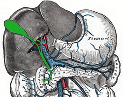
The portal vein or hepatic portal vein (HPV) is a blood vessel that carries blood from the gastrointestinal tract, gallbladder, pancreas and spleen to the liver. This blood contains nutrients and toxins extracted from digested contents. Approximately 75% of total liver blood flow is through the portal vein, with the remainder coming from the hepatic artery proper. The blood leaves the liver to the heart in the hepatic veins.

The left and right brachiocephalic veins are major veins in the upper chest, formed by the union of the ipsilateral internal jugular vein and subclavian vein behind the sternoclavicular joint. The left brachiocephalic vein is more than twice the length of the right brachiocephalic vein.

In human anatomy, the thoracic duct is the larger of the two lymph ducts of the lymphatic system. The thoracic duct usually begins from the upper aspect of the cisterna chyli, passing out of the abdomen through the aortic hiatus into first the posterior mediastinum and then the superior mediastinum, extending as high up as the root of the neck before descending to drain into the systemic (blood) circulation at the venous angle.

The pulmonary veins are the veins that transfer oxygenated blood from the lungs to the heart. The largest pulmonary veins are the four main pulmonary veins, two from each lung that drain into the left atrium of the heart. The pulmonary veins are part of the pulmonary circulation.

The azygos vein is a vein running up the right side of the thoracic vertebral column draining itself towards the superior vena cava. It connects the systems of superior vena cava and inferior vena cava and can provide an alternative path for blood to the right atrium when either of the venae cavae is blocked.

The external jugular vein receives the greater part of the blood from the exterior of the cranium and the deep parts of the face, being formed by the junction of the posterior division of the retromandibular vein with the posterior auricular vein.

The renal veins in the renal circulation, are large-calibre veins that drain blood filtered by the kidneys into the inferior vena cava. There is one renal vein draining each kidney. Each renal vein is formed by the convergence of the interlobar veins of one kidney.
The facial vein is a relatively large vein in the human face. It commences at the side of the root of the nose and is a direct continuation of the angular vein where it also receives a small nasal branch. It lies behind the facial artery and follows a less tortuous course. It receives blood from the external palatine vein before it either joins the anterior branch of the retromandibular vein to form the common facial vein, or drains directly into the internal jugular vein.
The maxillary vein or internal maxillary vein is a vein of the head. It is a short trunk which accompanies the maxillary artery. It is formed by a confluence of the veins of the pterygoid plexus. It and passes posterior-ward between the sphenomandibular ligament and the neck of the mandible to enter the parotid gland where unites with the superficial temporal vein to form the retromandibular vein.
The supraorbital vein is a vein of the forehead. It communicates with the frontal branch of the superficial temporal vein. It passes through the supraorbital notch, and merges with the angular vein to form the superior ophthalmic vein. The supraorbital vein helps to drain blood from the forehead, eyebrow, and upper eyelid.
The superficial temporal vein is a vein of the side of the head which collects venous blood from the region of the temple. It arises from an anastomosing venous plexus on the side and vertex of the head. The superficial temporal vein terminates within the substance of the parotid gland by uniting with the maxillary vein to form the retromandibular vein.
The retromandibular vein is a major vein of the face. It is formed within the parotid gland by the confluence of the maxillary vein, and superficial temporal vein. It descends in the gland and splits into two branches upon emerging from the gland. Its anterior branch then joins the (anterior) facial vein forming the common facial vein, while its posterior branch joins the posterior auricular vein forming the external jugular vein.

The internal pudendal veins are a set of veins in the pelvis. They are the venae comitantes of the internal pudendal artery. Internal pudendal veins are enclosed by pudendal canal, with internal pudendal artery and pudendal nerve.

The rectal venous plexus is the venous plexus surrounding the rectum. It consists of an internal and an external rectal plexus. It is drained by the superior, middle, and inferior rectal veins. It forms a portosystemic (portocaval) anastomosis. This allows rectally administered medications to bypassing first pass metabolism.

In human anatomy, the cerebral veins are blood vessels in the cerebral circulation which drain blood from the cerebrum of the human brain. They are divisible into external and internal groups according to the outer or inner parts of the hemispheres they drain into.

The vorticose veins, referred to clinically as the vortex veins, are veins that drain the choroid of the eye. There are usually 4-5 vorticose veins in each eye, with at least one vorticose vein per each quadrant of the eye. Vorticose veins drain into the superior ophthalmic vein, and inferior ophthalmic vein.

The intercostal arteries are a group of arteries passing within an intercostal space. There are 9 anterior and 11 posterior intercostal arteries on each side of the body. The anterior intercostal arteries are branches of the internal thoracic artery and its terminal branch - the musculophrenic artery. The posterior intercostal arteries are branches of the supreme intercostal artery and thoracic aorta.

In human male anatomy, the dorsal veins of the penis are blood vessels that drain the shaft, the skin and the glans of the human penis. They are typically located in the midline on the dorsal aspect of the penis and they comprise the superficial dorsal veinof the penis, that lies in the subcutaneous tissue of the shaft, and the deep dorsal veinof the penis, that lies beneath the deep fascia.