
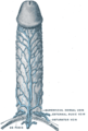
In male human anatomy, the glans penis or penile glans, commonly referred to as the glans, is the bulbous structure at the distal end of the human penis that is the human male's most sensitive erogenous zone and primary anatomical source of sexual pleasure. The glans penis is present in the male reproductive organs of humans and most other mammals where it may appear smooth, spiny, elongated or divided. It is externally lined with mucosal tissue, which creates a smooth texture and glossy appearance. In humans, the glans is located over the distal ends of the corpora cavernosa and is a continuation of the corpus spongiosum of the penis. At the summit appears the urinary meatus and at the base forms the corona glandis. An elastic band of tissue, known as the frenulum, runs on its ventral surface. In men who are not circumcised, it is completely or partially covered by a fold of skin called the foreskin. In adults, the foreskin can generally be retracted over and past the glans manually or sometimes automatically during an erection.

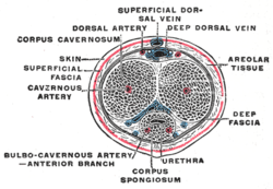
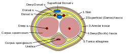
The corpus spongiosum is the mass of spongy tissue surrounding the male urethra within the penis. It is also called the corpus cavernosum urethrae in older texts.

The bulbospongiosus muscles are a subgroup of the superficial muscles of the perineum. They have a slightly different origin, insertion and function in males and females. In males, these muscles cover the bulb of the penis, while in females, they cover the vestibular bulbs.

The dartos fascia, dartos tunic or simply dartos is a layer of connective tissue found in the penile shaft, foreskin and scrotum. The penile portion is referred to as the superficial fascia of penis or the subcutaneous tissue of penis, while the scrotal part is the dartos proper. In addition to being continuous with itself between the scrotum and the penis, it is also continuous with Colles' fascia of the perineum and Scarpa's fascia of the abdomen.

The internal pudendal artery is one of the three pudendal arteries. It branches off the internal iliac artery, and provides blood to the external genitalia.

A corpus cavernosum penis (singular) is one of a pair of sponge-like regions of erectile tissue, which contain most of the blood in the penis during an erection.
The dorsal artery of the penis is an artery on the top surface of the penis. It is a branch of the internal pudendal artery. It runs forward on the dorsum of the penis to the glans, where it divides into two branches to the glans penis and the foreskin (prepuce).

The dorsal nerve of the penis is the deepest of three divisions of the pudendal nerve; it accompanies the internal pudendal artery along the ramus of the ischium; it then runs forward along the margin of the inferior ramus of the pubis, between the superior and inferior layers of the fascia of the urogenital diaphragm.

The prostatic plexus is continued from the lower part of the pelvic plexus. It lies within the fascial shell of the prostate.

The perineal membrane is an anatomical term for a fibrous membrane in the perineum. The term "inferior fascia of urogenital diaphragm", used in older texts, is considered equivalent to the perineal membrane.

The membranous urethra or intermediate part of male urethra is the shortest, least dilatable, and, with the exception of the urinary meatus, the narrowest part of the urethra.

The prostatic veins form a well-marked prostatic plexus which lies partly in the fascial sheath of the prostate and partly between the sheath and the prostatic capsule. It collects blood from the prostate, and the corpora cavernosa of penis. It communicates with the pudendal and vesical plexuses.
The pudendal venous plexus lies behind the arcuate pubic ligament and the lower part of the pubic symphysis, and in front of the bladder and prostate. Its chief tributary is the deep dorsal vein of the penis, but it also receives branches from the front of the bladder and prostate. It communicates with the vesical venous plexus and with the internal pudendal vein and drains into the vesical and hypogastric veins.

The following outline is provided as an overview of and topical guide to human anatomy:

The body or shaft of the penis is the free portion of the human penis that is located outside of the pelvic cavity. It is the continuation of the internal root, which is embedded in the pelvis and extends to the glans. It is made up of the two corpora cavernosa and the corpus spongiosum on the underside. The corpora cavernosa are intimately bound to one another with a dorsally fenestrated septum, which becomes a complete one before the penile crura. The body of the penis is homologous to the female clitoral body.

In human male anatomy, the radix or root of the penis is the internal and most proximal portion of the human penis that lies in the perineum. Unlike the pendulous body of the penis, which is suspended from the pubic symphysis, the root is attached to the pubic arch of the pelvis and is not visible externally. It is triradiate in form, consisting of three masses of erectile tissue; the two diverging crura, one on either side, and the median bulb of the penis or urethral bulb. Approximately one third to one half of the penis is embedded in the pelvis and can be felt through the scrotum and in the perineum.
The subcutaneous tissue of penis is continuous above with the fascia of Scarpa, and below with the dartos tunic of the scrotum and the fascia of Colles.
In human male anatomy, the septum of the penis or penile septum refers to the fibrous junction (septum) between the two corpora cavernosa of the human penis. The tunica albuginea of the penis forms a thick fibrous coat to the spongy tissue of the corpora cavernosa and corpus spongiosum. The two corpora cavernosa are surrounded by a strong fibrous envelope consisting of superficial and deep fibers. The superficial or outer fibers are longitudinal in direction, and form a single tube which encloses both corpora; the deep or inner fibers are arranged circularly around each corpus and meet in the center. By their junction in the median plane, the inner fibers form the intercavernous septum of the penis.

An erection is a physiological phenomenon in which the penis becomes firm, engorged, and enlarged. Penile erection is the result of a complex interaction of psychological, neural, vascular, and endocrine factors, and is often associated with sexual arousal, sexual attraction or libido, although erections can also be spontaneous. The shape, angle, and direction of an erection vary considerably between humans.
Penile ulltrasonography is medical ultrasonography of the penis. Ultrasound is an excellent method for the study of the penis, such as indicated in trauma, priapism, erectile dysfunction or suspected Peyronie's disease.


