
In male human anatomy, the glans penis or penile glans, commonly referred to as the glans, is the bulbous structure at the distal end of the human penis that is the human male's most sensitive erogenous zone and primary anatomical source of sexual pleasure. The glans penis is present in the male reproductive organs of humans and most other mammals where it may appear smooth, spiny, elongated or divided. It is externally lined with mucosal tissue, which creates a smooth texture and glossy appearance. In humans, the glans is located over the distal ends of the corpora cavernosa and is a continuation of the corpus spongiosum of the penis. At the summit appears the urinary meatus and at the base forms the corona glandis. An elastic band of tissue, known as the frenulum, runs on its ventral surface. In men who are not circumcised, it is completely or partially covered by a fold of skin called the foreskin. In adults, the foreskin can generally be retracted over and past the glans manually or sometimes automatically during an erection.

The femoral artery is a large artery in the thigh and the main arterial supply to the thigh and leg. The femoral artery gives off the deep femoral artery and descends along the anteromedial part of the thigh in the femoral triangle. It enters and passes through the adductor canal, and becomes the popliteal artery as it passes through the adductor hiatus in the adductor magnus near the junction of the middle and distal thirds of the thigh.

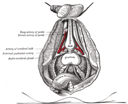
The bulbospongiosus muscles are a subgroup of the superficial muscles of the perineum. They have a slightly different origin, insertion and function in males and females. In males, these muscles cover the bulb of the penis, while in females, they cover the vestibular bulbs.

The internal pudendal artery is one of the three pudendal arteries. It branches off the internal iliac artery, and provides blood to the external genitalia.

In human anatomy, the inferior epigastric artery is an artery that arises from the external iliac artery. It is accompanied by the inferior epigastric vein; inferiorly, these two inferior epigastric vessels together travel within the lateral umbilical fold The inferior epigastric artery then traverses the arcuate line of rectus sheath to enter the rectus sheath, then anastomoses with the superior epigastric artery within the rectus sheath.

The lingual artery arises from the external carotid artery between the superior thyroid artery and facial artery. It can be located easily in the tongue.

The inferior rectal artery is an artery that supplies blood to the lower third of the anal canal below the pectinate line.

The middle colic artery is an artery of the abdomen; a branch of the superior mesenteric artery distributed to parts of the ascending and transverse colon. It usually divides into two terminal branches - a left one and a right one - which go on to form anastomoses with the left colic artery, and right colic artery (respectively), thus participating in the formation of the marginal artery of the colon.

The perineal nerve is a nerve of the pelvis. It arises from the pudendal nerve in the pudendal canal. It gives superficial branches to the skin, and a deep branch to muscles. It supplies the skin and muscles of the perineum. Its latency is tested with electrodes.

The internal pudendal veins are a set of veins in the pelvis. They are the venae comitantes of the internal pudendal artery. Internal pudendal veins are enclosed by pudendal canal, with internal pudendal artery and pudendal nerve.

The suspensory ligament of the penis is a triangular midline structure anchoring the penis to the pubic symphysis, holding the penis close to the pubic bone and supporting it during erection.

The superior rectal artery is an artery that descends into the pelvis to supply blood to the rectum.

The dorsal nasal artery is an artery of the face. It is one of the two terminal branches of the ophthalmic artery. It contributes arterial supply to the lacrimal sac, and outer surface of the nose.

A corpus cavernosum penis (singular) is one of a pair of sponge-like regions of erectile tissue, which contain most of the blood in the penis during an erection.
The dorsal nerve of the penis is the deepest of three divisions of the pudendal nerve; it accompanies the internal pudendal artery along the ramus of the ischium; it then runs forward along the margin of the inferior ramus of the pubis, between the superior and inferior layers of the fascia of the urogenital diaphragm.

The deep artery of the penis is a small collateral branch of the internal pudendal artery that supplies the corpus spongiosum. The artery enters the crus of penis at the crus' anterior extremity.

In human male anatomy, the dorsal veins of the penis are blood vessels that drain the shaft, the skin and the glans of the human penis. They are typically located in the midline on the dorsal aspect of the penis and they comprise the superficial dorsal veinof the penis, that lies in the subcutaneous tissue of the shaft, and the deep dorsal veinof the penis, that lies beneath the deep fascia.

The frenulum of the penis, often known simply as the frenulum or frenum, is a thin elastic strip of tissue on the underside of the glans and the neck of the human penis. In men who are not circumcised, it also connects the foreskin to the glans and the ventral mucosa. In adults, the frenulum is typically supple enough to allow manual movement of the foreskin over the glans and help retract the foreskin during erection. In flaccid state, it tightens to narrow the foreskin opening.

An erection is a physiological phenomenon in which the penis becomes firm, engorged, and enlarged. Penile erection is the result of a complex interaction of psychological, neural, vascular, and endocrine factors, and is often associated with sexual arousal, sexual attraction or libido, although erections can also be spontaneous. The shape, angle, and direction of an erection vary considerably between humans.

In male human anatomy, the foreskin, also known as the prepuce, is the double-layered fold of skin, mucosal and muscular tissue at the distal end of the human penis that covers the glans and the urinary meatus. The foreskin is attached to the glans by an elastic band of tissue, known as the frenulum. The outer skin of the foreskin meets with the inner preputial mucosa at the area of the mucocutaneous junction. The foreskin is mobile, fairly stretchable and sustains the glans in a moist environment. Except for humans, a similar structure known as a penile sheath appears in the male sexual organs of all primates and the vast majority of mammals.


