Left gastric artery

Last updated
Left gastric artery
Gray532.png
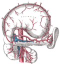
The left gastric artery and other branches of the celiac artery (stomach in situ ). Left gastric artery identified near lesser curvature.
Illu lymph chain08.jpg
Left gastric artery is at #2 -- the upper of the two arrows.
Details
Source Celiac artery
Identifiers
Latin arteria gastrica sinistra
TA98 A12.2.12.013
TA2 4212
FMA 14768
Anatomical terminology

In human anatomy, the left gastric artery arises from the celiac artery and runs along [1] the superior portion of[ citation needed ] the lesser curvature of the stomach before anastomosing with the right gastric artery (which runs right to left[ citation needed ]). It also issues esophageal branches [1] that supply lower esophagus and ascend through the esophageal hiatus to form anastomoses with the esophageal branches of thoracic part of aorta.[ citation needed ]

Contents

Anatomy

Origin

The LGA usually arises from (the superior aspect of) the coeliac trunk - sometimes as a terminal branch of a trifurcation, and more rarely as a side branch of the splenic artery or of common hepatic artery. [2]

Course

From the crus of diaphragm, the LGA arches obliquely anterior-ward and to the left to reach the left curvature of the stomach just inferior to the gastric cardia (thus erecting the gastropancreatic (peritoneal) fold). [2]

Fate

Upon reaching the cardia, the LGA splits into two terminal branches - a ventral one and a dorsal one - which anastomose with corresponding terminal branches of the right gastric artery, together providing arterial supply to the lesser curvature of the stomach. [2]

Branches

Besides its terminal ventral branch and dorsal branch, the LGA yields multiple side branches: the left lateral hepatic artery, posterior esophageal artery, anterior esophagocardiotuberous artery, branches to lymph nodes, and omental branches. [2]

Clinical significance

In terms of disease, the left gastric artery may be involved in peptic ulcer disease: if an ulcer erodes through the stomach mucosa into a branch of the artery, this can cause massive blood loss into the stomach, which may result in such symptoms as hematemesis or melaena.

Additional images

Related Research Articles

<span class="mw-page-title-main">Stomach</span> Digestive organ

The stomach is a muscular, hollow organ in the upper gastrointestinal tract of humans and many other animals, including several invertebrates. The stomach has a dilated structure and functions as a vital organ in the digestive system. The stomach is involved in the gastric phase of digestion, following the cephalic phase in which the sight and smell of food and the act of chewing are stimuli. In the stomach a chemical breakdown of food takes place by means of secreted digestive enzymes and gastric acid.

<span class="mw-page-title-main">Esophagus</span> Vertebrate organ through which food passes to the stomach

The esophagus or oesophagus, colloquially known also as the food pipe, food tube, or gullet, is an organ in vertebrates through which food passes, aided by peristaltic contractions, from the pharynx to the stomach. The esophagus is a fibromuscular tube, about 25 cm (10 in) long in adults, that travels behind the trachea and heart, passes through the diaphragm, and empties into the uppermost region of the stomach. During swallowing, the epiglottis tilts backwards to prevent food from going down the larynx and lungs. The word oesophagus is from Ancient Greek οἰσοφάγος (oisophágos), from οἴσω (oísō), future form of φέρω + ἔφαγον.

<span class="mw-page-title-main">Celiac plexus</span> Complex network of nerves located in the upper abdomen

The celiac plexus, also known as the solar plexus because of its radiating nerve fibers, is a complex network of nerves located in the abdomen, near where the celiac trunk, superior mesenteric artery, and renal arteries branch from the abdominal aorta. It is behind the stomach and the omental bursa, and in front of the crura of the diaphragm, on the level of the first lumbar vertebra.

<span class="mw-page-title-main">Abdominal aorta</span> Largest artery in the abdomen

In human anatomy, the abdominal aorta is the largest artery in the abdominal cavity. As part of the aorta, it is a direct continuation of the descending aorta.

<span class="mw-page-title-main">Splenic artery</span> Artery which supplies blood to the spleen

In human anatomy, the splenic artery or lienal artery, an older term, is the blood vessel that supplies oxygenated blood to the spleen. It branches from the celiac artery, and follows a course superior to the pancreas. It is known for its tortuous path to the spleen.

<span class="mw-page-title-main">Gastroduodenal artery</span>

In anatomy, the gastroduodenal artery is a small blood vessel in the abdomen. It supplies blood directly to the pylorus and proximal part of the duodenum. It also indirectly supplies the pancreatic head.

<span class="mw-page-title-main">Common hepatic artery</span> Artery

The common hepatic artery is a short blood vessel that supplies oxygenated blood to the liver, pylorus of the stomach, duodenum, pancreas, and gallbladder.

<span class="mw-page-title-main">Lesser omentum</span> Double layer of peritoneum in the abdomen

The lesser omentum is the double layer of peritoneum that extends from the liver to the lesser curvature of the stomach, and to the first part of the duodenum. The lesser omentum is usually divided into these two connecting parts: the hepatogastric ligament, and the hepatoduodenal ligament.

<span class="mw-page-title-main">Hepatic artery proper</span>

The hepatic artery proper is the artery that supplies the liver and gallbladder. It raises from the common hepatic artery, a branch of the celiac artery.

<span class="mw-page-title-main">Inferior phrenic arteries</span>

The inferior phrenic artery is a bilaterally paired artery of the abdominal cavity which represents the main source of arterial supply to the diaphragm. Each artery usually arises either from the coeliac trunk or the abdominal aorta, however, their origin is highly variable and the different sites of origin are different for the left artery and right artery. The superior suprarenal artery is a branch of the inferior phrenic artery.

<span class="mw-page-title-main">Right gastroepiploic artery</span>

The right gastroepiploic artery is one of the two terminal branches of the gastroduodenal artery. It runs from right to left along the greater curvature of the stomach, between the layers of the greater omentum, anastomosing with the left gastroepiploic artery, a branch of the splenic artery.

<span class="mw-page-title-main">Left gastroepiploic artery</span>

The left gastroepiploic artery, the largest branch of the splenic artery, runs from left to right about a finger's breadth or more from the greater curvature of the stomach, between the layers of the greater omentum, and anastomoses with the right gastroepiploic.

<span class="mw-page-title-main">Short gastric arteries</span> Small group of arteries from the splenic artery that supply the fundus of the stomach

The short gastric arteries are 5-7 small branches of the splenic artery that pass along part of the greater curvature of the stomach from left to right between the layers of the gastrolienal ligament, and are distributed to the greater curvature of the stomach.

<span class="mw-page-title-main">Right gastric artery</span> Blood vessel of the human digestive system

The right gastric artery usually arises from the proper hepatic artery. It descends to the pyloric end of the stomach before passing from right to left along its lesser curvature, supplying it with branches, and finally anastomosing with the left gastric artery.

<span class="mw-page-title-main">Superior pancreaticoduodenal artery</span> Artery

The superior pancreaticoduodenal artery is an artery that supplies blood to the duodenum and pancreas.

<span class="mw-page-title-main">Foregut</span> Anterior part of the gastrointestinal tract

The foregut in humans is the anterior part of the alimentary canal, from the distal esophagus to the first half of the duodenum, at the entrance of the bile duct. Beyond the stomach, the foregut is attached to the abdominal walls by mesentery. The foregut arises from the endoderm, developing from the folding primitive gut, and is developmentally distinct from the midgut and hindgut. Although the term “foregut” is typically used in reference to the anterior section of the primitive gut, components of the adult gut can also be described with this designation. Pain in the epigastric region, just below the intersection of the ribs, typically refers to structures in the adult foregut.

<span class="mw-page-title-main">Greater omentum</span> Fat sheath under abdominal wall

The greater omentum is a large apron-like fold of visceral peritoneum that hangs down from the stomach. It extends from the greater curvature of the stomach, passing in front of the small intestines and doubles back to ascend to the transverse colon before reaching to the posterior abdominal wall. The greater omentum is larger than the lesser omentum, which hangs down from the liver to the lesser curvature. The common anatomical term "epiploic" derives from "epiploon", from the Greek epipleein, meaning to float or sail on, since the greater omentum appears to float on the surface of the intestines. It is the first structure observed when the abdominal cavity is opened anteriorly.

In anatomy, arterial tree is used to refer to all arteries and/or the branching pattern of the arteries. This article regards the human arterial tree. Starting from the aorta:

<span class="mw-page-title-main">Anterior vagal trunk</span>

The anterior vagal trunk is one of the two divisions into which the vagus nerve splits as it passes through the esophageal hiatus to enter the abdominal cavity. The anterior and posterior vagal trunks represent the inferior continuation of the esophageal nervous plexus inferior to the diaphragm. The majority of nerve fibres in the anterior vagal trunk are derived from the left vagus nerve.

<span class="mw-page-title-main">Curvatures of the stomach</span> Anatomy of the human stomach

The curvatures of the stomach are the long, convex, lateral surface, and the shorter, concave, medial surface of the stomach, which are referred to as the greater and lesser curvatures, respectively. The greater curvature, which begins at the cardiac notch, and arches backwards, passing inferiorly to the left, is four or five times longer than the lesser curvature, which attaches to the hepatogastric ligament and is supplied by the left gastric artery and right gastric branch of the hepatic artery.

References

  1. 1 2 Lung, Kirsten; Lui, Forshing (2022), "Anatomy, Abdomen and Pelvis, Arteries", StatPearls, Treasure Island (FL): StatPearls Publishing, PMID   30247834 , retrieved 2023-01-14
  2. 1 2 3 4 "Artère gastrique gauche - Dictionnaire médical de l'Académie de Médecine". www.academie-medecine.fr. Retrieved 2024-05-09.
  3. Essential Clinical Anatomy. K.L. Moore & A.M. Agur. Lippincott, 2 ed. 2002. Page 150