Internal pudendal artery

Last updated
Internal pudendal artery
Internal iliac branches.PNG
Internal iliac artery with branches, including internal pudendal artery.
Gray542.png
The superficial branches of the internal pudendal artery.
Details
Source Internal iliac artery
Branches Inferior rectal artery and others
Vein Internal pudendal veins
Supplies External genitalia, perineum
Identifiers
Latin arteria pudenda interna
TA98 A12.2.15.038
TA2 4341
FMA 18835
Anatomical terminology

The internal pudendal artery is one of the three pudendal arteries. It branches off the internal iliac artery, and provides blood to the external genitalia.

Contents

Structure

The internal pudendal artery is the terminal branch of the anterior trunk of the internal iliac artery. [1] It is smaller in the female than in the male.

Path

It arises from the anterior division of internal iliac artery. It runs on the lateral pelvic wall. It exits the pelvic cavity through the greater sciatic foramen, inferior to the piriformis muscle, to enter the gluteal region.

It then curves around the sacrospinous ligament to enter the perineum through the lesser sciatic foramen.

It travels through the pudendal canal with the internal pudendal veins and the pudendal nerve.

Branches

The internal pudendal artery gives off the following branches:

In femalesIn malesDescription
Inferior rectal artery Inferior rectal artery to anal canal
Perineal artery Perineal artery supplies transversus perinei superficialis muscle
Posterior labial branches Posterior scrotal branches -
Artery of bulb of vestibule Artery of bulb of penis [1] supplies bulb of vestibule/bulb of penis
Dorsal artery of clitoris Dorsal artery of the penis [1] -
Deep artery of clitoris Deep artery of the penis [1] to corpus cavernosum penis/clitoridis

The deep artery of clitoris is a branch of the internal pudendal artery and supplies the clitoral crura. Another branch of the internal pudendal artery is the dorsal artery of clitoris.

Some sources consider the urethral artery a direct branch of the internal pudendal artery, [2] while others consider it a branch of the perineal artery.[ citation needed ]

In males, the internal pudendal artery also gives rise to the perforating arteries of the penis. [1]

Variation

Around 70% of men have an accessory internal pudendal artery. [1] This usually does not originate from the internal iliac artery, instead originating from the external iliac artery, the obturator artery, or the vesical arteries. [1]

Function

The internal pudendal artery supplies blood to the external genitalia.

Clinical significance

In women, the internal pudendal artery may be damaged during childbirth. [3] This may cause a haematoma, which usually resolves without treatment, but may form an infected abscess. [3]

Additional images

See also

Related Research Articles

<span class="mw-page-title-main">Pudendal nerve</span> Main nerve of the perineum

The pudendal nerve is the main nerve of the perineum. It is a mixed nerve and also conveys sympathetic autonomic fibers. It carries sensation from the external genitalia of both sexes and the skin around the anus and perineum, as well as the motor supply to various pelvic muscles, including the male or female external urethral sphincter and the external anal sphincter.

<span class="mw-page-title-main">Ischiocavernosus muscle</span> Sheet of fibrous tissue between the thigh and the front of the perineum

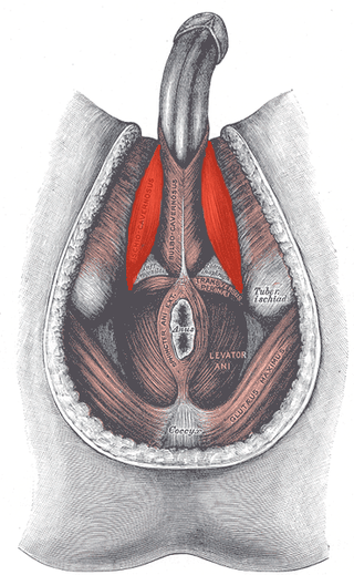
The ischiocavernosus muscle is a muscle just below the surface of the perineum, present in both men and women.

<span class="mw-page-title-main">Bulbospongiosus muscle</span> Superficial muscle of the perineum

The bulbospongiosus muscles are a subgroup of the superficial muscles of the perineum. They have a slightly different origin, insertion and function in males and females. In males, these muscles cover the bulb of the penis, while in females, they cover the vestibular bulbs.

<span class="mw-page-title-main">External iliac artery</span> Arteries of the pelvis

The external iliac arteries are two major arteries which bifurcate off the common iliac arteries anterior to the sacroiliac joint of the pelvis.

<span class="mw-page-title-main">Internal iliac artery</span> Main artery of the pelvis

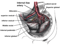
The internal iliac artery is the main artery of the pelvis.

<span class="mw-page-title-main">Inferior epigastric artery</span> Blood vessel

In human anatomy, the inferior epigastric artery is an artery that arises from the external iliac artery. It is accompanied by the inferior epigastric vein; inferiorly, these two inferior epigastric vessels together travel within the lateral umbilical fold The inferior epigastric artery then traverses the arcuate line of rectus sheath to enter the rectus sheath, then anastomoses with the superior epigastric artery within the rectus sheath.

<span class="mw-page-title-main">Sacrotuberous ligament</span>

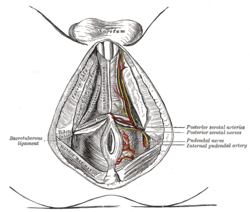
The sacrotuberous ligament is situated at the lower and back part of the pelvis. It is flat, and triangular in form; narrower in the middle than at the ends.

<span class="mw-page-title-main">Sacrospinous ligament</span>

The sacrospinous ligament is a thin, triangular ligament in the human pelvis. The base of the ligament is attached to the outer edge of the sacrum and coccyx, and the tip of the ligament attaches to the spine of the ischium, a bony protuberance on the human pelvis. Its fibres are intermingled with the sacrotuberous ligament.

<span class="mw-page-title-main">Inferior gluteal artery</span>

The inferior gluteal artery is a terminal branch of the anterior trunk of the internal iliac artery. It exits the pelvis through the greater sciatic foramen. It is distributed chiefly to the buttock and the back of the thigh.

<span class="mw-page-title-main">Inferior rectal artery</span>

The inferior rectal artery is an artery that supplies blood to the lower third of the anal canal below the pectinate line.

<span class="mw-page-title-main">Perineal nerve</span> Nerve of the perineum

The perineal nerve is a nerve of the pelvis. It arises from the pudendal nerve in the pudendal canal. It gives superficial branches to the skin, and a deep branch to muscles. It supplies the skin and muscles of the perineum. Its latency is tested with electrodes.

<span class="mw-page-title-main">Internal pudendal veins</span> Set of veins in the pelvis enclosed by pudendal canal

The internal pudendal veins are a set of veins in the pelvis. They are the venae comitantes of the internal pudendal artery. Internal pudendal veins are enclosed by pudendal canal, with internal pudendal artery and pudendal nerve.

<span class="mw-page-title-main">Ischioanal fossa</span> Wedge-shaped space located lateral to the anal canal and inferior to the pelvic diaphragm

The ischioanal fossa is the fat-filled wedge-shaped space located lateral to the anal canal and inferior to the pelvic diaphragm. It is somewhat prismatic in shape, with its base directed to the surface of the perineum and its apex at the line of meeting of the obturator and anal fasciae.

<span class="mw-page-title-main">Superior rectal artery</span>

The superior rectal artery is an artery that descends into the pelvis to supply blood to the rectum.

<span class="mw-page-title-main">Internal iliac vein</span> Large blood vessel of the pelvis

The internal iliac vein begins near the upper part of the greater sciatic foramen, passes upward behind and slightly medial to the internal iliac artery and, at the brim of the pelvis, joins with the external iliac vein to form the common iliac vein.

<span class="mw-page-title-main">Inferior anal nerves</span>

The inferior rectal nerves usually branch from the pudendal nerve but occasionally arises directly from the sacral plexus; they cross the ischiorectal fossa along with the inferior rectal artery and veins, toward the anal canal and the lower end of the rectum, and is distributed to the sphincter ani externus and to the integument (skin) around the anus.

<span class="mw-page-title-main">Dorsal nerve of the penis</span> Deepest division of the pudendal nerve

The dorsal nerve of the penis is the deepest of three divisions of the pudendal nerve; it accompanies the internal pudendal artery along the ramus of the ischium; it then runs forward along the margin of the inferior ramus of the pubis, between the superior and inferior layers of the fascia of the urogenital diaphragm.

<span class="mw-page-title-main">Superficial perineal pouch</span>

The superficial perineal pouch is a compartment of the perineum.

<span class="mw-page-title-main">Deep perineal pouch</span> Anatomic space enclosed partly by the perineum

The deep perineal pouch is the anatomic space enclosed in part by the perineum and located superior to the perineal membrane.

<span class="mw-page-title-main">Dorsal nerve of the clitoris</span>

The dorsal nerve of the clitoris is a nerve in females that branches off the pudendal nerve to innervate the clitoris. The nerve is important for female sexual pleasure, and it may play a role in clitoral erections.

References

  1. 1 2 3 4 5 6 7 Keegan, Kirk A.; Penson, David F. (2013-01-01), Creager, Mark A.; Beckman, Joshua A.; Loscalzo, Joseph (eds.), "Chapter 28 - Vasculogenic Erectile Dysfunction", Vascular Medicine: A Companion to Braunwald's Heart Disease (Second Edition), Philadelphia: W.B. Saunders, pp. 341–348, ISBN   978-1-4377-2930-6 , retrieved 2021-01-14
  2. "Internal pudendal artery" at Dorland's Medical Dictionary
  3. 1 2 Gilbert, Robert O.; Cable, Christina; Fubini, Susan L.; Steiner, Adrian (2017-01-01), Fubini, Susan L.; Ducharme, Norm G. (eds.), "Chapter 16 - Surgery of the Bovine Reproductive System and Urinary Tract", Farm Animal Surgery (Second Edition), W.B. Saunders, pp. 439–503, ISBN   978-0-323-31665-1 , retrieved 2021-01-14