
The urethra is a tube that connects the mammalian urinary bladder to the urinary meatus in the glans penis or vulval vestibule. Male and female placental mammals release urine through the urethra during urination, but males also release semen through the urethra during ejaculation.

In male human anatomy, the glans penis or penile glans, commonly referred to as the glans, is the bulbous structure at the distal end of the human penis that is the human male's most sensitive erogenous zone and primary anatomical source of sexual pleasure. The glans penis is present in the male reproductive organs of humans and most other mammals where it may appear smooth, spiny, elongated or divided. It is externally lined with mucosal tissue, which creates a smooth texture and glossy appearance. In humans, the glans is located over the distal ends of the corpora cavernosa and is a continuation of the corpus spongiosum of the penis. At the summit appears the urinary meatus and at the base forms the corona glandis. An elastic band of tissue, known as the frenulum, runs on its ventral surface. In men who are not circumcised, it is completely or partially covered by a fold of skin called the foreskin. In adults, the foreskin can generally be retracted over and past the glans manually or sometimes automatically during an erection.
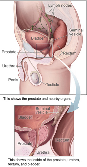
The prostate is both an accessory gland of the male reproductive system and a muscle-driven mechanical switch between urination and ejaculation. It is found in all male mammals. It differs between species anatomically, chemically, and physiologically. Anatomically, the prostate is found below the bladder, with the urethra passing through it. It is described in gross anatomy as consisting of lobes and in microanatomy by zone. It is surrounded by an elastic, fibromuscular capsule and contains glandular tissue, as well as connective tissue.

The pudendal nerve is the main nerve of the perineum. It is a mixed nerve and also conveys sympathetic autonomic fibers. It carries sensation from the external genitalia of both sexes and the skin around the anus and perineum, as well as the motor supply to various pelvic muscles, including the male or female external urethral sphincter and the external anal sphincter.

The corpus spongiosum is the mass of spongy tissue surrounding the male urethra within the penis. It is also called the corpus cavernosum urethrae in older texts.

The bulbospongiosus muscles are a subgroup of the superficial muscles of the perineum. They have a slightly different origin, insertion and function in males and females. In males, these muscles cover the bulb of the penis, while in females, they cover the vestibular bulbs.

The internal pudendal artery is one of the three pudendal arteries. It branches off the internal iliac artery, and provides blood to the external genitalia.

The external anal sphincter is an oval tube of skeletal muscle fibers. Distally, it is adherent to the skin surrounding the margin of the anus. It exhibits a resting state of tonical contraction and also contracts during the bulbospongiosus reflex.

The external sphincter muscle of male urethra, also sphincter urethrae membranaceae, sphincter urethrae externus, surrounds the whole length of the membranous urethra, and is enclosed in the fascia of the urogenital diaphragm.

The internal pudendal veins are a set of veins in the pelvis. They are the venae comitantes of the internal pudendal artery. Internal pudendal veins are enclosed by pudendal canal, with internal pudendal artery and pudendal nerve.

A corpus cavernosum penis (singular) is one of a pair of sponge-like regions of erectile tissue, which contain most of the blood in the penis during an erection.

The artery of bulb of penis is a short artery of large caliber which arises from the internal pudendal artery between the two layers of fascia of the urogenital diaphragm. It passes medialward, pierces the inferior fascia of the urogenital diaphragm and gives off branches which ramify in the bulb of the urethra and in the posterior part of the corpus spongiosum.
The deep artery of the penis is a branch of the internal pudendal artery that supplies the corpus spongiosum. The artery enters the crus of penis at its anterior extremity.
The perineal membrane is an anatomical term for a fibrous membrane in the perineum. The term "inferior fascia of urogenital diaphragm", used in older texts, is considered equivalent to the perineal membrane.

The urogenital triangle is the anterior part of the perineum. In female mammals, it contains the vagina and associated parts of the internal genitalia.

The spongy urethra is the longest part of the male urethra, and is contained in the corpus spongiosum of the penis.

The membranous urethra or intermediate part of male urethra is the shortest, least dilatable, and, with the exception of the urinary meatus, the narrowest part of the urethra.
In human male anatomy, the dorsal veins of the penis are blood vessels that drain the shaft, the skin and the glans of the human penis. They are typically located in the midline on the dorsal aspect of the penis and they comprise the superficial dorsal veinof the penis, that lies in the subcutaneous tissue of the shaft, and the deep dorsal veinof the penis, that lies beneath the deep fascia.
The urethral sphincters are two muscles used to control the exit of urine in the urinary bladder through the urethra. The two muscles are either the male or female external urethral sphincter and the internal urethral sphincter. When either of these muscles contracts, the urethra is sealed shut.

The external sphincter muscle of female urethra is a muscle which controls urination in females. The muscle fibers arise on either side from the margin of the inferior ramus of the pubis. They are directed across the pubic arch in front of the urethra, and pass around it to blend with the muscular fibers of the opposite side, between the urethra and vagina.
