Hepatic artery proper

Last updated
Hepatic artery proper
Common and proper hepatic artery.png
The hepatic artery proper branches from the common hepatic artery.
Gray1086-liver.PNG
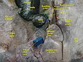
Inferior surface of the liver. (Entrance for hepatic artery labeled at bottom center.)
Details
Source Common hepatic artery
Identifiers
Latin arteria hepatica propria
TA98 A12.2.12.029
TA2 4227
FMA 14772
Anatomical terminology

The hepatic artery proper (also proper hepatic artery) is the artery that supplies the liver and gallbladder. It raises from the common hepatic artery, a branch of the celiac artery.

Contents

Structure

The hepatic artery proper arises from the common hepatic artery and runs alongside the portal vein and the common bile duct to form the portal triad. A branch of the common hepatic artery –the gastroduodenal artery gives off the small supraduodenal artery to the duodenal bulb. Then the right gastric artery comes off and runs to the left along the lesser curvature of the stomach to meet the left gastric artery, which is a branch of the celiac trunk. It subsequently bifurcates into the right and left hepatic arteries.

Variant anatomy

Of note, the right and left hepatic arteries may demonstrate variant anatomy. A misplaced right hepatic artery may arise from the superior mesenteric artery (SMA) and a misplaced left hepatic artery may arise from the left gastric artery. The cystic artery generally comes from the right hepatic artery. [1]

Other variants of right hepatic artery includes: arising directly from the proximal or middle part of common hepatic artery, gastroduodenal artery, superior mesenteric artery, celiac axis, aorta, splenic artery, or left gastric artery instead of arising from proper hepatic artery. [2]

Additional images

Related Research Articles

<span class="mw-page-title-main">Portal vein</span> Short thick vein formed by the union of the superior mesenteric vein and the splenic vein

The portal vein or hepatic portal vein (HPV) is a blood vessel that carries blood from the gastrointestinal tract, gallbladder, pancreas and spleen to the liver. This blood contains nutrients and toxins extracted from digested contents. Approximately 75% of total liver blood flow is through the portal vein, with the remainder coming from the hepatic artery proper. The blood leaves the liver to the heart in the hepatic veins.

<span class="mw-page-title-main">Celiac plexus</span> Complex network of nerves located in the upper abdomen

The celiac plexus, also known as the solar plexus because of its radiating nerve fibers, is a complex network of nerves located in the abdomen, near where the celiac trunk, superior mesenteric artery, and renal arteries branch from the abdominal aorta. It is behind the stomach and the omental bursa, and in front of the crura of the diaphragm, on the level of the first lumbar vertebra.

<span class="mw-page-title-main">Abdominal aorta</span> Largest artery in the abdomen

In human anatomy, the abdominal aorta is the largest artery in the abdominal cavity. As part of the aorta, it is a direct continuation of the descending aorta.

<span class="mw-page-title-main">Celiac artery</span> First major branch of the abdominal aorta

The celiacartery, also known as the celiac trunk or truncus coeliacus, is the first major branch of the abdominal aorta. It is about 1.25 cm in length. Branching from the aorta at thoracic vertebra 12 (T12) in humans, it is one of three anterior/ midline branches of the abdominal aorta.

<span class="mw-page-title-main">Superior mesenteric artery</span> Artery which supplies blood to the intestines and pancreas

In human anatomy, the superior mesenteric artery (SMA) is an artery which arises from the anterior surface of the abdominal aorta, just inferior to the origin of the celiac trunk, and supplies blood to the intestine from the lower part of the duodenum through two-thirds of the transverse colon, as well as the pancreas.

<span class="mw-page-title-main">Splenic artery</span> Artery which supplies blood to the spleen

In human anatomy, the splenic artery or lienal artery, an older term, is the blood vessel that supplies oxygenated blood to the spleen. It branches from the celiac artery, and follows a course superior to the pancreas. It is known for its tortuous path to the spleen.

<span class="mw-page-title-main">Gastroduodenal artery</span>

In anatomy, the gastroduodenal artery is a small blood vessel in the abdomen. It supplies blood directly to the pylorus and proximal part of the duodenum. It also indirectly supplies the pancreatic head.

<span class="mw-page-title-main">Common hepatic artery</span> Artery

The common hepatic artery is a short blood vessel that supplies oxygenated blood to the liver, pylorus of the stomach, duodenum, pancreas, and gallbladder.

<span class="mw-page-title-main">Lesser omentum</span> Double layer of peritoneum in the abdomen

The lesser omentum is the double layer of peritoneum that extends from the liver to the lesser curvature of the stomach, and to the first part of the duodenum. The lesser omentum is usually divided into these two connecting parts: the hepatogastric ligament, and the hepatoduodenal ligament.

<span class="mw-page-title-main">Left gastric artery</span> Artery

In human anatomy, the left gastric artery arises from the celiac artery and runs along the superior portion of the lesser curvature of the stomach before anastomosing with the right gastric artery. It also issues esophageal branches that supply lower esophagus and ascend through the esophageal hiatus to form anastomoses with the esophageal branches of thoracic part of aorta.

<span class="mw-page-title-main">Cystic artery</span>

The cystic artery is (usually) a branch of the right hepatic artery that provides arterial supply to the gallbladder and contributes arterial supply to the extrahepatic bile ducts.

<span class="mw-page-title-main">Inferior phrenic arteries</span>

The inferior phrenic artery is a bilaterally paired artery of the abdominal cavity which represents the main source of arterial supply to the diaphragm. Each artery usually arises either from the coeliac trunk or the abdominal aorta, however, their origin is highly variable and the different sites of origin are different for the left artery and right artery. The superior suprarenal artery is a branch of the inferior phrenic artery.

<span class="mw-page-title-main">Right gastroepiploic artery</span>

The right gastroepiploic artery is one of the two terminal branches of the gastroduodenal artery. It runs from right to left along the greater curvature of the stomach, between the layers of the greater omentum, anastomosing with the left gastroepiploic artery, a branch of the splenic artery.

<span class="mw-page-title-main">Left gastroepiploic artery</span>

The left gastroepiploic artery, the largest branch of the splenic artery, runs from left to right about a finger's breadth or more from the greater curvature of the stomach, between the layers of the greater omentum, and anastomoses with the right gastroepiploic.

<span class="mw-page-title-main">Middle colic artery</span> Artery supplying the colon

The middle colic artery is an artery of the abdomen; a branch of the superior mesenteric artery distributed to parts of the ascending and transverse colon. It usually divides into two terminal branches - a left one and a right one - which go on to form anastomoses with the left colic artery, and right colic artery (respectively), thus participating in the formation of the marginal artery of the colon.

<span class="mw-page-title-main">Right gastric artery</span> Blood vessel of the human digestive system

The right gastric artery usually arises from the proper hepatic artery. It descends to the pyloric end of the stomach before passing from right to left along its lesser curvature, supplying it with branches, and finally anastomosing with the left gastric artery.

<span class="mw-page-title-main">Superior pancreaticoduodenal artery</span> Artery

The superior pancreaticoduodenal artery is an artery that supplies blood to the duodenum and pancreas.

<span class="mw-page-title-main">Greater omentum</span> Fat sheath under abdominal wall

The greater omentum is a large apron-like fold of visceral peritoneum that hangs down from the stomach. It extends from the greater curvature of the stomach, passing in front of the small intestines and doubles back to ascend to the transverse colon before reaching to the posterior abdominal wall. The greater omentum is larger than the lesser omentum, which hangs down from the liver to the lesser curvature. The common anatomical term "epiploic" derives from "epiploon", from the Greek epipleein, meaning to float or sail on, since the greater omentum appears to float on the surface of the intestines. It is the first structure observed when the abdominal cavity is opened anteriorly.

<span class="mw-page-title-main">Hepatic plexus</span>

The hepatic plexus is a sympathetic and parasympathetic nerve plexus that provides innervation to the parenchyma of the liver as well as contributing innervation to some other abdominal structures.

<span class="mw-page-title-main">Uncinate process of pancreas</span>

The uncinate process is a small part of the pancreas. The uncinate process is the formed prolongation of the angle of junction of the lower and left lateral borders in the head of the pancreas. The word "uncinate" comes from the Latin "uncinatus", meaning "hooked".

References

  1. Balija, M; Huis, M; Nikolic, V; Stulhofer, M (1999). "Laparoscopic visualization of the cystic artery anatomy". World Journal of Surgery. 23 (7): 703–7, discussion 707. doi:10.1007/pl00012372. PMID   10390590. S2CID   23174989.
  2. Choi TW, Chung JW, Kim HC, Lee M, Choi JW, Jae HJ, Hur S (August 2021). "Anatomic Variations of the Hepatic Artery in 5625 Patients". Radiology: Cardiothoracic Imaging. 3 (4): e210007. doi:10.1148/ryct.2021210007. PMC   8415139 . PMID   34498005.