Superior mesenteric artery

Last updated
Superior mesenteric artery
Superior mesenteric a.gif
Frontal view of the superior mesenteric artery and its branches. The large vessel (blue) beside the SMA is the superior mesenteric vein. A considerable number of different branching patterns exist.
3D rendered CT of abdominal aortic branches and kidneys.svg
3D-rendered computed tomography of abdominal aortic branches, showing exit of superior mesenteric artery between the kidneys.
Details
Precursor Vitelline arteries
Source Abdominal aorta
Branches Inferior pancreaticoduodenal
middle colic
right colic
intestinal branches (jejunal, ileal)
ileocolic Marginal artery of the colon
Vein Superior mesenteric vein
Supplies Intestine
Identifiers
Latin arteria mesenterica superior
MeSH D017538
TA98 A12.2.12.053
TA2 4252
FMA 14749
Anatomical terminology

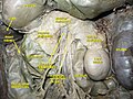
In human anatomy, the superior mesenteric artery (SMA) is an artery which arises from the anterior surface of the abdominal aorta, just inferior to the origin of the celiac trunk, and supplies blood to the intestine from the lower part of the duodenum through two-thirds of the transverse colon, as well as the pancreas.

Contents

Structure

Origin

In the adult, the SMA arises anterior to inferior border of vertebra L1 (transpyloric plane). It is usually 1 cm lower than the celiac trunk.

Course and relations

It initially travels in an anterior/inferior direction, passing behind/under the neck of the pancreas and the splenic vein. Located under this portion of the superior mesenteric artery, between it and the aorta, are the following:

The SMA typically runs to the left of its associated vein, the superior mesenteric vein. After passing the neck of the pancreas it starts giving off its branches.

Branches

BranchSupplies
inferior pancreaticoduodenal artery head of the pancreas and to the ascending and inferior parts of the duodenum (proximal loop)
intestinal arteries branches to ileum, branches to jejunum (proximal loop)
ileocolic artery supplies last part of ileum, cecum, and appendix (distal loop)
right colic artery to ascending colon (distal loop)
middle colic artery to the transverse colon (distal loop)

The number of arterial arcades in the ileum is more than the number of arcades in the jejunum. [1]

The middle, right, and ileocecal branches anastomose with each other to form a marginal artery along the inner border of the colon. This artery is completed by branches of the left colic which is a branch of the inferior mesenteric artery.

Clinical significance

Additional images

Related Research Articles

<span class="mw-page-title-main">Duodenum</span> First section of the small intestine

The duodenum is the first section of the small intestine in most higher vertebrates, including mammals, reptiles, and birds. In mammals it may be the principal site for iron absorption. The duodenum precedes the jejunum and ileum and is the shortest part of the small intestine.

<span class="mw-page-title-main">Abdominal aorta</span> Largest artery in the abdomen

In human anatomy, the abdominal aorta is the largest artery in the abdominal cavity. As part of the aorta, it is a direct continuation of the descending aorta.

<span class="mw-page-title-main">Renal artery</span> Vessel supplying blood to kidney

The renal arteries are paired arteries that supply the kidneys with blood. Each is directed across the crus of the diaphragm, so as to form nearly a right angle.

<span class="mw-page-title-main">Renal vein</span> Short thick veins which return blood from the kidneys to the vena cava

The renal veins in the renal circulation, are large-calibre veins that drain blood filtered by the kidneys into the inferior vena cava. There is one renal vein draining each kidney. Each renal vein is formed by the convergence of the interlobar veins of one kidney.

<span class="mw-page-title-main">Celiac artery</span> First major branch of the abdominal aorta

The celiacartery, also known as the celiac trunk or truncus coeliacus, is the first major branch of the abdominal aorta. It is about 1.25 cm in length. Branching from the aorta at thoracic vertebra 12 (T12) in humans, it is one of three anterior/ midline branches of the abdominal aorta.

The Kocher manoeuvre is a surgical procedure to expose structures in the retroperitoneum behind the duodenum and pancreas. In vascular surgery, it is described as a method to expose the abdominal aorta. It usually has been in contrast to midline laparotomy and right retroperitoneal space dissection. These two procedures have been used for diverse cases, but have approximately equivalent outcomes.

<span class="mw-page-title-main">Inferior mesenteric artery</span>

In human anatomy, the inferior mesenteric artery (IMA) is the third main branch of the abdominal aorta and arises at the level of L3, supplying the large intestine from the distal transverse colon to the upper part of the anal canal. The regions supplied by the IMA are the descending colon, the sigmoid colon, and part of the rectum.

<span class="mw-page-title-main">Gastroduodenal artery</span>

In anatomy, the gastroduodenal artery is a small blood vessel in the abdomen. It supplies blood directly to the pylorus and proximal part of the duodenum. It also indirectly supplies the pancreatic head.

<span class="mw-page-title-main">Superior mesenteric vein</span> Vein which drains blood from the small intestine

In human anatomy, the superior mesenteric vein (SMV) is a blood vessel that drains blood from the small intestine. Behind the neck of the pancreas, the superior mesenteric vein combines with the splenic vein to form the portal vein that carries blood to the liver. The superior mesenteric vein lies to the right of the similarly named artery, the superior mesenteric artery, which originates from the abdominal aorta.

<span class="mw-page-title-main">Internal iliac artery</span> Main artery of the pelvis

The internal iliac artery is the main artery of the pelvis.

<span class="mw-page-title-main">Hepatic artery proper</span>

The hepatic artery proper is the artery that supplies the liver and gallbladder. It raises from the common hepatic artery, a branch of the celiac artery.

<span class="mw-page-title-main">Middle colic artery</span> Artery supplying the colon

The middle colic artery is an artery of the abdomen; a branch of the superior mesenteric artery distributed to parts of the ascending and transverse colon. It usually divides into two terminal branches - a left one and a right one - which go on to form anastomoses with the left colic artery, and right colic artery (respectively), thus participating in the formation of the marginal artery of the colon.

<span class="mw-page-title-main">Left colic artery</span> Artery

The left colic artery is a branch of the inferior mesenteric artery distributed to the descending colon, and left part of the transverse colon. It ends by dividing into an ascending branch and a descending branch; the terminal branches of the two branches go on to form anastomoses with the middle colic artery, and a sigmoid artery (respectively).

<span class="mw-page-title-main">Ileocolic artery</span> Artery in the abdomen

The ileocolic artery is the lowest branch arising from the concavity of the superior mesenteric artery. It supplies the cecum, ileum, and appendix.

<span class="mw-page-title-main">Superior pancreaticoduodenal artery</span> Artery

The superior pancreaticoduodenal artery is an artery that supplies blood to the duodenum and pancreas.

<span class="mw-page-title-main">Inferior pancreaticoduodenal artery</span> Branch of the superior mesenteric artery that supplies parts of the pancreas and the duodenum

The inferior pancreaticoduodenal artery is a branch of the superior mesenteric artery. It supplies the head of the pancreas, and the ascending and inferior parts of the duodenum. Rarely, it may have an aneurysm.

<span class="mw-page-title-main">Greater omentum</span> Fat sheath under abdominal wall

The greater omentum is a large apron-like fold of visceral peritoneum that hangs down from the stomach. It extends from the greater curvature of the stomach, passing in front of the small intestines and doubles back to ascend to the transverse colon before reaching to the posterior abdominal wall. The greater omentum is larger than the lesser omentum, which hangs down from the liver to the lesser curvature. The common anatomical term "epiploic" derives from "epiploon", from the Greek epipleein, meaning to float or sail on, since the greater omentum appears to float on the surface of the intestines. It is the first structure observed when the abdominal cavity is opened anteriorly.

<span class="mw-page-title-main">Testicular vein</span>

The testicular vein, the male gonadal vein, carries deoxygenated blood from its corresponding testis to the inferior vena cava or one of its tributaries. It is the male equivalent of the ovarian vein, and is the venous counterpart of the testicular artery.

<span class="mw-page-title-main">Superior mesenteric artery syndrome</span> Medical condition

Superior mesenteric artery (SMA) syndrome is a gastro-vascular disorder in which the third and final portion of the duodenum is compressed between the abdominal aorta (AA) and the overlying superior mesenteric artery. This rare, potentially life-threatening syndrome is typically caused by an angle of 6–25° between the AA and the SMA, in comparison to the normal range of 38–56°, due to a lack of retroperitoneal and visceral fat. In addition, the aortomesenteric distance is 2–8 millimeters, as opposed to the typical 10–20. However, a narrow SMA angle alone is not enough to make a diagnosis, because patients with a low BMI, most notably children, have been known to have a narrow SMA angle with no symptoms of SMA syndrome.

The superior mesenteric vessels are composed of the superior mesenteric artery and the superior mesenteric vein.

References

  1. Conley, Dylan; Hurst, Peter R.; Stringer, Mark D. (March 2010). "An investigation of human jejunal and ileal arteries". Anatomical Science International. 85 (1): 23–30. doi: 10.1007/s12565-009-0047-9 . ISSN   1447-6959. PMID   19488686. S2CID   7926213.
  2. Redaelli CA, Schilling MK, Büchler MW (1998). "Intraoperative laser Doppler flowmetry: a predictor of ischemic injury in acute mesenteric infarction". Digestive Surgery. 15 (1): 55–9. doi:10.1159/000018587. ISSN   0253-4886. PMID   9845564. S2CID   46785333.