
In human anatomy, the superior mesenteric artery (SMA) arises from the anterior surface of the abdominal aorta, just inferior to the origin of the celiac trunk, and supplies the intestine from the lower part of the duodenum through two-thirds of the transverse colon, as well as the pancreas.

In human anatomy, the inferior mesenteric artery, often abbreviated as IMA, is the third main branch of the abdominal aorta and arises at the level of L3, supplying the large intestine from the left colic flexure to the upper part of the rectum, which includes the descending colon, the sigmoid colon, and part of the rectum. Proximally, its territory of distribution overlaps with the middle colic artery, and therefore the superior mesenteric artery. The SMA and IMA anastomose via the marginal artery of the colon and via Riolan's arcade. The territory of distribution of the IMA is more or less equivalent to the embryonic hindgut.

In anatomy, the gastroduodenal artery is a small blood vessel in the abdomen. It supplies blood directly to the pylorus and proximal part of the duodenum, and indirectly to the pancreatic head.

The superior mesenteric vein is a blood vessel that drains blood from the small intestine. At its termination behind the neck of the pancreas, the superior mesenteric vein combines with the splenic vein to form the hepatic portal vein. The superior mesenteric vein lies to the right of the similarly named artery, the superior mesenteric artery, which originates from the abdominal aorta.

The splenic vein is a blood vessel that drains blood from the spleen, the stomach fundus and part of the pancreas. It is part of the hepatic portal system.
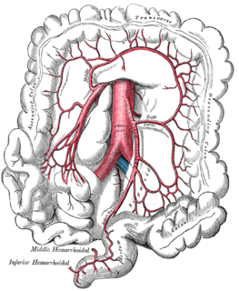
In human anatomy, the marginal artery of the colon, also known as the marginal artery of Drummond and artery of Drummond is an artery that connects the inferior mesenteric artery with the superior mesenteric artery. It is sometimes absent, as an anatomical variant.

In human anatomy, inferior epigastric artery refers to the artery that arises from the external iliac artery and anastomoses with the superior epigastric artery. Along its course, it is accompanied by a similarly named vein, the inferior epigastric vein. These epigastric vessels form the lateral border of Hesselbach's triangle, which outlines the area through which direct inguinal hernias protrude.
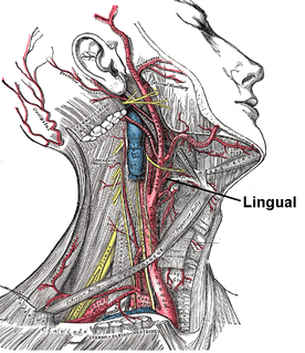
The lingual artery arises from the external carotid between the superior thyroid artery and facial artery. It can be located easily in the tongue.

The middle rectal artery is an artery in the pelvis that supplies blood to the rectum.
The right gastroepiploic artery is one of the two terminal branches of the gastroduodenal artery. It runs from right to left along the greater curvature of the stomach, between the layers of the greater omentum, anastomosing with the left gastroepiploic artery, a branch of the splenic artery.
The sigmoid arteries, two or three in number, run obliquely downward and to the left behind the peritoneum and in front of the psoas major, ureter, and internal spermatic vessels. They originate from the inferior mesenteric artery branch of the abdominal aorta.
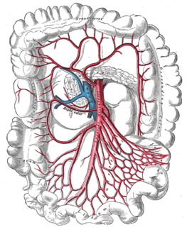
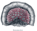
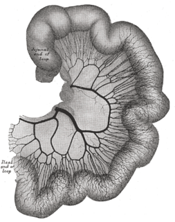
The right colic artery arises from about the middle of the concavity of the superior mesenteric artery, or from a stem common to it and the ileocolic.

The middle colic artery is a branch of the superior mesenteric artery that mostly supplies the transverse colon. It arises just below the pancreas. It passes inferiorly and anteriorly between the layers of the transverse mesocolon, and divides into left and right branches. The right branch anastomoses with the right colic artery, and the left anastomoses with the left colic artery, a branch of the inferior mesenteric artery. This sequence of anastomses are frequently referred to as the marginal artery of the colon.
The left colic artery is a branch of the inferior mesenteric artery that runs to the left behind the peritoneum and in front of the psoas major muscle, and after a short, but variable, course divides into an ascending and a descending branch; the stem of the artery or its branches cross the left ureter and left internal spermatic vessels.

The middle suprarenal arteries are two small vessels which arise, one from either side of the abdominal aorta, opposite the superior mesenteric artery.

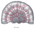
The ileocolic artery is the lowest branch arising from the concavity of the superior mesenteric artery.
The superior rectal artery is an artery that descends into the pelvis to supply blood to the rectum.

The pancreatic branches or pancreatic arteries are numerous small vessels derived from the splenic artery as it runs behind the upper border of the pancreas, supplying its body and tail.
The public domain consists of all the creative work to which no exclusive intellectual property rights apply. Those rights may have expired, been forfeited, expressly waived, or may be inapplicable.

Gray's Anatomy is an English language textbook of human anatomy originally written by Henry Gray and illustrated by Henry Vandyke Carter. Earlier editions were called Anatomy: Descriptive and Surgical, Anatomy of the Human Body and Gray's Anatomy: Descriptive and Applied, but the book's name is commonly shortened to, and later editions are titled, Gray's Anatomy. The book is widely regarded as an extremely influential work on the subject, and has continued to be revised and republished from its initial publication in 1858 to the present day. The latest edition of the book, the 41st, was published in September 2015.


