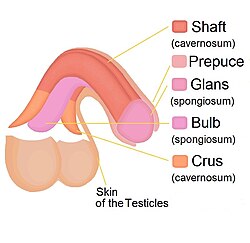
In male human anatomy, the glans penis or penile glans, commonly referred to as the glans, is the bulbous structure at the distal end of the human penis that is the human male's most sensitive erogenous zone and primary anatomical source of sexual pleasure. The glans penis is present in the male reproductive organs of humans and most other mammals where it may appear smooth, spiny, elongated or divided. It is externally lined with mucosal tissue, which creates a smooth texture and glossy appearance. In humans, the glans is located over the distal ends of the corpora cavernosa and is a continuation of the corpus spongiosum of the penis. At the summit appears the urinary meatus and at the base forms the corona glandis. An elastic band of tissue, known as the frenulum, runs on its ventral surface. In men who are not circumcised, it is completely or partially covered by a fold of skin called the foreskin. In adults, the foreskin can generally be retracted over and past the glans manually or sometimes automatically during an erection.

The corpus spongiosum is the mass of spongy tissue surrounding the male urethra within the penis. It is also called the corpus cavernosum urethrae in older texts.
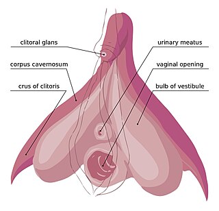
In female anatomy, the vestibular bulbs, bulbs of the vestibule or clitoral bulbs are two elongated masses of erectile tissue typically described as being situated on either side of the vaginal opening. They are united to each other in front by a narrow median band. Some research indicates that they do not surround the vaginal opening, and are more closely related to the clitoris than to the vestibule. They constitute the root of the clitoris along with the crura.

A corpus cavernosum penis (singular) is one of a pair of sponge-like regions of erectile tissue, which contain most of the blood in the penis during an erection.

The spongy urethra is the longest part of the male urethra, and is contained in the corpus spongiosum of the penis.
In human male anatomy, the dorsal veins of the penis are blood vessels that drain the shaft, the skin and the glans of the human penis. They are typically located in the midline on the dorsal aspect of the penis and they comprise the superficial dorsal veinof the penis, that lies in the subcutaneous tissue of the shaft, and the deep dorsal veinof the penis, that lies beneath the deep fascia.
Venous leak, also called venogenic erectile dysfunction and penile venous insufficiency, is one category of vasculogenic impotence — a cause of erectile dysfunction in males. It affects all ages, being particularly awkward in young men. Much about venous leaks has not reached a consensus among the medical community, and many aspects of the condition, particularly its treatment strategies, are controversial. The prevalence of the condition is still unknown, although some sources claim it to be a common cause of erectile dysfunction.
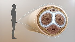
The tunica albuginea is the fibrous envelope that extends the length of the corpus cavernosum penis and corpus spongiosum penis. It is a bi-layered structure that includes an outer longitudinal layer and an inner circular layer.
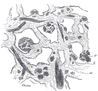
From the internal surface of the fibrous envelope of the corpora cavernosa penis, as well as from the sides of the septum, numerous bands or cords are given off, which cross the interior of these corpora cavernosa in all directions, subdividing them into a number of separate compartments, and giving the entire structure a spongy appearance.
Buck's fascia is a layer of deep fascia covering the three erectile bodies of the penis.

In human anatomy, the penis is an external male sex organ that additionally serves as the urinary duct. The main parts are the root, body, the epithelium of the penis including the shaft skin, and the foreskin covering the glans. The body of the penis is made up of three columns of tissue: two corpora cavernosa on the dorsal side and corpus spongiosum between them on the ventral side. The urethra passes through the prostate gland, where it is joined by the ejaculatory duct, and then through the penis. The urethra goes across the corpus spongiosum and ends at the tip of the glans as the opening, the urinary meatus. It is a passage both for excretion of urine and the ejaculation of semen.
The development of the reproductive system is the part of embryonic growth that results in the sex organs and contributes to sexual differentiation. Due to its large overlap with development of the urinary system, the two systems are typically described together as the genitourinary system.

In human male anatomy, the radix or root of the penis is the internal and most proximal portion of the human penis that lies in the perineum. Unlike the pendulous body of the penis, which is suspended from the pubic symphysis, the root is attached to the pubic arch of the pelvis and is not visible externally. It is triradiate in form, consisting of three masses of erectile tissue; the two diverging crura, one on either side, and the median bulb of the penis or urethral bulb. Approximately one third to one half of the penis is embedded in the pelvis and can be felt through the scrotum and in the perineum.
In human male anatomy, the septum of the penis or penile septum refers to the fibrous junction (septum) between the two corpora cavernosa of the human penis. The tunica albuginea of the penis forms a thick fibrous coat to the spongy tissue of the corpora cavernosa and corpus spongiosum. The two corpora cavernosa are surrounded by a strong fibrous envelope consisting of superficial and deep fibers. The superficial or outer fibers are longitudinal in direction, and form a single tube which encloses both corpora; the deep or inner fibers are arranged circularly around each corpus and meet in the center. By their junction in the median plane, the inner fibers form the intercavernous septum of the penis.

The corona of glans penis or penis crown refers to the rounded projecting border or flare that forms at the base of the glans in human males. The corona overhangs a mucosal surface, known as the neck of the penis, which separates the shaft and the glans. The deep retro-glandular coronal sulcus forms between the corona and the neck of the penis. The two sides of the corona merge on the ventral midline forming the septum glandis. The circumference of the corona is richly innervated and is described as a highly erogenous area of the glans.

An erection is a physiological phenomenon in which the penis becomes firm, engorged, and enlarged. Penile erection is the result of a complex interaction of psychological, neural, vascular, and endocrine factors, and is often associated with sexual arousal, sexual attraction or libido, although erections can also be spontaneous. The shape, angle, and direction of an erection vary considerably between humans.

Clitoral erection is a physiological phenomenon where the clitoris becomes enlarged and firm.

In male human anatomy, the foreskin, also known as the prepuce, is the double-layered fold of skin, mucosal and muscular tissue at the distal end of the human penis that covers the glans and the urinary meatus. The foreskin is attached to the glans by an elastic band of tissue, known as the frenulum. The outer skin of the foreskin meets with the inner preputial mucosa at the area of the mucocutaneous junction. The foreskin is mobile, fairly stretchable and sustains the glans in a moist environment. Except for humans, a similar structure known as a penile sheath appears in the male sexual organs of all primates and the vast majority of mammals.
Penile ulltrasonography is medical ultrasonography of the penis. Ultrasound is an excellent method for the study of the penis, such as indicated in trauma, priapism, erectile dysfunction or suspected Peyronie's disease.

Glans insufficiency syndrome, also known as the soft glans, cold glans, or glans insufficiency, is a medical condition that affects male individuals. This condition is characterized by the persistent inability of the glans penis to achieve and maintain an erect or turgid state during sexual arousal, remaining soft and cold. This condition can have an impact on a person's sexual function, including decreased sensitivity, difficulty in maintaining an erection, and overall quality of life.