Inflammatory demyelinating diseases of the central nervous system

Last updated

Inflammatory demyelinating diseases (IDDs), sometimes called Idiopathic (IIDDs) due to the unknown etiology of some of them, are a heterogenous group of demyelinating diseases - conditions that cause damage to myelin, the protective sheath of nerve fibers - that occur against the background of an acute or chronic inflammatory process. [1] [2] [3] IDDs share characteristics with and are often grouped together under Multiple Sclerosis. They are sometimes considered different diseases from Multiple Sclerosis, [4] [5] but considered by others to form a spectrum differing only in terms of chronicity, severity, and clinical course. [6] [7]

Contents

Multiple sclerosis for some people is a syndrome more than a single disease. [8] As of 2019, three auto-antibodies have been found in atypical MS giving birth to separate diseases: Anti-AQP4 diseases, Anti-MOG and Anti-NF spectrums. [9] A LHON-associated MS [10] has also been reported, and in addition there have been inconclusive reports of TNF-α blockers inducing demyelinating disorders. [11]

The subject is under intense research and the list of MS autoantibodies is expected to grow in the near future. [12] [13] [14]

Separated variants

As of 2014 several previous MS variants had been separated from MS after the discovery of a specific auto-antibody. Those autoantibodies were anti-AQP4, anti-MOG and some anti-Neurofascins. [15]

The pathogenic mechanism is usually not related to the clinical course. Therefore, one given pathogenic underlying condition can yield several clinical diseases, and one disease can be produced by several pathogenic conditions.[ citation needed ]

These conditions can appear as Neuromyelitis optica (NMO), and its associated "spectrum of disorders" (NMOSD), currently considered a common syndrome for several separated diseases [16] but with some still idiopathic subtypes. Some researchers think that there could exist an overlapping between Anti-NMDA receptor encephalitis cases and neuromyelitis optica or acute disseminated encephalomyelitis. [17]

Anti-AQP4 spectrum

See Anti-AQP4 diseases

Originally found in neuromyelitis optica, this autoantibody has been associated with other conditions. Its current spectrum is as following:

Anti-MOG spectrum

See Anti-MOG associated encephalomyelitis

Anti-MOG associated spectrum, often clinically presented as an anti-MOG autoimmune encephalomyelitis, [21] [22] but can also appear as negative NMO or atypical multiple sclerosis. [23]

The presence of anti-MOG autoantibodies has been associated with the following conditions [24]

The anti-mog spectrum in children is equally variated: Out of a sample of 41 children with MOG-antibodies 29 had clinical NMOSD (17 relapsing), 8 had ADEM (4 relapsing with ADEM-ON), 3 had a single clinical event CIS, and 1 had a relapsing tumefactive disorder. Longitudinal myelitis was evident on MRI in 76[percent]. It has also been noted that percentage of children with anti-mog antibodies respect a demyelinating sample is higher than for adults [32]

Some NMO patients present double positive for autoantibodies to AQP4 and MOG. These patients have MS-like brain lesions, multifocal spine lesions and retinal and optic nerves atrophy. [33]

Anti-neurofascin spectrum

See Anti-neurofascin demyelinating diseases

Some anti-neurofascin demyelinating diseases were previously considered a subtype of Multiple Sclerosis but now they are considered a separate entity, as it happened before to anti-MOG and anti-AQP4 cases. Around 10% of MS cases are now thought to be anti-Neurofascin disease in reality. [34]

Anti-neurofascin autoantibodies have been reported in atypical cases of MS and CIDP, and a whole spectrum of Anti-neurofascin demyelinating diseases has been proposed. [35]

Some cases of CIDP are reported to be produced by auto-antibodies against several neurofascin proteins. These proteins are present in the neurons and four of them have been reported to produce disease: NF186, NF180, NF166 and NF155. [35]

Antibodies against Neurofascins NF-155 can also appear in MS [36] and NF-186 could be involved in subtypes of MS [37] yielding an intersection between both conditions.

Summarising, autoantibodies against several neurofascins can produce MS: neurofascin186 (NF186), neurofascin155 (NF155), contactin 1 (CNTN1), contactin associated protein 1 (CASPR1) and gliomedin. All of them nodal and paranodal proteins. [35]

Demyelination associated with anti-TNF therapy

Several anti-TNF drugs like adalimumab [38] [39] are commonly prescribed by a number of autoimmune conditions. Some of them have been reported to produce a CNS-demyelination compatible with standard MS. [11] [40] [41] Several other monoclonal antibodies like pembrolizumab, [42] nivolumab [43] and infliximab [44] have been also reported to produce MS as an adverse event. [45] [11] Nevertheless, it is not so similar as reported in the previous references. [46] [ improper synthesis? ][ clarification needed ]

The reactions following Anti-TNF therapy have been diverse according to the source of the disease. [40] [41] [43] [ failed verification ] Some of these cases can be classify as ADEM, using the confluent demyelination as barrier between both conditions. [47] [ non-primary source needed ]

In most cases, the damage fulfills all pathological diagnostic criteria of MS and can therefore be classified as MS in its own right.[ medical citation needed ] The lesions were classified as pattern II in the Lassman/Lucchinetti system. Some lesions also showed Dawson fingers, [41] [ improper synthesis? ] which is supposed to be a MS-only feature.

These recent problems with artificial anti-TNF-α autoimmunity also point to the possibility of tumor necrosis factor alpha involvement in some multiple sclerosis variants.[ medical citation needed ]

LHON-associated MS

Also a previous subtype of MS associated to LHON has been described (LHON-MS) [10] It is a presentation of LHON with MS-like CNS damage.

It used to satisfy McDonalds definition for MS, but after demonstration that LHON can produce this kind of lesions, the "no better explanation" requirement does not hold anymore. It is not due to auto-antibodies, but to defective mitochondria instead. [48]

The symptoms of this higher form of the disease include loss of the brain's ability to control the movement of muscles, tremors, and cardiac arrhythmia. [49] and the lack of muscular control. [50]

Relapsing anti-NMDAR encephalitis

Atypical Anti-NMDA receptor encephalitis (a kind of Autoimmune encephalitis) can appear in the form of relapsing optic neuritis. [51] [ failed verification ][ non-primary source needed ]

Variants still idiopathic

Apart from the previously cited spectrums (Anti-AQP4 diseases, Anti-MOG, and Anti-NF) there is a long list of MS variants, with possibly different pathogenesis, which are still idiopathic and considered inside the MS-spectrum.

Pseudotumefactive variants

Most atypical variants appear as tumefactive or pseudotumefactive variants (lesions whose size is more than 2 cm (0.79 in), with mass effect, oedema and/or ring enhancement) [52] [53] Some cases of the following have shown anti-MOG auto-antibodies and therefore they represent MS cases only partially.[ citation needed ]

Atypical lesion location variants

Also the location of the lesions can be used to classify variants:

Myelocortical multiple sclerosis

Myelocortical multiple sclerosis (MCMS), proposed variant with demyelination of spinal cord and cerebral cortex but not of cerebral white matter [61] Several atypical cases could belong here. See the early reports of MCMS< [62] [63]

AQP4-negative Optic-spinal MS

Real Optic-spinal MS (OSMS) without anti-AQP4 antibodies, has been consistently reported, and it is classified into the MS spectrum. [64] [65]

OSMS has its own specific immunological biomarkers [66] The whole picture is under construction and several reports exists about overlapping conditions.

Pure spinal MS

Pure spinal multiple sclerosis: Patients with clinical and paraclinical features suggestive of cord involvement of multiple sclerosis (MS)-type albeit not rigidly fulfilling the McDonald criteria [67] Some inflammatory conditions are associated with the presence of scleroses in the CNS. [68] Optic neuritis (monophasic and recurrent) and Transverse myelitis (monophasic and recurrent)

LHON associated MS

LHON associated MS (LHON-MS), a presentation of LHON with MS-like CNS damage, and therefore a subtype of MS according to McDonalds definition. [10]

Atypical OCB variants

Also different classifications by body fluid biomarkers is possible:

Radiologically atypical variants

Inside well defined MS (Lesions disseminated in time and space with no other explanation) there are atypical cases based in radiological or metabolic criteria. A four-groups classification has been proposed: [76]

Other radiological classification of atypical lesions proposes the following four subtypes: [78]

Atypical clinical courses

In 1996, the US National Multiple Sclerosis Society (NMSS) Advisory Committee on Clinical Trials in Multiple Sclerosis (ACCTMS) standardized four clinical courses for MS (Remitent-Recidivant, Secondary Progressive, Progressive-Relapsing and Primary progressive). Later, [79] some reports state that those "types" were artificially made up trying to classify RRMS as a separate disease so that the number of patients was low enough to get the interferon approved by the FDA under the orphan drugs act. [79] Revisions in 2013 and 2017 removed the Progressive-Relapsing course and introduced CIS as a variety/course/status of MS, establishing the actual classification (CIS, RRMS, SPMS and PPMS). Nevertheless, these types are not enough to predict the responses to medications and several regulatory agencies use additional types in their recommendations lide Highly active MS, Malignant MS, Aggressive MS or Rapidly progressive MS. [80]

Highly Active MS

As of 2019, HAMS is defined as an RRMS phenotype with one or more of the following characteristics: [80]

  1. DSS scale of 4 points at 5 years of onset of the disease
  2. Multiple relapses (two or more) with incomplete recovery in the ongoing year
  3. More than 2 brain magnetic resonance imaging (MRI) studies demonstrating new lesions or increase in the size of the lesions in T2, or lesions that enhance with gadolinium despite treatment (Clinical case 1 and 2).
  4. No response to treatment with one or more DMTs for at least one year.

There is a group of patients who have defined clinical and radiological risk factors that predict a behavior of greater risk of conversion to HAMS, without having the diagnostic criteria of HAMS in a first clinical attack have predictors of high risk.

Some other previous authors have used other definitions like:

  • High activity according to 2017 definition of activity
  • Rapid accumulation of physical and cognitive deficit, despite treatment with DMT's.
  • Being eligible for immunoablative therapy followed by autologous haematopoietic stem cell transplantation (aHSCT) because of a) the failure of conventional treatment, b) frequent and severe (disabling) relapses, or c) MRI activity (new T2 or gadolinium-enhancing lesions).

Malignant MS

See malignant multiple sclerosis

Occasionally, the term ‘malignant’ MS (MMS) has been used to describe aggressive phenotypes of MS, but this is another ambiguous term that—despite wide usage—means different things to different people.

In 1996, the US National MS Society (NMSS) Advisory Committee on Clinical Trials in Multiple Sclerosis, “malignant MS” was also included, namely, “disease with a rapid progressive course, leading to significant disability in multiple neurologic systems or death in a relatively short time after disease onset.”[ full citation needed ]

Many authors[ who? ] reserve the term malignant for fulminant forms of MS that deteriorate so rapidly from the outset as to be almost monophasic, and result in death within months to a few years. One such example is the Marburg variant of MS, which is classically characterized by extensive necrotic and/or tumefactive lesions with mass effect.

Despite recent (and increasing) emphasis on early detection of patients with aggressive MS, the original definition of MMS was not modified by the NMSS Advisory Committee in its latest publication in 2013 (Lublin et al., 2014).[ full citation needed ]

Aggressive MS

Common to all definitions is the early, unexpected acquisition of disability followed by frequent relapses and highly active disease seen on MRI.

One definition can be based on EDSS score and the time to develop secondary progressive MS (SPMS) (Menon et al., 2013).[ full citation needed ]

No consensus exists on the speed of progression or degree of disability sufficient for aggressive MS, but some assume that reaching an EDSS score of 6 points probably represents an upper limit beyond which the risk-benefit ratio for an aggressive treatment is unfavourable.[ citation needed ]

Pragmatically, AMS has been defined as any type of MS that is associated with repeated severe attacks and accelerated accrual of disability—put more simply, "rapidly progressive" MS.

Rapidly progressive multiple sclerosis

This kind of MS was previously reported to behave different that the standard progressive course, [81] being linked to Connexin 43 autoantibodies with pattern III lesions (distal oligodendrogliopathy) [82] and being responsive to plasma exchange [83]

In very rapidly progressive multiple sclerosis the use of immunosuppressive therapy (mitoxantrone/cyclophosphamide), rituximab, autologous haematopoietic stem cell therapy or combination therapy should be considered carefully. [84]

Under research

Some auto-antibodies have been found consistently across different MS cases but there is still no agreement on their relevance:

Other auto-antibodies can be used to stablish a differential diagnosis from very different diseases like Sjögren syndrome which can be separated by Anti–Calponin-3 autoantibodies. [101]

The correlation between this genetic mutation and MS was challenged but in 2018 has been replicated by an independent team. [102] Notice that this results do not refer to general MS.

In general, NMO-like spectrum without known auto-antibodies is considered MS. Principal component analysis of these cases show 3 different kinds of antibody-negative patients. The metabolite discriminators of RRMS and Ab-NMOSD suggest that these groupings have some pathogenic meaning. [103]

As MS is an active field for research, the list of auto-antibodies is not closed nor definitive. For example, some diseases like Autoimmune GFAP Astrocytopathy or variants of CIDP that affects the CNS (CIDP is the chronic counterpart of Guillain–Barré syndrome) could be included. Autoimmune variants peripheral neuropathies or progressive inflammatory neuropathy could be in the list assuming the autoimmune model for MS, together with a rare demyelinating lesional variant of trigeminal neuralgia [104] [ failed verification ] and some NMDAR Anti-NMDA receptor encephalitis. [54]

Venous induced demyelination has also been proposed as a hypothetical MS variant produced by CCSVI, Susac's syndrome and Neuro-Behçet's disease (MS has an important vascular component [105] ), myalgic encephalomyelitis (aka chronic fatigue syndrome). [106]

Also leukoaraiosis can produce lesions disseminated in time and space, condition usually sufficient in the MS definition. Maybe two sub-conditions of Leukodystrophy: Adrenoleukodystrophy and Adrenomyeloneuropathy could be in the list. Specially interesting is X-linked adrenoleukodystrophy (X-ALD or CALD). [107]

Genetic types

Different behaviour has been reported according to the presence of different HLA genes.

HLA DRB3*02:02 patients

In HLA DRB3 cases, autoimmune reactions against the enzyme GDP-L-fucose synthase has been reported [108] [109] The same report points that the autoimmune problem could derive from the gut microbiota.

HLA-DRB1*15:01 has the strongest association with MS. [110]

HLA-DRB1*04:05, HLA-B*39:01, and HLA-B*15:01 are associated with independent MS susceptibility and HLA-DQβ1 position 9 with phenylalanine had the strongest effect on MS susceptibility. [110]

Another possible type is one with auto-antibodies against GDP-L-fucose synthase. In HLA-DRB3*02:02 patients, autoimmune reactions against the enzyme GDP-L-fucose synthase has been reported [108] [109] The same report points that the autoimmune problem could derive from the gut microbiota.

Rapidly progressive multiple sclerosis

See malignant multiple sclerosis

This is a specially aggressive clinical course of progressive MS [111] that has been found to be caused by a special genetic variant. It is due to a mutation inside the gene NR1H3, an arginine to glutamine mutation in the position p.Arg415Gln, in an area that codifies the protein LXRA. [112]

Primary progressive variants

Some researchers propose to separate primary progressive MS from other clinical courses. PPMS, after recent findings seem to point that it is pathologically a very different disease. [113] [114] [115] [116] [ non-primary source needed ]

Some authors think since long ago that primary progressive MS should be considered a disease different from standard MS, [117] [118] and it was also proposed that PPMS could be heterogeneous [119]

Clinical variants have been described. For example, Late Onset MS. [120] [111] It is due to a mutation inside the gene NR1H3, an arginine to glutamine mutation in the position p.Arg415Gln, in an area that codifies the protein LXRA.


For the rest of the progressive cases, it has been found that the lesions are diffuse instead of the normal focal ones, [121] and are different under MR spectroscopy. [122] RRMS and PPMS patients also show differences on the retinal layers yields examined under OCT. [123]

Some authors have proposed a dual classification of PPMS, according to the shape of edges of the scars, in MS-like and ADEM-like [124] Proteomic analysis have shown that two proteins, Secretogranin II and Protein 7B2, in CSF can be used to separate RRMS from PPMS [125]

Recently, the hypothesis of PPMS being apart from RRMS/SPMS is taken further credibility due that it was shown that CSF from PPMS patients can carry the disease to other animals, producing neurodegeneration in mice [113] and that Normal Appearing White Matter (NAWM) structure is also different [126] [ non-primary source needed ]

The predominant lesions in PPMS are slowly expanding lesions with T cells, microglial, and macrophage-associated demyelination in close similar to pattern I demyelination [127]

As of 2019 it has been found that the profile of T-cells is different in PPMS and SPMS [128] [ non-primary source needed ]

Clinical situations inside standard MS

MS can be considered among the acquired demyelinating syndromes with a multiphasic instead of monophasic behaviour. [129] Multiple sclerosis has a prodromal stage in which an unknown underlying condition, able to damage the brain, is present, but no lesion has still developed.

MS is usually classified in clinical types, though they are unrelated to the underlying pathology. Some critical reports say that the current "types" were artificially made up, just to treat RRMS as a separate disease. In this way the number of patients was low enough to enter the orphan drugs act, and get the interferon approved by the FDA under this schema. [79] Recent reviews state that all types are a mixture of inflammation and neurodegeneration, and that all types should be considered the same disease. [130]

Other possible clinical courses are:

Preclinical MS: CIS and CDMS

The first manifestation of MS is the so-called Clinically isolated syndrome, or CIS, which is the first isolated attack. The current diagnosis criteria for MS do not allow doctors to give an MS diagnosis until a second attack takes place. Therefore, the concept of "clinical MS", for an MS that can be diagnosed, has been developed. Until MS diagnosis has been established, nobody can tell whether the disease one is dealing with is MS.[ citation needed ]

Cases of MS before the CIS are sometimes found during other neurological inspections and are referred to as subclinical MS. [131] Preclinical MS refers to cases after the CIS but before the confirming second attack. [132] After the second confirming attack the situation is referred to as CDMS (clinically defined multiple sclerosis). [133]

CIS itself is sometimes considered itself as a disease entity, different from MS. Even if they share the same underlying condition CIS is not MS given that it lacks the presence of lesions. [134] Approximately 84% of the subjects with CIS experience a second clinical demyelinating event and are diagnosed with clinically definite MS (CDMS) within 20 years.

RIS, subclinical, silent and prodromal MS

See also Radiologically isolated syndrome

Silent MS has been found in autopsies before the existence of MRI [135] showing that the so-called "clinical definitions" cannot be applied to around 25% of the MS cases. [136] Currently a distinction is made between "silent" and subclinical.

In absence of attacks, sometimes a radiological finding suggestive of demyelination (T2 hyperintensities [137] ) can be used to establish a pre-diagnosis of MS. This is often named "Radiologically Isolated Syndrome" (RIS). Cases before the first attack or CIS are subclinical in the sense that they do not produce clinical situations.

If a second radiological event appears without clinical consequences, the clinical situation is named "Silent MS" (Okuda criteria). [138] Anyway, it is reported that all MS cases have an active subclinical phase before the CIS [139]

It has been noted that some aspects of the MS underlying condition are present in otherwise healthy MS patients' relatives, [140] suggesting a wider scope for the "silent MS" term.

In these cases Interleukin-8 is a risk for clinical conversion. [141] It has also been proposed that always exists a subclinical phase in the beginning of every MS case, during which the permeability of the BBB can be used for diagnosis [142]

It is also under investigation whether MS has a prodrome, i.e., a preliminary stage in which the disease exists with non-specific symptoms. Some reports point to a prodrome of several years for RRMS and decades for PPMS. [143] [144] [145]

Aggressive multiple sclerosis

Relapsing-Remitting MS is considered aggressive when the frequency of exacerbations is not less than 3 during 2 years. Special treatment is often considered for this subtype. [146] According to these definition aggressive MS would be a subtype of RRMS.

Other authors disagree and define aggressive MS by the accumulation of disability, considering it as a rapidly disabling disease course [147] and therefore inside PPMS.

The aggressive course is associated to grey matter damage and meningeal inflammation, and presents a special intrathecal (meninges and CSF) inflammatory profile. [148]

After the 2016 revision of the MS phenotypes, it is called Highly active multiple sclerosis [80]

Mitoxantrone was approved for this special clinical course. Some reports point to Alemtuzumal being beneficial. [149]

Pediatric and pubertal MS

MS cases are rare before puberty, but they can happen. Whether they constitute a separate disease is still an open subject. Anyway, even this pubertal MS could be more than one disease, because early-onset and late-onset have different demyelination patterns. [150]

Pediatric MS patients tend to have active inflammatory disease course with a tendency to have brainstem / cerebellar presentations at onset. Due to efficient repair mechanisms at early life, pediatric MS patients tend to have longer time to reach EDSS 6 but reach it at earlier age. [151]

An iron-responsive variant of MS has been reported in children. [152]

Controversy for the definition

Given that the etiology of MS is unknown, the current definitions of MS are all based on its appearance.[ medical citation needed ] The most commonly used definition, the McDonald criteria, requires just the presence of demyelinating lesions separated in space and time, together with the exclusion of every known demyelinating condition.[ medical citation needed ] However as of 2023 most current MRI machines only pick up 60% of MS lesions, and even 7T machines only pick up 85%. The MS of MS people not discovered by MRI is found only in autopsy. [153]

This unspecific definition has been criticized. For some people this has turned MS into a heterogeneous condition with several underlying problems. [154] Besides, the complementary problem also exists. Given that McDonalds-MS is based just in the distribution of the lesions, even twins with the same underlying condition can be classified different [155]

Finally, the "exclusion of every other known disease" condition also creates problems. Rightfully classified MS patients can be rightfully classified out of the spectrum when their particular underlying problem is discovered. For example, neuromyelitis optica was previously considered MS and currently is not, even if it appears that the MS definition has not changed.[ medical citation needed ]

Currently there is no single diagnosis test for MS that is 100% sensitive and specific. [156] [157]

Pathological and clinical definitions

McDonald criteria propose a clinical diagnosis based on a pathological definition, saying that the focus for diagnosis "remains on the objective demonstration of dissemination of lesions in both time and space" (DIT and DIS). But given that other diseases produce similar lesions, it is also required that those lesions cannot be explained by any other known disease.[ citation needed ]

This open definition present problems. [158] For example, before the discovery of anti-AQP4 in 2006, most optic-spinal MS patients were classified rightfully as MS. Currently they are classified as NMO. Both diagnosis are correct even though the definition has not (apparently) changed.

According to some pathologists, a pathological definition is required because clinical definitions have problems with differential diagnosis [43] and they always use a pathological definition on articles about post-mortem retrospective diagnosis, but for practitioners that need a diagnosis as soon as possible MS is often regarded as a pure clinical entity, defined simply by a positive result in the standard clinical case definition being then named "clinically definite MS" (CDMS, Poser) or simply "MS" (McDonald). [159]

Both definitions lead to different results. For example, confluent subpial cortical lesions are the most specific finding for MS, being exclusively present in MS patients. [160] but can only be detected post-mortem by an autopsy [161] Therefore, any other diagnosis method will have false positives.

Other meanings of MS

There is no known etiology for MS and therefore no etiology-based definition is possible. Comparison to a post-mortem retrospective diagnosis is possible, but useless to practitioners and short-term researchers, and it is not usually done. Therefore, all meanings for the words "Multiple Sclerosis" are somehow diffuse.[ citation needed ]

The pathological definition based on proven dissemination in time and space has problems. For example, it leaves situations like RIS (radiologically isolated syndrome) outside the MS spectrum because the lack of proof, even in the case that this condition later could shown the same pathogenic conditions than MS cases. [162]

Besides, usually the term "multiple sclerosis" is used to refer to the presence of the unknown underlying condition that produces the MS lesions instead to the mere presence of the lesions. The term MS is also used to refers to the process of developing the lesions. [163]

Some authors instead speak about the biological disease vs. its clinical presentation. [164]

Anyway, the precise meaning in each case can be normally deduced from the context.

Handling several clinical definitions

Given that several definitions of MS coexist, some authors are referring to them using whether CDMS for Poser positives, or McDonalds-MS with a prefix for McDonalds positives, including the release year in the prefix. [165]

CIS and conversion to MS

The 2010 revision of the McDonald criteria [166] allows the diagnosis of MS with only one proved lesion (CIS). Consistently, the later revision for the MS phenotypes in 2013 was forced to consider CIS as one of the MS phenotypes. [167]

Therefore, the former concept of "Conversion from CIS to MS", that was declared when a patient had a second MS attack, does not apply anymore. More accurate is now to speak about conversions from the CIS phenotype to other MS phenotype. [168]

See also

Related Research Articles

<span class="mw-page-title-main">Acute disseminated encephalomyelitis</span> Autoimmune disease

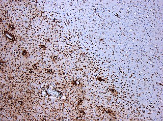
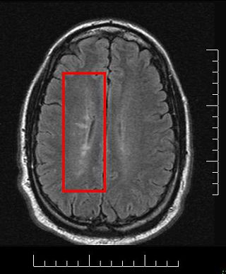
Acute disseminated encephalomyelitis (ADEM), or acute demyelinating encephalomyelitis, is a rare autoimmune disease marked by a sudden, widespread attack of inflammation in the brain and spinal cord. As well as causing the brain and spinal cord to become inflamed, ADEM also attacks the nerves of the central nervous system and damages their myelin insulation, which, as a result, destroys the white matter. The cause is often a trigger such as from viral infection or vaccinations.

<span class="mw-page-title-main">Optic neuritis</span> Medical condition

Optic neuritis describes any condition that causes inflammation of the optic nerve; it may be associated with demyelinating diseases, or infectious or inflammatory processes.

<span class="mw-page-title-main">Multiple sclerosis</span> Disease that damages the myelin sheaths around nerves

Multiple sclerosis (MS) is an autoimmune disease in which the insulating covers of nerve cells in the brain and spinal cord are damaged. This damage disrupts the ability of parts of the nervous system to transmit signals, resulting in a range of signs and symptoms, including physical, mental, and sometimes psychiatric problems. Specific symptoms can include double vision, vision loss, eye pain, muscle weakness, and loss of sensation or coordination. MS takes several forms, with new symptoms either occurring in isolated attacks or building up over time. In the relapsing forms of MS, between attacks, symptoms may disappear completely, although some permanent neurological problems often remain, especially as the disease advances. In the progressive forms of MS, bodily function slowly deteriorates and disability worsens once symptoms manifest and will steadily continue to do so if the disease is left untreated.

Encephalomyelitis is inflammation of the brain and spinal cord. Various types of encephalomyelitis include:

Neuromyelitis optica spectrum disorders (NMOSD) are a spectrum of autoimmune diseases characterized by acute inflammation of the optic nerve and the spinal cord (myelitis). Episodes of ON and myelitis can be simultaneous or successive. A relapsing disease course is common, especially in untreated patients.

<span class="mw-page-title-main">Myelin oligodendrocyte glycoprotein</span>

Myelin oligodendrocyte glycoprotein (MOG) is a glycoprotein believed to be important in the myelination of nerves in the central nervous system (CNS). In humans this protein is encoded by the MOG gene. It is speculated to serve as a necessary "adhesion molecule" to provide structural integrity to the myelin sheath and is known to develop late on the oligodendrocyte.

Experimental autoimmune encephalomyelitis, sometimes experimental allergic encephalomyelitis (EAE), is an animal model of brain inflammation. It is an inflammatory demyelinating disease of the central nervous system (CNS). It is mostly used with rodents and is widely studied as an animal model of the human CNS demyelinating diseases, including multiple sclerosis (MS) and acute disseminated encephalomyelitis (ADEM). EAE is also the prototype for T-cell-mediated autoimmune disease in general.

<span class="mw-page-title-main">Pathophysiology of multiple sclerosis</span>

Multiple sclerosis is an inflammatory demyelinating disease of the CNS in which activated immune cells invade the central nervous system and cause inflammation, neurodegeneration, and tissue damage. The underlying cause is currently unknown. Current research in neuropathology, neuroimmunology, neurobiology, and neuroimaging, together with clinical neurology, provide support for the notion that MS is not a single disease but rather a spectrum.

<span class="mw-page-title-main">Lesional demyelinations of the central nervous system</span>

Multiple sclerosis and other demyelinating diseases of the central nervous system (CNS) produce lesions and glial scars or scleroses. They present different shapes and histological findings according to the underlying condition that produces them.

<span class="mw-page-title-main">Balo concentric sclerosis</span> Medical condition

Baló's concentric sclerosis is a disease in which the white matter of the brain appears damaged in concentric layers, leaving the axis cylinder intact. It was described by József Mátyás Baló who initially named it "leuko-encephalitis periaxialis concentrica" from the previous definition, and it is currently considered one of the borderline forms of multiple sclerosis.

<span class="mw-page-title-main">Marburg acute multiple sclerosis</span> Medical condition

Marburg acute multiple sclerosis, also known as Marburg multiple sclerosis or acute fulminant multiple sclerosis, is considered one of the multiple sclerosis borderline diseases, which is a collection of diseases classified by some as MS variants and by others as different diseases. Other diseases in this group are neuromyelitis optica (NMO), Balo concentric sclerosis, and Schilder's disease. The graver course is one form of malignant multiple sclerosis, with patients reaching a significant level of disability in less than five years from their first symptoms, often in a matter of months.

Research in multiple sclerosis may find new pathways to interact with the disease, improve function, curtail attacks, or limit the progression of the underlying disease. Many treatments already in clinical trials involve drugs that are used in other diseases or medications that have not been designed specifically for multiple sclerosis. There are also trials involving the combination of drugs that are already in use for multiple sclerosis. Finally, there are also many basic investigations that try to understand better the disease and in the future may help to find new treatments.

<span class="mw-page-title-main">Tumefactive multiple sclerosis</span> Medical condition

Tumefactive multiple sclerosis is a condition in which the central nervous system of a person has multiple demyelinating lesions with atypical characteristics for those of standard multiple sclerosis (MS). It is called tumefactive as the lesions are "tumor-like" and they mimic tumors clinically, radiologically and sometimes pathologically.

Malignant multiple sclerosis is used to describe MS patients who reach significant level of disability in a short period of time. Malignant MS cases are not common, less than 5% of patients with MS experience this type of progression.

<span class="mw-page-title-main">Multiple sclerosis diagnosis</span>

Current standards for diagnosing multiple sclerosis (MS) are based on the 2018 revision of McDonald criteria. They rely on MRI detection of demyelinating lesions in the CNS, which are distributed in space (DIS) and in time (DIT). It is also a requirement that any possible known disease that produces demyelinating lesions is ruled out before applying McDonald's criteria.

<span class="mw-page-title-main">Pathology of multiple sclerosis</span> Pathologic overview

Multiple sclerosis (MS) can be pathologically defined as the presence of distributed glial scars (scleroses) in the central nervous system that must show dissemination in time (DIT) and in space (DIS) to be considered MS lesions.

MOG antibody disease (MOGAD) or MOG antibody-associated encephalomyelitis (MOG-EM) is an inflammatory demyelinating disease of the central nervous system. Serum anti-myelin oligodendrocyte glycoprotein antibodies are present in up to half of patients with an acquired demyelinating syndrome and have been described in association with a range of phenotypic presentations, including acute disseminated encephalomyelitis, optic neuritis, transverse myelitis, and neuromyelitis optica.

Anti-neurofascin demyelinating diseases refers to health conditions engendered by auto-antibodies against neurofascins, which can produce both central and peripheral demyelination. Some cases of combined central and peripheral demyelination (CCPD) could be produced by them.

Anti-AQP4 diseases, are a group of diseases characterized by auto-antibodies against aquaporin 4.

Brenda Banwell is Chief of the Division of Neurology and Co-Director of the Neuroscience Center, and Professor of Neurology at Children's Hospital of Philadelphia and holder of the Grace R. Loeb Endowed Chair in Neurosciences. She also holds the title of Professor of Pediatrics and Neurology at the Perelman School of Medicine at the University of Pennsylvania.

References

  1. Höftberger R, Lassmann H (2018). "Inflammatory demyelinating diseases of the central nervous system". Neuropathology. Handbook of Clinical Neurology. Vol. 145. pp. 263–283. doi:10.1016/B978-0-12-802395-2.00019-5. ISBN   9780128023952. PMC   7149979 . PMID   28987175.
  2. "Find out more about demyelinating diseases, including multiple sclerosis". Mayo Clinic. Retrieved 2022-06-23.
  3. Fontaine B (September 2001). "[Borderline forms of multiple sclerosis]". Revue Neurologique (in French). 157 (8-9 Pt 2): 929–934. PMID   11787357.
  4. Wingerchuk DM, Lucchinetti CF (June 2007). "Comparative immunopathogenesis of acute disseminated encephalomyelitis, neuromyelitis optica, and multiple sclerosis". Current Opinion in Neurology. 20 (3): 343–350. doi:10.1097/WCO.0b013e3280be58d8. PMID   17495631. S2CID   17386506.
  5. Poser CM, Brinar VV (October 2007). "Disseminated encephalomyelitis and multiple sclerosis: two different diseases - a critical review". Acta Neurologica Scandinavica. 116 (4): 201–206. doi: 10.1111/j.1600-0404.2007.00902.x . PMID   17824894. S2CID   44411472.
  6. Weinshenke B, Miller D (1999-01-13). "Multiple sclerosis: one disease or many?". In Siva A, Kesselring J, Thompson AJ (eds.). Frontiers in Multiple Sclerosis, II. Taylor & Francis. pp. 37–46. ISBN   978-1-85317-506-0.
  7. Hartung HP, Grossman RI (May 2001). "ADEM: distinct disease or part of the MS spectrum?". Neurology. 56 (10): 1257–1260. doi:10.1212/WNL.56.10.1257. PMID   11376169. S2CID   219199163.
  8. Zabad RK, Stewart R, Healey KM (October 2017). "Pattern Recognition of the Multiple Sclerosis Syndrome". Brain Sciences. 7 (10): 138. doi: 10.3390/brainsci7100138 . PMC   5664065 . PMID   29064441.
  9. Fujihara K (June 2019). "Neuromyelitis optica spectrum disorders: still evolving and broadening". Current Opinion in Neurology. 32 (3): 385–394. doi:10.1097/WCO.0000000000000694. PMC   6522202 . PMID   30893099.
  10. 1 2 3 Bargiela D, Chinnery PF (September 2019). "Mitochondria in neuroinflammation - Multiple sclerosis (MS), leber hereditary optic neuropathy (LHON) and LHON-MS" (PDF). Neuroscience Letters. 710: 132932. doi:10.1016/j.neulet.2017.06.051. PMID   28668384. S2CID   38817368.
  11. 1 2 3 Kemanetzoglou E, Andreadou E (2017). "CNS Demyelination with TNF-α Blockers". Current Neurology and Neuroscience Reports. 17 (4): 36. doi:10.1007/s11910-017-0742-1. ISSN   1528-4042. PMC   5364240 . PMID   28337644.
  12. Lang K, Prüss H (September 2017). "Frequencies of neuronal autoantibodies in healthy controls: Estimation of disease specificity". Neurology. 4 (5): e386. doi:10.1212/NXI.0000000000000386. PMC   5515597 . PMID   28761905.
  13. Kusunoki S (December 2013). "Autoantibodies in neuroimmunological diseases; relevance of fine specificity". Experimental Neurology. 250: 219–220. doi:10.1016/j.expneurol.2013.10.009. PMID   24120752. S2CID   45173537.
  14. Seay M, Galetta S (September 2018). "Glial Fibrillary Acidic Protein Antibody: Another Antibody in the Multiple Sclerosis Diagnostic Mix". Journal of Neuro-Ophthalmology. 38 (3): 281–284. doi: 10.1097/WNO.0000000000000689 . PMID   29923872. S2CID   49310332.
  15. Sato DK, Callegaro D, Lana-Peixoto MA, Waters PJ, de Haidar Jorge FM, Takahashi T, et al. (February 2014). "Distinction between MOG antibody-positive and AQP4 antibody-positive NMO spectrum disorders". Neurology. 82 (6): 474–481. doi:10.1212/WNL.0000000000000101. PMC   3937859 . PMID   24415568.
  16. Weinshenker BG, Wingerchuk DM (February 2014). "The two faces of neuromyelitis optica". Neurology. 82 (6): 466–467. doi:10.1212/WNL.0000000000000114. PMID   24415570. S2CID   30860834.
  17. 1 2 Gahr M, Lauda F, Wigand ME, Connemann BJ, Rosenbohm A, Tumani H, et al. (April 2015). "Periventricular white matter lesion and incomplete MRZ reaction in a male patient with anti-N-methyl-D-aspartate receptor encephalitis presenting with dysphoric mania". BMJ Case Reports. 2015: bcr2014209075. doi:10.1136/bcr-2014-209075. PMC   4422915 . PMID   25917068.
  18. Li Y, Xie P, Lv F, Mu J, Li Q, Yang Q, et al. (October 2008). "Brain magnetic resonance imaging abnormalities in neuromyelitis optica". Acta Neurologica Scandinavica. 118 (4): 218–225. doi:10.1111/j.1600-0404.2008.01012.x. PMID   18384459. S2CID   22270592.
  19. Wingerchuk D (2006). "Neuromyelitis Optica (Devic's Syndrome)" (PDF). 2006 Rare Neuroimmunologic Disorders Symposium. Archived from the original (PDF) on 2006-09-25. Retrieved 2007-01-05.
  20. Ikeda K, Ito H, Hidaka T, Takazawa T, Sekine T, Yoshii Y, et al. (2011). "Repeated non-enhancing tumefactive lesions in a patient with a neuromyelitis optica spectrum disorder". Internal Medicine. 50 (9): 1061–1064. doi: 10.2169/internalmedicine.50.4295 . PMID   21532234.
  21. Pröbstel AK, Rudolf G, Dornmair K, Collongues N, Chanson JB, Sanderson NS, et al. (March 2015). "Anti-MOG antibodies are present in a subgroup of patients with a neuromyelitis optica phenotype". Journal of Neuroinflammation. 12 (1): 46. doi: 10.1186/s12974-015-0256-1 . PMC   4359547 . PMID   25889963.
  22. 1 2 Spadaro M, Gerdes LA, Mayer MC, Ertl-Wagner B, Laurent S, Krumbholz M, et al. (March 2015). "Histopathology and clinical course of MOG-antibody-associated encephalomyelitis". Annals of Clinical and Translational Neurology. 2 (3): 295–301. doi:10.1002/acn3.164. PMC   4369279 . PMID   25815356.
  23. 1 2 Spadaro M, Gerdes LA, Krumbholz M, Ertl-Wagner B, Thaler FS, Schuh E, et al. (October 2016). "Autoantibodies to MOG in a distinct subgroup of adult multiple sclerosis". Neurology. 3 (5): e257. doi:10.1212/NXI.0000000000000257. PMC   4949775 . PMID   27458601.
  24. 1 2 3 4 Reindl M, Di Pauli F, Rostásy K, Berger T (August 2013). "The spectrum of MOG autoantibody-associated demyelinating diseases". Nature Reviews. Neurology. 9 (8): 455–461. doi:10.1038/nrneurol.2013.118. PMID   23797245. S2CID   7219279.
  25. Baumann M, Hennes EM, Schanda K, Karenfort M, Bajer-Kornek B, Diepold K, et al. (2015). "OP65 – 3006: Clinical characteristics and neuroradiological findings in children with multiphasic demyelinating encephalomyelitis and MOG antibodies". European Journal of Paediatric Neurology. 19: S21. doi:10.1016/S1090-3798(15)30066-0.
  26. Jarius S, Metz I, König FB, Ruprecht K, Reindl M, Paul F, et al. (October 2016). "Screening for MOG-IgG and 27 other anti-glial and anti-neuronal autoantibodies in 'pattern II multiple sclerosis' and brain biopsy findings in a MOG-IgG-positive case". Multiple Sclerosis. 22 (12): 1541–1549. doi:10.1177/1352458515622986. PMID   26869529. S2CID   1387384.
  27. Kitagawa S, Osada T, Kaneko K, Takahashi T, Suzuki N, Nakahara J (December 2018). "[Clinical analysis of opticospinal multiple sclerosis (OSMS) presentation detecting anti-myelin oligodendrocyte glycoprotein (MOG) antibody]". Rinsho Shinkeigaku = Clinical Neurology. 58 (12): 737–744. doi: 10.5692/clinicalneurol.cn-001184 . PMID   30487359.
  28. Chalmoukou K, Stathopoulos P, Alexopoulos H, Akrivou S, Dalakas M (2015). "Recurrent Optic Neuritis (rON) is characterised by Anti-MOG Antibodies: A follow-up study" . Neurology. 84 (14 Suppl P5): 274. doi:10.1212/WNL.84.14_supplement.P5.274. S2CID   70688931.
  29. Rossman I (2015). "An 8 Year Old Girl with Chronic Inflammatory Optic Neuritis (CRION): the Youngest Reported Case to Date". Neurology. 84 (14): Supplement P7.015. doi:10.1212/WNL.84.14_supplement.P7.015. S2CID   54126682.
  30. Chalmoukou K, Alexopoulos H, Akrivou S, Stathopoulos P, Reindl M, Dalakas MC (August 2015). "Anti-MOG antibodies are frequently associated with steroid-sensitive recurrent optic neuritis". Neurology. 2 (4): e131. doi:10.1212/NXI.0000000000000131. PMC   4496630 . PMID   26185777.
  31. Cantó LN, Boscá SC, Vicente CA, Gil-Perontín S, Pérez-Miralles F, Villalba JC, et al. (2019). "Brain Atrophy in Relapsing Optic Neuritis Is Associated With Crion Phenotype". Frontiers in Neurology. 10: 1157. doi: 10.3389/fneur.2019.01157 . PMC   6838209 . PMID   31736862.
  32. Tenembaum S, Waters P, Leite M, Woodhall M, Princich J, Segura M, et al. (2015-04-06). "Spectrum of MOG Autoantibody-Associated Inflammatory Diseases in Pediatric Patients (I4-3A)". Neurology. 84 (14 Supplement). ISSN   0028-3878.
  33. Yan Y, Li Y, Fu Y, Yang L, Su L, Shi K, et al. (December 2016). "Autoantibody to MOG suggests two distinct clinical subtypes of NMOSD". Science China Life Sciences. 59 (12): 1270–1281. doi:10.1007/s11427-015-4997-y. PMC   5101174 . PMID   26920678.
  34. Goncalves MV, Fragoso YD (April 2019). "The involvement of anti-neurofascin 155 antibodies in central and peripheral demyelinating diseases". Neuroimmunology and Neuroinflammation. 8: 6. doi: 10.20517/2347-8659.2019.08 . S2CID   146038130.
  35. 1 2 3 Kira JI, Yamasaki R, Ogata H (November 2019). "Anti-neurofascin autoantibody and demyelination". Neurochemistry International. 130: 104360. doi: 10.1016/j.neuint.2018.12.011 . PMID   30582947.
  36. Stich O, Perera S, Berger B, Jarius S, Wildemann B, Baumgartner A, Rauer S (May 2016). "Prevalence of neurofascin-155 antibodies in patients with multiple sclerosis". Journal of the Neurological Sciences. 364: 29–32. doi:10.1016/j.jns.2016.03.004. PMID   27084211. S2CID   29204735.
  37. "Progressive MS: a new perspective | Multiple Sclerosis Society UK". www.mssociety.org.uk. Archived from the original on 2007-06-21.
  38. Engel S, Luessi F, Mueller A, Schopf RE, Zipp F, Bittner S (2020). "PPMS onset upon adalimumab treatment extends the spectrum of anti-TNF-α therapy-associated demyelinating disorders". Therapeutic Advances in Neurological Disorders. 13: 1756286419895155. doi:10.1177/1756286419895155. PMC   6940603 . PMID   31921355.
  39. Alnasser Alsukhni R, Jriekh Z, Aboras Y (2016). "Adalimumab Induced or Provoked MS in Patient with Autoimmune Uveitis: A Case Report and Review of the Literature". Case Reports in Medicine. 2016: 1423131. doi: 10.1155/2016/1423131 . PMC   5093248 . PMID   27840642.
  40. 1 2 Vicente LG, Viqueira BR, de las Peñas MJ, Cobos RG, Moreno JP, Gonzalez RA (2016). "Relapse in a paucisymptomatic form of multiple sclerosis in a patient treated with nivolumab". Neuro Oncol. 18 (Suppl 4): iv25. doi: 10.1093/neuonc/now188.085 . PMC   5782583 .
  41. 1 2 3 Höftberger R, Leisser M, Bauer J, Lassmann H (December 2015). "Autoimmune encephalitis in humans: how closely does it reflect multiple sclerosis ?". Acta Neuropathologica Communications. 3 (1): 80. doi: 10.1186/s40478-015-0260-9 . PMC   4670499 . PMID   26637427.
  42. Romeo MA, Garassino MC, Moiola L, Galli G, Comi G, Martinelli V, Filippi M (December 2019). "Multiple sclerosis associated with pembrolizumab in a patient with non-small cell lung cancer". Journal of Neurology. 266 (12): 3163–3166. doi:10.1007/s00415-019-09562-z. PMID   31586260. S2CID   203654410.
  43. 1 2 3 Lassmann H (February 2010). "Acute disseminated encephalomyelitis and multiple sclerosis". Brain. 133 (Pt 2): 317–319. doi: 10.1093/brain/awp342 . PMID   20129937.
  44. Kalinowska-Lyszczarz A, Fereidan-Esfahani M, Guo Y, Lucchinetti CF, Tobin WO (August 2020). "Pathological findings in central nervous system demyelination associated with infliximab". Multiple Sclerosis. 26 (9): 1124–1129. doi:10.1177/1352458519894710. PMC   7297659 . PMID   31845616.
  45. Garcia CR, Jayswal R, Adams V, Anthony LB, Villano JL (10 February 2019). "Multiple Sclerosis Outcomes after Cancer Immunotherapy". Clinical & Translational Oncology. 21 (10): 1336–1342. doi:10.1007/s12094-019-02060-8. ISSN   1699-048X. PMC   6702101 . PMID   30788836.
  46. Williams I, Uhlig HH (December 2020). "Demyelination After Anti-TNF Therapy: Who is at Risk?". Journal of Crohn's & Colitis. 14 (12): 1651–1652. doi: 10.1093/ecco-jcc/jjaa144 . PMID   33026456.
  47. Young NP, Weinshenker BG, Parisi JE, Scheithauer B, Giannini C, Roemer SF, et al. (February 2010). "Perivenous demyelination: association with clinically defined acute disseminated encephalomyelitis and comparison with pathologically confirmed multiple sclerosis". Brain. 133 (Pt 2): 333–348. doi:10.1093/brain/awp321. PMC   2822631 . PMID   20129932.
  48. Nikoskelainen EK, Marttila RJ, Huoponen K, Juvonen V, Lamminen T, Sonninen P, Savontaus ML (August 1995). "Leber's "plus": neurological abnormalities in patients with Leber's hereditary optic neuropathy". Journal of Neurology, Neurosurgery, and Psychiatry. 59 (2): 160–164. doi:10.1136/jnnp.59.2.160. PMC   485991 . PMID   7629530.
  49. "Cardiac arrythmia". health-cares.net. Archived from the original on 6 March 2016.
  50. "Multiple sclerosis - Symptoms and causes". Mayo Clinic. Retrieved 2022-06-24.
  51. Ma C, Wang C, Zhang Q, Lian Y (2019). "Emerging role of prodromal headache in patients with anti-N-methyl-D-aspartate receptor encephalitis". Journal of Pain Research. 12: 519–526. doi: 10.2147/JPR.S189301 . PMC   6365221 . PMID   30787630.
  52. Lucchinetti CF, Gavrilova RH, Metz I, Parisi JE, Scheithauer BW, Weigand S, et al. (July 2008). "Clinical and radiographic spectrum of pathologically confirmed tumefactive multiple sclerosis". Brain. 131 (Pt 7): 1759–1775. doi:10.1093/brain/awn098. PMC   2442427 . PMID   18535080.
  53. Given CA, Stevens BS, Lee C (January 2004). "The MRI appearance of tumefactive demyelinating lesions". AJR. American Journal of Roentgenology. 182 (1): 195–199. doi:10.2214/ajr.182.1.1820195. PMID   14684539.
  54. 1 2 Hardy TA, Reddel SW, Barnett MH, Palace J, Lucchinetti CF, Weinshenker BG (August 2016). "Atypical inflammatory demyelinating syndromes of the CNS". The Lancet. Neurology. 15 (9): 967–981. doi:10.1016/S1474-4422(16)30043-6. PMID   27478954. S2CID   26341166.
  55. Jiménez Arango JA, Uribe Uribe CS, Toro González G (March 2015). "Lesser-known myelin-related disorders: focal tumour-like demyelinating lesions". Neurologia. 30 (2): 97–105. doi: 10.1016/j.nrl.2013.06.004 . PMID   24094691.
  56. Garrido C, Levy-Gomes A, Teixeira J, Temudo T (2004). "[Schilder's disease: two new cases and a review of the literature]" [Schilder's disease: two new cases and a review of the literature]. Revista de Neurología (in Spanish). 39 (8): 734–738. doi:10.33588/rn.3908.2003023. PMID   15514902.
  57. Afifi AK, Bell WE, Menezes AH, Moore SA (October 1994). "Myelinoclastic diffuse sclerosis (Schilder's disease): report of a case and review of the literature". Journal of Child Neurology. 9 (4): 398–403. CiteSeerX   10.1.1.1007.559 . doi:10.1177/088307389400900412. PMID   7822732. S2CID   38765870.
  58. Schmalstieg WF, Keegan BM, Weinshenker BG (February 2012). "Solitary sclerosis: progressive myelopathy from solitary demyelinating lesion". Neurology. 78 (8): 540–544. doi:10.1212/WNL.0b013e318247cc8c. PMID   22323754. S2CID   52859541.
  59. Lattanzi S (July 2012). "Solitary sclerosis: Progressive myelopathy from solitary demyelinating lesion". Neurology. 79 (4): 393, author reply 393. doi: 10.1212/01.wnl.0000418061.10382.f7 . PMID   22826546.
  60. Ayrignac X, Carra-Dalliere C, Homeyer P, Labauge P (December 2013). "Solitary sclerosis: progressive myelopathy from solitary demyelinating lesion. A new entity?". Acta Neurologica Belgica. 113 (4): 533–534. doi:10.1007/s13760-013-0182-x. PMID   23358965. S2CID   17631796.
  61. Trapp BD, Vignos M, Dudman J, Chang A, Fisher E, Staugaitis SM, et al. (October 2018). "Cortical neuronal densities and cerebral white matter demyelination in multiple sclerosis: a retrospective study". The Lancet. Neurology. 17 (10): 870–884. doi:10.1016/S1474-4422(18)30245-X. PMC   6197820 . PMID   30143361.
  62. Vignos MC (2016). A histopathological and magnetic resonance imaging assessment of myelocortical multiple sclerosis: A new pathological variant (Ph.D. thesis). Kent State University. Archived from the original on 2022-10-29. Retrieved 2022-06-24.
  63. Hendrickson M, Chang A, Fisher E (September 30, 2013). "ECTRIMS 2013: Posters: Myelocortical multiple sclerosis: a subgroup of multiple sclerosis patients with spinal cord and cortical demyelination". Multiple Sclerosis Journal. 19 (11_suppl): 74–558. doi:10.1177/1352458513502429. hdl: 20.500.11940/2342 . ISSN   1352-4585. S2CID   208628537.
  64. Meneguette NS, Almeida KM, Figueiredo MT, de Araújo E, Araújo AC, Alvarenga MP, et al. (November 2021). "Optic neuritis in Asian type opticospinal multiple sclerosis (OSMS-ON) in a non-Asian population: A functional-structural paradox" (PDF). Multiple Sclerosis and Related Disorders. 56: 103260. doi:10.1016/j.msard.2021.103260. PMID   34562767. S2CID   237636213.
  65. Matsushita T, Isobe N, Matsuoka T, Shi N, Kawano Y, Wu XM, et al. (July 2009). "Aquaporin-4 autoimmune syndrome and anti-aquaporin-4 antibody-negative opticospinal multiple sclerosis in Japanese". Multiple Sclerosis. 15 (7): 834–847. doi:10.1177/1352458509104595. PMID   19465451. S2CID   19372571.
  66. Mercan Ö, Ülger Z, Handan Mısırlı C, Türkoğlu R (2018). "Opticospinal Multiple Sclerosis Clinical Course and Immunological Parameters". Experimed. 8 (2): 47–51. doi: 10.26650/experimed.2018.18002 .
  67. Schee JP, Viswanathan S (May 17, 2018). "Pure spinal multiple sclerosis: A possible novel entity within the multiple sclerosis disease spectrum" . Multiple Sclerosis Journal. 25 (8): 1189–1195. doi:10.1177/1352458518775912. ISSN   1352-4585. PMID   29771191. S2CID   21709554.
  68. O'Connor P, Marriott J (2010). "Differential Diagnosis and Diagnostic Criteria for Multiple Sclerosis: Application and Pitfalls" (PDF). In Hohlfeld R, Lucchinetti CF (eds.). Multiple sclerosis 3 (1st ed.). Philadelphia: Saunders Elsevier. ISBN   978-1-4160-6068-0. Archived from the original (PDF) on 2015-08-24. Retrieved 2015-01-14.
  69. Mihalova T, Kwong JL, Alachkar H (2019-12-01). "214 Analysis of CSF IgG index, IgG synthesis and clinical presentation among CSF OCBs negative patients". Journal of Neurology, Neurosurgery & Psychiatry. 90 (12): e54.1–e54. doi:10.1136/jnnp-2019-ABN-2.181. ISSN   0022-3050. S2CID   214428195.
  70. Link H, Huang YM (November 2006). "Oligoclonal bands in multiple sclerosis cerebrospinal fluid: an update on methodology and clinical usefulness". Journal of Neuroimmunology. 180 (1–2): 17–28. doi:10.1016/j.jneuroim.2006.07.006. PMID   16945427. S2CID   22724352.
  71. Imrell K, Landtblom AM, Hillert J, Masterman T (September 2006). "Multiple sclerosis with and without CSF bands: clinically indistinguishable but immunogenetically distinct". Neurology. 67 (6): 1062–1064. doi:10.1212/01.wnl.0000237343.93389.35. PMID   17000979. S2CID   21855273.
  72. Zeman AZ, Kidd D, McLean BN, Kelly MA, Francis DA, Miller DH, et al. (January 1996). "A study of oligoclonal band negative multiple sclerosis". Journal of Neurology, Neurosurgery, and Psychiatry. 60 (1): 27–30. doi:10.1136/jnnp.60.1.27. PMC   486185 . PMID   8558146.
  73. Li R, Patterson KR, Bar-Or A (July 2018). "Reassessing B cell contributions in multiple sclerosis". Nature Immunology. 19 (7): 696–707. doi:10.1038/s41590-018-0135-x. PMID   29925992. S2CID   49347759.
  74. Casanova B, Lacruz L, Villar ML, Domínguez JA, Gadea MC, Gascón F, et al. (August 2018). "Different clinical response to interferon beta and glatiramer acetate related to the presence of oligoclonal IgM bands in CSF in multiple sclerosis patients". Neurological Sciences. 39 (8): 1423–1430. doi:10.1007/s10072-018-3442-y. PMID   29882169. S2CID   46954452.
  75. Graner M, Pointon T, Manton S, Green M, Dennison K, Davis M, et al. (21 February 2020). "Oligoclonal IgG antibodies in multiple sclerosis target patient-specific peptides". PLOS ONE. 15 (2): e0228883. Bibcode:2020PLoSO..1528883G. doi: 10.1371/journal.pone.0228883 . PMC   7034880 . PMID   32084151.
  76. Codjia P, Ayrignac X, Carra-Dalliere C, Cohen M, Charif M, Lippi A, et al. (SFSEP OFSEP) (February 2019). "Multiple sclerosis with atypical MRI presentation: Results of a nationwide multicenter study in 57 consecutive cases". Multiple Sclerosis and Related Disorders. 28: 109–116. doi:10.1016/j.msard.2018.12.022. PMID   30592992. S2CID   58550590.
  77. 1 2 Ayrignac X, Menjot de Champfleur N, Menjot de Champfleur S, Carra-Dallière C, Deverdun J, Corlobe A, Labauge P (June 2016). "Brain magnetic resonance imaging helps to differentiate atypical multiple sclerosis with cavitary lesions and vanishing white matter disease". European Journal of Neurology. 23 (6): 995–1000. doi:10.1111/ene.12931. PMID   26727496. S2CID   3917307.
  78. Wallner-Blazek M, Rovira A, Fillipp M, Rocca MA, Miller DH, Schmierer K, et al. (August 2013). "Atypical idiopathic inflammatory demyelinating lesions: prognostic implications and relation to multiple sclerosis". Journal of Neurology. 260 (8): 2016–2022. doi:10.1007/s00415-013-6918-y. PMID   23620065. S2CID   28377856.
  79. 1 2 3 Dobson R, Giovannoni G (January 2019). "Multiple sclerosis - a review". European Journal of Neurology. 26 (1): 27–40. doi: 10.1111/ene.13819 . PMC   1589931 . PMID   30300457.
  80. 1 2 3 Díaz C, Zarco LA, Rivera DM (May 2019). "Highly active multiple sclerosis: An update". Multiple Sclerosis and Related Disorders. 30: 215–224. doi:10.1016/j.msard.2019.01.039. PMID   30822617. S2CID   73460639.
  81. Morcos Y, Lee SM, Levin MC (August 2003). "A role for hypertrophic astrocytes and astrocyte precursors in a case of rapidly progressive multiple sclerosis". Multiple Sclerosis. 9 (4): 332–341. doi:10.1191/1352458503ms931oa. PMID   12926837. S2CID   43651011.
  82. Masaki K, Suzuki SO, Matsushita T, Matsuoka T, Imamura S, Yamasaki R, et al. (2013). "Connexin 43 astrocytopathy linked to rapidly progressive multiple sclerosis and neuromyelitis optica". PLOS ONE. 8 (8): e72919. Bibcode:2013PLoSO...872919M. doi: 10.1371/journal.pone.0072919 . PMC   3749992 . PMID   23991165.
  83. Seidi O (January 27, 2015). The Role of Therapeutic Plasma Exchange in Clinical Neurology (Report). Archived from the original on June 21, 2021. Retrieved July 13, 2016 via khartoumspace.uofk.edu.
  84. Broadley SA, Barnett MH, Boggild M, Brew BJ, Butzkueven H, Heard R, et al. (August 2015). "A new era in the treatment of multiple sclerosis". The Medical Journal of Australia. 203 (3): 139–41, 141e.1. doi:10.5694/mja14.01218. hdl: 10536/DRO/DU:30080418 . PMID   26224184. S2CID   10805065.
  85. Wekerle H (March 2022). "Epstein-Barr virus sparks brain autoimmunity in multiple sclerosis". Nature. 603 (7900): 230–232. Bibcode:2022Natur.603..230W. doi:10.1038/d41586-022-00382-2. PMID   35169323. S2CID   246866517.
  86. Srivastava R, Aslam M, Kalluri SR, Schirmer L, Buck D, Tackenberg B, et al. (July 2012). "Potassium channel KIR4.1 as an immune target in multiple sclerosis". The New England Journal of Medicine. 367 (2): 115–123. doi:10.1056/NEJMoa1110740. PMC   5131800 . PMID   22784115.
  87. Schneider R (2013). "Autoantibodies to Potassium Channel KIR4.1 in Multiple Sclerosis". Frontiers in Neurology. 4: 125. doi: 10.3389/fneur.2013.00125 . PMC   3759297 . PMID   24032025.
  88. Ayoglu B, Häggmark A, Khademi M, Olsson T, Uhlén M, Schwenk JM, Nilsson P (September 2013). "Autoantibody profiling in multiple sclerosis using arrays of human protein fragments". Molecular & Cellular Proteomics. 12 (9): 2657–2672. doi: 10.1074/mcp.M112.026757 . PMC   3769337 . PMID   23732997.
  89. Ayoglu B, Mitsios N, Kockum I, Khademi M, Zandian A, Sjöberg R, et al. (February 2016). "Anoctamin 2 identified as an autoimmune target in multiple sclerosis". Proceedings of the National Academy of Sciences of the United States of America. 113 (8): 2188–2193. Bibcode:2016PNAS..113.2188A. doi: 10.1073/pnas.1518553113 . PMC   4776531 . PMID   26862169.
  90. Tengvall K, Huang J, Hellström C, Kammer P, Biström M, Ayoglu B, et al. (August 2019). "Molecular mimicry between Anoctamin 2 and Epstein-Barr virus nuclear antigen 1 associates with multiple sclerosis risk". Proceedings of the National Academy of Sciences of the United States of America. 116 (34): 16955–16960. Bibcode:2019PNAS..11616955T. doi: 10.1073/pnas.1902623116 . PMC   6708327 . PMID   31375628.
  91. Belova AN, Grygorieva VN, Rasteryaeva MV, Ruina EA, Belova EM, Solovieva VS, Boyko AN (2019). "[Anti-NMDAR encephalitis associated with relapsing optic neuritis: a case report and differential diagnosis]". Zhurnal Nevrologii I Psikhiatrii Imeni S.S. Korsakova. 119 (10. Vyp. 2): 137–146. doi:10.17116/jnevro201911910137. PMID   31934999. S2CID   210193928.
  92. Baheerathan A, Brownlee WJ, Chard DT, Shields K, Gregory R, Trip SA (February 2017). "Antecedent anti-NMDA receptor encephalitis in two patients with multiple sclerosis". Multiple Sclerosis and Related Disorders. 12: 20–22. doi:10.1016/j.msard.2016.12.009. PMID   28283100.
  93. Mariotto S, Gajofatto A, Alberti D, Lederer S, Monaco S, Ferrari S, Hoeftberger R (April 2019). "Flotillin-1/2 autoantibodies in patients with inflammatory disorders of the CNS". Neurology. 92 (15 Supplement): P2.2-072(P2. 2-072). doi:10.1212/WNL.92.15_supplement.P2.2-072. S2CID   199036968. Archived from the original on 2019-04-23. Retrieved 2019-04-23.
  94. Koutsis G, Breza M, Velonakis G, Tzartos J, Kasselimis D, Kartanou C, et al. (February 2019). "X linked Charcot-Marie-Tooth disease and multiple sclerosis: emerging evidence for an association". Journal of Neurology, Neurosurgery, and Psychiatry. 90 (2): 187–194. doi:10.1136/jnnp-2018-319014. PMID   30196252. S2CID   52175657.
  95. Yu-Wai-Man P, Spyropoulos A, Duncan HJ, Guadagno JV, Chinnery PF (September 2016). "A multiple sclerosis-like disorder in patients with OPA1 mutations". Annals of Clinical and Translational Neurology. 3 (9): 723–729. doi:10.1002/acn3.323. PMC   5018584 . PMID   27656661.
  96. Vojdani A, Mukherjee PS, Berookhim J, Kharrazian D (2015). "Detection of Antibodies against Human and Plant Aquaporins in Patients with Multiple Sclerosis". Autoimmune Diseases. 2015: 905208. doi: 10.1155/2015/905208 . PMC   4529886 . PMID   26290755.
  97. Baranova SV, Mikheeva EV, Buneva VN, Nevinsky GA (November 2019). "Antibodies from the Sera of Multiple Sclerosis Patients Efficiently Hydrolyze Five Histones". Biomolecules. 9 (11): 741. doi: 10.3390/biom9110741 . PMC   6920934 . PMID   31731780.
  98. Tzartos JS, Stergiou C, Kilidireas K, Zisimopoulou P, Thomaidis T, Tzartos SJ (2013). "Anti-aquaporin-1 autoantibodies in patients with neuromyelitis optica spectrum disorders". PLOS ONE. 8 (9): e74773. Bibcode:2013PLoSO...874773T. doi: 10.1371/journal.pone.0074773 . PMC   3781161 . PMID   24086369.
  99. Robers M, Tokhie H, Borazanci A (2018-04-10). "N-Type Calcium Channel Antibody Encephalitis Coexisting With Multiple Sclerosis (P5.397)". Neurology. 90 (15 Supplement). doi:10.1212/WNL.90.15_supplement.P5.397. ISSN   0028-3878. S2CID   266111836.
  100. Faergeman SL, Evans H, Attfield KE, Desel C, Kuttikkatte SB, Sommerlund M, et al. (May 2020). "A novel neurodegenerative spectrum disorder in patients with MLKL deficiency". Cell Death & Disease. 11 (5): 303. doi:10.1038/s41419-020-2494-0. PMC   7195448 . PMID   32358523.
  101. Birnbaum J, Hoke A, Lalji A, Calabresi P, Bhargava P, Casciola-Rosen L (October 2018). "Brief Report: Anti-Calponin 3 Autoantibodies: A Newly Identified Specificity in Patients With Sjögren's Syndrome". Arthritis & Rheumatology. 70 (10): 1610–1616. doi:10.1002/art.40550. PMID   29749720. S2CID   13693467.
  102. Zhang Y, Wang L, Jia H, Liao M, Chen X, Xu J, et al. (July 2018). "Genetic variants regulate NR1H3 expression and contribute to multiple sclerosis risk". Journal of the Neurological Sciences. 390: 162–165. doi:10.1016/j.jns.2018.04.037. PMID   29801879. S2CID   44082278.
  103. Yeo T, Probert F, Jurynczyk M, Sealey M, Cavey A, Claridge TD, et al. (November 2019). "Classifying the antibody-negative NMO syndromes: Clinical, imaging, and metabolomic modeling". Neurology. 6 (6): e626. doi:10.1212/NXI.0000000000000626. PMC   6865851 . PMID   31659123.
  104. Tohyama S, Hung PS, Cheng JC, Zhang JY, Halawani A, Mikulis DJ, et al. (May 2020). "Trigeminal neuralgia associated with a solitary pontine lesion: clinical and neuroimaging definition of a new syndrome". Pain. 161 (5): 916–925. doi:10.1097/j.pain.0000000000001777. PMC   7170433 . PMID   31842151.
  105. Minagar A, Jy W, Jimenez JJ, Alexander JS (April 2006). "Multiple sclerosis as a vascular disease". Neurological Research. 28 (3): 230–235. doi:10.1179/016164106X98080. PMID   16687046. S2CID   16896871.
  106. "Chronic Fatigue Syndrome - Symptoms, Diagnosis, Treatment of Chronic Fatigue Syndrome". Health Information. New York Times. The following test results... are seen consistently...: Brain MRI showing swelling in the brain or destruction of part of the nerve cells (demyelination)
  107. Bergner CG, Genc N, Hametner S, Franz J, van der Meer F, Mitkovski M, et al. (October 2021). "Concurrent axon and myelin destruction differentiates X-linked adrenoleukodystrophy from multiple sclerosis". Glia. 69 (10): 2362–2377. doi: 10.1002/glia.24042 . PMID   34137074.
  108. 1 2 "Link Between Gut Flora and Multiple Sclerosis Discovered". Neuroscience News. 2018-10-11. Retrieved 2022-06-29.
  109. 1 2 Planas R, Santos R, Tomas-Ojer P, Cruciani C, Lutterotti A, Faigle W, et al. (October 2018). "GDP-l-fucose synthase is a CD4+ T cell-specific autoantigen in DRB3*02:02 patients with multiple sclerosis". Science Translational Medicine. 10 (462): eaat4301. doi:10.1126/scitranslmed.aat4301. PMID   30305453. S2CID   52959112.
  110. 1 2 Ogawa K, Okuno T, Hosomichi K, Hosokawa A, Hirata J, Suzuki K, et al. (August 2019). "Next-generation sequencing identifies contribution of both class I and II HLA genes on susceptibility of multiple sclerosis in Japanese". Journal of Neuroinflammation. 16 (1): 162. doi: 10.1186/s12974-019-1551-z . PMC   6683481 . PMID   31382992.
  111. 1 2 Wang Z, Sadovnick AD, Traboulsee AL, Ross JP, Bernales CQ, Encarnacion M, et al. (June 2016). "Nuclear Receptor NR1H3 in Familial Multiple Sclerosis". Neuron. 90 (5): 948–954. doi:10.1016/j.neuron.2016.04.039. PMC   5092154 . PMID   27253448.
  112. Hain HS, Hakonarson H (June 2016). "The Added Value of Family Material in the Discovery of Multiple Sclerosis Genes". Neuron. 90 (5): 905–906. doi: 10.1016/j.neuron.2016.05.027 . PMID   27253441.
  113. 1 2 Cristofanilli M, Rosenthal H, Cymring B, Gratch D, Pagano B, Xie B, Sadiq SA (November 2014). "Progressive multiple sclerosis cerebrospinal fluid induces inflammatory demyelination, axonal loss, and astrogliosis in mice". Experimental Neurology. 261: 620–632. doi:10.1016/j.expneurol.2014.07.020. PMID   25111532. S2CID   21263405.
  114. Ulloa B, Alahiri M, Liu Y, Sadiq S (2015). "Cerebrospinal Fluid Haptoglobin (Hp) Levels are elevated in MS patients with progressive disease". Neurology. 84 (14): 213. doi:10.1212/WNL.84.14_supplement.P5.213. S2CID   58984399.
  115. Elkjaer ML, Nawrocki A, Kacprowski T, Lassen P, Simonsen AH, Marignier R, et al. (2020). "Molecular markers in the CSF proteome differentiate neuroinflammatory diseases". Research Square. doi:10.21203/rs.3.rs-19710/v1. S2CID   236819936.
  116. Vernejoul J, Cali F, Lin J, Sadiq S (2020-04-14). "Single Cell Analysis of MS CSF B-Cells Show Significant Differences in Expansion Patterns and Gene Family Usage Between MS Subtypes (4673)". Neurology. 94 (15 Supplement). doi:10.1212/WNL.94.15_supplement.4673. ISSN   0028-3878. S2CID   266118700.
  117. Vukusic S, Confavreux C (February 2003). "Primary and secondary progressive multiple sclerosis". Journal of the Neurological Sciences. 206 (2): 153–155. doi:10.1016/S0022-510X(02)00427-6. PMID   12559503. S2CID   19215860.
  118. Dressel A, Kolb AK, Elitok E, Bitsch A, Bogumil T, Kitze B, et al. (December 2006). "Interferon-beta1b treatment modulates cytokines in patients with primary progressive multiple sclerosis". Acta Neurologica Scandinavica. 114 (6): 368–373. doi: 10.1111/j.1600-0404.2006.00700.x . PMID   17083335. S2CID   71465403.
  119. Villar LM, Casanova B, Ouamara N, Comabella M, Jalili F, Leppert D, et al. (August 2014). "Immunoglobulin M oligoclonal bands: biomarker of targetable inflammation in primary progressive multiple sclerosis". Annals of Neurology. 76 (2): 231–240. doi:10.1002/ana.24190. PMID   24909126. S2CID   16397501.
  120. Curbelo M, Martinez A, Steinberg J, Carra A (April 2016). "LOMS vs. Other Diseases: The Consequence of "To Be or Not To Be"". Neurology. 86 (16 Supplement): P3.081. doi:10.1212/WNL.86.16_supplement.P3.081. S2CID   78885337. Archived from the original on 2022-01-27. Retrieved 2022-01-27.
  121. Zwemmer JN, Bot JC, Jelles B, Barkhof F, Polman CH (April 2008). "At the heart of primary progressive multiple sclerosis: three cases with diffuse MRI abnormalities only". Multiple Sclerosis. 14 (3): 428–430. doi:10.1177/1352458507084591. PMID   18208890. S2CID   27178351.
  122. Reinke SN, Broadhurst DL, Sykes BD, Baker GB, Catz I, Warren KG, Power C (September 2014). "Metabolomic profiling in multiple sclerosis: insights into biomarkers and pathogenesis". Multiple Sclerosis. 20 (10): 1396–1400. doi:10.1177/1352458513516528. PMID   24468817. S2CID   24226475.
  123. Balk LJ, Tewarie P, Killestein J, Polman CH, Uitdehaag B, Petzold A (August 2014). "Disease course heterogeneity and OCT in multiple sclerosis". Multiple Sclerosis. 20 (9): 1198–1206. doi:10.1177/1352458513518626. PMID   24402036. S2CID   7737278.
  124. Poser CM, Brinar VV (June 2004). "The nature of multiple sclerosis". Clinical Neurology and Neurosurgery. 106 (3): 159–171. doi:10.1016/j.clineuro.2004.02.005. PMID   15177764. S2CID   23522920.
  125. Liguori M, Qualtieri A, Tortorella C, Direnzo V, Bagalà A, Mastrapasqua M, et al. (2014). "Proteomic profiling in multiple sclerosis clinical courses reveals potential biomarkers of neurodegeneration". PLOS ONE. 9 (8): e103984. Bibcode:2014PLoSO...9j3984L. doi: 10.1371/journal.pone.0103984 . PMC   4123901 . PMID   25098164.
  126. Ramos I, Rehman IU, Sharrack B, Woodroofe N (2017-04-18). "Revealing underlying differences of NAWM from primary and secondary progressive MS (P4.404)". Neurology. 88 (16 Supplement). doi:10.1212/WNL.88.16_supplement.P4.404. ISSN   0028-3878. S2CID   81874161.
  127. Abdelhak A, Weber MS, Tumani H (2017). "Primary Progressive Multiple Sclerosis: Putting Together the Puzzle". Frontiers in Neurology. 8: 234. doi: 10.3389/fneur.2017.00234 . PMC   5449443 . PMID   28620346.
  128. Acquaviva M, Bassani C, Sarno N, Dalla Costa G, Romeo M, Sangalli F, et al. (2019). "Loss of Circulating CD8+ CD161high T Cells in Primary Progressive Multiple Sclerosis". Frontiers in Immunology. 10: 1922. doi: 10.3389/fimmu.2019.01922 . PMC   6702304 . PMID   31474991.
  129. Quintana FJ, Patel B, Yeste A, Nyirenda M, Kenison J, Rahbari R, et al. (December 2014). "Epitope spreading as an early pathogenic event in pediatric multiple sclerosis". Neurology. 83 (24): 2219–2226. doi:10.1212/WNL.0000000000001066. PMC   4277672 . PMID   25381299.
  130. Lassmann H (2019). "Pathogenic Mechanisms Associated With Different Clinical Courses of Multiple Sclerosis". Frontiers in Immunology. 9: 3116. doi: 10.3389/fimmu.2018.03116 . PMC   6335289 . PMID   30687321.
  131. Hakiki B, Goretti B, Portaccio E, Zipoli V, Amato MP (August 2008). "'Subclinical MS': follow-up of four cases". European Journal of Neurology. 15 (8): 858–861. doi:10.1111/j.1468-1331.2008.02155.x. PMID   18507677. S2CID   27212599.
  132. Lebrun C, Bensa C, Debouverie M, De Seze J, Wiertlievski S, Brochet B, et al. (February 2008). "Unexpected multiple sclerosis: follow-up of 30 patients with magnetic resonance imaging and clinical conversion profile". Journal of Neurology, Neurosurgery, and Psychiatry. 79 (2): 195–198. doi:10.1136/jnnp.2006.108274. PMID   18202208. S2CID   11750372.
  133. Frisullo G, Nociti V, Iorio R, Patanella AK, Marti A, Mirabella M, et al. (December 2008). "The persistency of high levels of pSTAT3 expression in circulating CD4+ T cells from CIS patients favors the early conversion to clinically defined multiple sclerosis". Journal of Neuroimmunology. 205 (1–2): 126–134. doi:10.1016/j.jneuroim.2008.09.003. PMID   18926576. S2CID   27303451.
  134. Hou Y, Jia Y, Hou J (July 2018). "Natural Course of Clinically Isolated Syndrome: A Longitudinal Analysis Using a Markov Model". Scientific Reports. 8 (1): 10857. Bibcode:2018NatSR...810857H. doi:10.1038/s41598-018-29206-y. PMC   6052069 . PMID   30022111.
  135. Mackay RP, Hirano A (December 1967). "Forms of benign multiple sclerosis. Report of two "clinically silent" cases discovered at autopsy". Archives of Neurology. 17 (6): 588–600. doi:10.1001/archneur.1967.00470300030007. PMID   6054893.
  136. Engell T (May 1989). "A clinical patho-anatomical study of clinically silent multiple sclerosis". Acta Neurologica Scandinavica. 79 (5): 428–430. doi: 10.1111/j.1600-0404.1989.tb03811.x . PMID   2741673. S2CID   21581253.
  137. Lebrun C, Kantarci OH, Siva A, Pelletier D, Okuda DT (February 2018). "Anomalies Characteristic of Central Nervous System Demyelination: Radiologically Isolated Syndrome". Neurologic Clinics. 36 (1): 59–68. doi:10.1016/j.ncl.2017.08.004. PMID   29157404.
  138. Gabelic T, Ramasamy DP, Weinstock-Guttman B, Hagemeier J, Kennedy C, Melia R, et al. (January 2014). "Prevalence of radiologically isolated syndrome and white matter signal abnormalities in healthy relatives of patients with multiple sclerosis". AJNR. American Journal of Neuroradiology. 35 (1): 106–112. doi: 10.3174/ajnr.A3653 . PMC   7966501 . PMID   23886745.
  139. Banwell B (June 2019). "Are children with multiple sclerosis really "old" adults". Multiple Sclerosis. 25 (7): 888–890. doi: 10.1177/1352458519841505 . PMID   30945591.
  140. Gabelic T, Weinstock-Guttman B, Melia R, Lincoff N, Masud MW, Kennedy C, et al. (December 2013). "Retinal nerve fiber thickness and MRI white matter abnormalities in healthy relatives of multiple sclerosis patients". Clinical Neurology and Neurosurgery. 115 (Suppl 1): S49–S54. doi:10.1016/j.clineuro.2013.09.021. PMID   24321155. S2CID   41756033.
  141. Rossi S, Motta C, Studer V, Macchiarulo G, Germani G, Finardi A, et al. (October 2015). "Subclinical central inflammation is risk for RIS and CIS conversion to MS". Multiple Sclerosis. 21 (11): 1443–1452. doi:10.1177/1352458514564482. PMID   25583841. S2CID   22377941.
  142. Cramer SP, Modvig S, Simonsen HJ, Frederiksen JL, Larsson HB (September 2015). "Permeability of the blood-brain barrier predicts conversion from optic neuritis to multiple sclerosis". Brain. 138 (Pt 9): 2571–2583. doi:10.1093/brain/awv203. PMC   4547053 . PMID   26187333.
  143. Cortese M (2017-12-18). Marianna, The timing of environmental risk factors and prodromal signs of multiple sclerosis (Ph.D. thesis).
  144. Makhani N, Tremlett H (August 2021). "The multiple sclerosis prodrome". Nature Reviews. Neurology. 17 (8): 515–521. doi:10.1038/s41582-021-00519-3. PMC   8324569 . PMID   34155379.
  145. Marrie RA (December 2019). "Mounting evidence for a multiple sclerosis prodrome". Nature Reviews. Neurology. 15 (12): 689–690. doi:10.1038/s41582-019-0283-0. PMID   31654040. S2CID   204887642.
  146. Sazonov DV, Malkova NA, Bulatova EV, Riabukhina OV (2009). "[Combined therapy of aggressive remitted multiple sclerosis with mitoxantrone in combination with copaxone]". Zhurnal Nevrologii I Psikhiatrii Imeni S.S. Korsakova. 109 (12): 76–79. PMID   20037526.
  147. Rush CA, MacLean HJ, Freedman MS (July 2015). "Aggressive multiple sclerosis: proposed definition and treatment algorithm". Nature Reviews. Neurology. 11 (7): 379–389. doi:10.1038/nrneurol.2015.85. PMID   26032396. S2CID   205516534.
  148. Magliozzi R, Howell OW, Nicholas R, Cruciani C, Castellaro M, Romualdi C, et al. (April 2018). "Inflammatory intrathecal profiles and cortical damage in multiple sclerosis" (PDF). Annals of Neurology. 83 (4): 739–755. doi:10.1002/ana.25197. PMID   29518260. S2CID   5042430.
  149. Notas K, Papadaki E, Orologas A, Moschou M, Kimiskidis VK (February 2020). "Switching from fingolimod to alemtuzumab in patients with highly active relapsing-remitting multiple sclerosis: Α case series". Multiple Sclerosis and Related Disorders. 38: 101517. doi:10.1016/j.msard.2019.101517. PMID   31751858. S2CID   208228516.
  150. Chabas D, Castillo-Trivino T, Mowry EM, Strober JB, Glenn OA, Waubant E (September 2008). "Vanishing MS T2-bright lesions before puberty: a distinct MRI phenotype?". Neurology. 71 (14): 1090–1093. CiteSeerX   10.1.1.594.9445 . doi:10.1212/01.wnl.0000326896.66714.ae. PMID   18824673. S2CID   24065442.
  151. Alroughani R, Boyko A (March 2018). "Pediatric multiple sclerosis: a review". BMC Neurology. 18 (1): 27. doi: 10.1186/s12883-018-1026-3 . PMC   5845207 . PMID   29523094.
  152. van Rensburg SJ, Peeters AV, van Toorn R, Schoeman J, Moremi KE, van Heerden CJ, Kotze MJ (June 2019). "Identification of an iron-responsive subtype in two children diagnosed with relapsing-remitting multiple sclerosis using whole exome sequencing". Molecular Genetics and Metabolism Reports. 19: 100465. doi:10.1016/j.ymgmr.2019.100465. PMC   6434495 . PMID   30963028.
  153. Will Brown, Cambridge University, 7.00 min, https://www.youtube.com/watch?v=EQxQoCqifRM&t=206s
  154. Minagar A (2014). "Multiple Sclerosis: An Overview of Clinical Features, Pathophysiology, Neuroimaging, and Treatment Options". Colloquium Series on Integrated Systems Physiology: From Molecule to Function. 6 (4): 1–117. doi:10.4199/C00116ED1V01Y201408ISP055. S2CID   71379237.
  155. Susman E (2016). "News from the ECTRIMS Congress". Neurology Today. 16 (21): 18–19. doi:10.1097/01.NT.0000508402.13786.97. S2CID   78597245.
  156. Rovira À, Wattjes MP, Tintoré M, Tur C, Yousry TA, Sormani MP, et al. (August 2015). "Evidence-based guidelines: MAGNIMS consensus guidelines on the use of MRI in multiple sclerosis-clinical implementation in the diagnostic process". Nature Reviews. Neurology. 11 (8): 471–482. doi: 10.1038/nrneurol.2015.106 . hdl: 11567/818152 . PMID   26149978.
  157. Mistry N, Dixon J, Tallantyre E, Tench C, Abdel-Fahim R, Jaspan T, et al. (May 2013). "Central veins in brain lesions visualized with high-field magnetic resonance imaging: a pathologically specific diagnostic biomarker for inflammatory demyelination in the brain". JAMA Neurology. 70 (5): 623–628. doi:10.1001/jamaneurol.2013.1405. PMID   23529352. S2CID   23435315.
  158. Schippling S (November 2017). "MRI for multiple sclerosis diagnosis and prognosis". Neurodegenerative Disease Management. 7 (6s): 27–29. doi:10.2217/nmt-2017-0038. PMID   29143579.
  159. McDonald WI, Compston A, Edan G, Goodkin D, Hartung HP, Lublin FD, et al. (July 2001). "Recommended diagnostic criteria for multiple sclerosis: guidelines from the International Panel on the diagnosis of multiple sclerosis". Annals of Neurology. 50 (1): 121–127. CiteSeerX   10.1.1.466.5368 . doi:10.1002/ana.1032. PMID   11456302. S2CID   13870943.
  160. Lassmann H (December 2014). "Multiple sclerosis: lessons from molecular neuropathology". Experimental Neurology. 262 (Pt A): 2–7. doi:10.1016/j.expneurol.2013.12.003. PMID   24342027. S2CID   25337149.
  161. Kutzelnigg A, Faber-Rod JC, Bauer J, Lucchinetti CF, Sorensen PS, Laursen H, et al. (January 2007). "Widespread demyelination in the cerebellar cortex in multiple sclerosis". Brain Pathology. 17 (1): 38–44. doi: 10.1111/j.1750-3639.2006.00041.x . PMC   8095596 . PMID   17493036. S2CID   38379112.
  162. Lebrun C, Kantarci OH, Siva A, Pelletier D, Okuda DT, et al. (RISConsortium) (February 2018). "Anomalies Characteristic of Central Nervous System Demyelination: Radiologically Isolated Syndrome". Neurologic Clinics. 36 (1): 59–68. doi:10.1016/j.ncl.2017.08.004. PMID   29157404.
  163. Dutta R, Trapp BD (June 2006). "[Pathology and definition of multiple sclerosis]". La Revue du Praticien (in French). 56 (12): 1293–1298. PMID   16948216.
  164. Quintana FJ, Patel B, Yeste A, Nyirenda M, Kenison J, Rahbari R, et al. (Canadian Pediatric Demyelinating Disease Network) (December 2014). "Epitope spreading as an early pathogenic event in pediatric multiple sclerosis". Neurology. 83 (24): 2219–2226. doi:10.1212/WNL.0000000000001066. PMC   4277672 . PMID   25381299.
  165. Dalla Costa G, Martinelli V, Sangalli F, Moiola L, Colombo B, Radaelli M, et al. (February 2019). "Prognostic value of serum neurofilaments in patients with clinically isolated syndromes". Neurology. 92 (7): e733–e741. doi:10.1212/WNL.0000000000006902. PMC   6382362 . PMID   30635483.
  166. Polman CH, Reingold SC, Banwell B, Clanet M, Cohen JA, Filippi M, et al. (February 2011). "Diagnostic criteria for multiple sclerosis: 2010 revisions to the McDonald criteria". Annals of Neurology. 69 (2): 292–302. doi:10.1002/ana.22366. PMC   3084507 . PMID   21387374.
  167. Lublin FD, Reingold SC, Cohen JA, Cutter GR, Sørensen PS, Thompson AJ, et al. (July 2014). "Defining the clinical course of multiple sclerosis: the 2013 revisions". Neurology. 83 (3): 278–286. doi:10.1212/WNL.0000000000000560. PMC   4117366 . PMID   24871874.
  168. Dalton CM, Brex PA, Miszkiel KA, Hickman SJ, MacManus DG, Plant GT, et al. (July 2002). "Application of the new McDonald criteria to patients with clinically isolated syndromes suggestive of multiple sclerosis". Annals of Neurology. 52 (1): 47–53. doi:10.1002/ana.10240. PMID   12112046. S2CID   24039151.