McDonald criteria

Last updated
McDonald criteria
Monthly multiple sclerosis anim cropped no text.gif
Animation showing dissemination of multiple sclerosis lesions in time and space as demonstrated by monthly MRI studies along a year
PurposeDiagnosis of MS

The McDonald criteria are diagnostic criteria for multiple sclerosis (MS). These criteria are named after neurologist W. Ian McDonald who directed an international panel in association with the National Multiple Sclerosis Society (NMSS) of America and recommended revised diagnostic criteria for MS in April 2001. These new criteria intended to replace the Poser criteria and the older Schumacher criteria. They have undergone revisions in 2005, [1] 2010 [2] and 2017. [3]

Contents

They maintain the Poser requirement to demonstrate "dissemination of lesions in space and time" (DIS and DIT) [2] but they discourage the previously used Poser terms such as "clinically definite" and "probable MS", and propose as diagnostic either "MS", "possible MS", or "not MS". [4]

The McDonald criteria maintained a scheme for diagnosing MS based solely on clinical grounds but also proposed for the first time that when clinical evidence is lacking, magnetic resonance imaging (MRI) findings can serve as surrogates for dissemination in space (DIS) and/or time (DIT) to diagnose MS. [5] The criteria try to prove the existence of demyelinating lesions, by image or by their effects, showing that they occur in different areas of the nervous system (DIS) and that they accumulate over time (DIT). The McDonald criteria facilitate the diagnosis of MS in patients who present with their first demyelinating attack and significantly increase the sensitivity for diagnosing MS without compromising the specificity. [5]

The McDonald criteria for the diagnosis of multiple sclerosis were revised first in 2005 to clarify exactly what is meant by an "attack", "dissemination" and a "positive MRI", etc. [1] Later they were revised again in 2017. [6]

McDonald criteria are the standard clinical case definition for MS and the 2010 version is regarded as the gold standard test for MS diagnosis.[ citation needed ]

Diagnostic Criteria

Clinical PresentationAdditional Data Needed
* 2 or more attacks (relapses)
* 2 or more objective clinical lesions
None; clinical evidence will suffice (additional evidence desirable but must be consistent with MS)
* 2 or more attacks
* 1 objective clinical lesion
Dissemination in space, demonstrated by:
* MRI
* or further clinical attack involving different site
* 1 attack
* 2 or more objective clinical lesions
Dissemination in time, demonstrated by:
* MRI
* or second clinical attack
* or CSF-specific oligoclonal bands
* 1 attack
* 1 objective clinical lesion
(monosymptomatic presentation)
Dissemination in space demonstrated by:
* MRI
* or an additional clinical attack implicating a different CNS site
and
Dissemination in time demonstrated by:
* MRI
* or second clinical attack
* or CSF-specific oligoclonal bands
Insidious neurological progression
suggestive of MS
(primary progressive MS)
One year of disease progression (retrospectively or prospectively determined) and

Two of the following:

a. Positive brain MRI (nine T2 lesions or four or more T2 lesions with positive VEP)
b. Positive spinal cord MRI (two focal T2 lesions)
c. Positive CSF

They discourage the previously used terms such as "clinically definite" and "probable MS", and propose as diagnostic variants like "MS", "possible MS", or "not MS", [4] though these terms change between revisions. As of 2017 revision The term ‘possible MS’ was added for people with a typical clinically isolated syndrome who did not meet the criteria. [7]

Criticism

Pathology is generally regarded as the gold standard in defining different forms of inflammatory demyelinating diseases. [8]

Specificity of the McDonald criteria is low due to the fact that the nature of the lesions is not considered, but only their dissemination. None of the criteria are MS-specific. In order to reduce false positives, McDonald et al. propose that their criteria should be applied only after any other disease has been ruled out. [4] In 2008 a consensus was developed for differential diagnosis. [9]

Another criticism of the McDonald criteria is that the definition of "lesions typical of MS" is unclear; a 2013 review identified the following characteristics: specific cell morphology shown by hematoxylin, demyelination shown by Luxol fast blue, macrophage appearance by KiM1P or CD68, damage to the axons shown by Bielschowsky stain, astrocytopathy shown by glial fibrillary acidic protein, and different lymphocyte subtypes, reacting to CD3, CD4, CD8, CD20 and CD138. [10]

The sensitivity of McDonald criteria is low with regard to pathologically defined MS because around 25% of MS cases are silent MS cases. [11]

McDonald criteria have been shown to have a low sensitivity and specificity (with respect to the pathological presence of lesions) in Asiatic populations. [12] [13] They have good predictive quality (with respect to CIS [clinically isolated syndrome] to CDMS [Clinically Definite Multiple Sclerosis] conversion) when evaluated in non-selected populations. [14]

The reliance of McDonald criteria on MRI results is problematic. As of 2023 most current MRI machines only pick up 60% of MS lesions, and even 7T machines only pick up 85%. The MS of MS people not discovered by MRI is found only in autopsy. [15]

Comparison of McDonald versions

Currently there is not too much information comparing the sensibility and specificity of different McDonald versions against autopsy. Some reports have used Poser "CDMS" delayed diagnosis (during a two-year follow-up) as milestone to evaluate these parameters.

It seems that 2017 revision has higher sensitivity (85 vs. 30% and 85 vs. 41%) and lower specificity (33 vs. 63% and 63 vs. 85%) compared to the 2010 revisions and Poser CDMS, [16] at two years follow-up.

2010 Revisions

In 2010, the International Panel on Diagnosis of MS met in Dublin, Ireland for a third time to discuss and revise the McDonald diagnostic criteria. [2] Reasons for revisions to the criteria included the simplification of demonstration of CNS lesions in space and time via imaging, and to address criticisms that the previous criteria did not appropriately apply to Asian populations. [2]

One study has suggested that the new criteria allow a faster diagnosis, but with slight sacrifice in accuracy. [17]

Revised Diagnostic Criteria (2010)

Clinical PresentationAdditional Data Needed
* 2 or more attacks (relapses)
* 2 or more objective clinical lesions
None; clinical evidence will suffice (additional evidence desirable but must be consistent with MS)
* 2 or more attacks
* 1 objective clinical lesion
Dissemination in space, demonstrated by:
* MRI
* or further clinical attack involving different site.
New criteria: Dissemination in Space (DIS) can be demonstrated by the presence of 1 or more T2 lesions in at least 2 of 4 of the following areas of the CNS: Periventricular, Juxtacortical, Infratentorial, or Spinal Cord.
* 1 attack
* 2 or more objective clinical lesions
Dissemination in time (DIT), demonstrated by:
* MRI
* or second clinical attack
New criteria: No longer a need to have separate MRIs run; Dissemination in time, demonstrated by: Simultaneous presence of asymptomatic gadolinium-enhancing

and nonenhancing lesions at any time; or A new T2 and/or gadolinium-enhancing lesion(s) on follow-up MRI, irrespective of its timing with reference to a baseline scan; or Await a second clinical attack. [This allows for quicker diagnosis without sacrificing specificity, while improving sensitivity.]

* 1 attack
* 1 objective clinical lesion
(clinically isolated syndrome)
New criteria: Dissemination in space and time, demonstrated by:

For DIS: 1 or more T2 lesion in at least 2 of 4 MS-typical regions of the CNS (periventricular, juxtacortical, infratentorial, or spinal cord); or Await a second clinical attack implicating a different CNS site; and For DIT: Simultaneous presence of asymptomatic gadolinium-enhancing and nonenhancing lesions at any time; or A new T2 and/or gadolinium-enhancing lesion(s) on follow-up MRI, irrespective of its timing with reference to a baseline scan; or Await a second clinical attack.

Insidious neurological progression
suggestive of MS
(primary progressive MS)
New criteria: One year of disease progression (retrospectively or prospectively determined) and

two or three of the following:
1. Evidence for DIS in the brain based on 1 or more T2 lesions in the MS-characteristic (periventricular, juxtacortical, or infratentorial) regions
2. Evidence for DIS in the spinal cord based on 2 or more T2 lesions in the cord
3. Positive CSF (isoelectric focusing evidence of oligoclonal bands and/or elevated IgG index)

2017 revision

The last revision (as of 2018) is the 2017 revision. [6] It has been reported to improve sensibility up to an 82% (respect around 8 years CIS to MS conversion, retrospectively evaluated). The 2017 revision predicted 86.8% of positives in the follow-up using as reference the 2010 criteria after a follow-up of 3.8 ± 2.9 years. [18] No reduction in specificity was reported.

The 2017 revision tries to accelerate the diagnosis without risking specificity. The new recommendations include: [6]

Future directions

Improvements in imaging technology

MRI results do not pick up all MS lesions. [19] The European group MAGNIMS periodically publishes guidelines for using MRI in the diagnosis of MS that are updated as MRI technology evolves. [20] [ third-party source needed ] Moreover, new MRI techniques, such as double inversion recovery imaging or phase sensitive inversion recovery, can be used to identify more lesions in MS which, if further validated, could be included in future criteria. Another promising MRI technique is magnetic transfer imaging, which will allow the detection of damage in normal-appearing brain tissue away from focal lesions. Finally, high resolution spectral domain optical coherence tomography could prove to be a very promising and sensitive way of identifying optic neuritis in the future. [5]

Improvements in biomarkers

Four biomarkers were identified for further study by the 2010 revisions of McDonald Criteria: The CSF, [21] the serum anti-GAGA4 [22] and protein signatures [23] and finally the circulating microRNA [24] Some blood tests have been proposed based in circulating neurofilament light chain (NFL), in RNA profiling [25] or in the MRZ reaction.

Addressing subclinical disease

Another issue of great clinical significance that is not addressed by 2010 McDonald criteria is subclinical disease. There are some patients who were incidentally found to have brain lesions with appearance and location consistent with MS who are now classified as having a radiologically isolated syndrome (RIS). Some of these people will develop MS even after several years. Because early initiation of MS disease-modifying therapy is associated with better clinical outcomes, it is important to identify individuals in the subclinical stage of disease and determine if initiation of treatment at this stage is beneficial. More research is currently being conducted to clarify this issue and address which RIS patients will progress to definite MS. Depending on the findings of this research, future criteria might address this controversial but highly important issue of MS care. [5]

Related Research Articles

<span class="mw-page-title-main">Acute disseminated encephalomyelitis</span> Autoimmune disease

Acute disseminated encephalomyelitis (ADEM), or acute demyelinating encephalomyelitis, is a rare autoimmune disease marked by a sudden, widespread attack of inflammation in the brain and spinal cord. As well as causing the brain and spinal cord to become inflamed, ADEM also attacks the nerves of the central nervous system and damages their myelin insulation, which, as a result, destroys the white matter. The cause is often a trigger such as from viral infection or vaccinations.

<span class="mw-page-title-main">Multiple sclerosis</span> Disease that damages the myelin sheaths around nerves

Multiplesclerosis (MS) is an autoimmune disease in which the insulating covers of nerve cells in the brain and spinal cord are damaged. This damage disrupts the ability of parts of the nervous system to transmit signals, resulting in a range of signs and symptoms, including physical, mental, and sometimes psychiatric problems. Specific symptoms can include double vision, vision loss, eye pain, muscle weakness, and loss of sensation or coordination. MS takes several forms, with new symptoms either occurring in isolated attacks or building up over time. In the relapsing forms of MS, between attacks, symptoms may disappear completely, although some permanent neurological problems often remain, especially as the disease advances. In the progressive forms of MS, bodily function slowly deteriorates and disability worsens once symptoms manifest and will steadily continue to do so if the disease is left untreated.

Neuromyelitis optica spectrum disorders (NMOSD), including neuromyelitis optica (NMO), are autoimmune diseases characterized by acute inflammation of the optic nerve and the spinal cord (myelitis). Episodes of ON and myelitis can be simultaneous or successive. A relapsing disease course is common, especially in untreated patients. In more than 80% of cases, NMO is caused by immunoglobulin G autoantibodies to aquaporin 4 (anti-AQP4), the most abundant water channel protein in the central nervous system. A subset of anti-AQP4-negative cases is associated with antibodies against myelin oligodendrocyte glycoprotein (anti-MOG). Rarely, NMO may occur in the context of other autoimmune diseases or infectious diseases. In some cases, the etiology remains unknown.

Interferon beta-1b is a cytokine in the interferon family used to treat the relapsing-remitting and secondary-progressive forms of multiple sclerosis (MS). It is approved for use after the first MS event. Closely related is interferon beta 1a, also indicated for MS, with a very similar drug profile.

<span class="mw-page-title-main">Lesional demyelinations of the central nervous system</span>

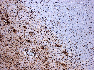
Multiple sclerosis and other demyelinating diseases of the central nervous system (CNS) produce lesions and glial scars or scleroses. They present different shapes and histological findings according to the underlying condition that produces them.

Inflammatory demyelinating diseases (IDDs), sometimes called Idiopathic (IIDDs) due to the unknown etiology of some of them, are a heterogenous group of demyelinating diseases - conditions that cause damage to myelin, the protective sheath of nerve fibers - that occur against the background of an acute or chronic inflammatory process. IDDs share characteristics with and are often grouped together under Multiple Sclerosis. They are sometimes considered different diseases from Multiple Sclerosis, but considered by others to form a spectrum differing only in terms of chronicity, severity, and clinical course.

<span class="mw-page-title-main">Marburg acute multiple sclerosis</span> Medical condition

Marburg acute multiple sclerosis, also known as Marburg multiple sclerosis or acute fulminant multiple sclerosis, is considered one of the multiple sclerosis borderline diseases, which is a collection of diseases classified by some as MS variants and by others as different diseases. Other diseases in this group are neuromyelitis optica (NMO), Balo concentric sclerosis, and Schilder's disease. The graver course is one form of malignant multiple sclerosis, with patients reaching a significant level of disability in less than five years from their first symptoms, often in a matter of months.

<span class="mw-page-title-main">Laquinimod</span> Chemical compound

Laquinimod is an experimental immunomodulator developed by Active Biotech and Teva. It is being investigated as an oral treatment for multiple sclerosis (MS).

Research in multiple sclerosis may find new pathways to interact with the disease, improve function, curtail attacks, or limit the progression of the underlying disease. Many treatments already in clinical trials involve drugs that are used in other diseases or medications that have not been designed specifically for multiple sclerosis. There are also trials involving the combination of drugs that are already in use for multiple sclerosis. Finally, there are also many basic investigations that try to understand better the disease and in the future may help to find new treatments.

A clinically isolated syndrome (CIS) is a clinical situation of an individual's first neurological episode, caused by inflammation or demyelination of nerve tissue. An episode may be monofocal, in which symptoms present at a single site in the central nervous system, or multifocal, in which multiple sites exhibit symptoms. CIS with enough paraclinical evidence can be considered as a clinical stage of multiple sclerosis (MS). It can also be retrospectively diagnosed as a kind of MS when more evidence is available.

<span class="mw-page-title-main">Tumefactive multiple sclerosis</span> Medical condition

Tumefactive multiple sclerosis is a condition in which the central nervous system of a person has multiple demyelinating lesions with atypical characteristics for those of standard multiple sclerosis (MS). It is called tumefactive as the lesions are "tumor-like" and they mimic tumors clinically, radiologically and sometimes pathologically.

Poser criteria are diagnostic criteria for multiple sclerosis (MS). They replaced the older Schumacher criteria, and are now considered obsolete as McDonald criteria have superseded them. Nevertheless, some of the concepts introduced have remained in MS research, like CDMS, and newer criteria are often calibrated against them. The criteria were unveiled in the Annals of Neurology in 1983 by a team led by Dr. Charles M. Poser.

Schumacher criteria are diagnostic criteria that were previously used for identifying multiple sclerosis (MS). Multiple sclerosis, understood as a central nervous system (CNS) condition, can be difficult to diagnose since its signs and symptoms may be similar to other medical problems. Medical organizations have created diagnostic criteria to ease and standardize the diagnostic process especially in the first stages of the disease. Schumacher criteria were the first internationally recognized criteria for diagnosis, and introduced concepts still in use, as CDMS.

Malignant multiple sclerosis is used to describe MS patients who reach significant level of disability in a short period of time. Malignant MS cases are not common, less than 5% of patients with MS experience this type of progression.

<span class="mw-page-title-main">Multiple sclerosis diagnosis</span>

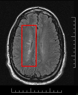
Current standards for diagnosing multiple sclerosis (MS) are based on the 2018 revision of McDonald criteria. They rely on MRI detection of demyelinating lesions in the CNS, which are distributed in space (DIS) and in time (DIT). It is also a requirement that any possible known disease that produces demyelinating lesions is ruled out before applying McDonald's criteria.

Clinical Electrophysiological Testing is based on techniques derived from electrophysiology used for the clinical diagnosis of patients. There are many processes that occur in the body which produce electrical signals that can be detected. Depending on the location and the source of these signals, distinct methods and techniques have been developed to properly target them.

<span class="mw-page-title-main">Pathology of multiple sclerosis</span> Pathologic overview

Multiple sclerosis (MS) can be pathologically defined as the presence of distributed glial scars (scleroses) in the central nervous system that must show dissemination in time (DIT) and in space (DIS) to be considered MS lesions.

Radiologically isolated syndrome (RIS) is a clinical situation in which a person has white matter lesions suggestive of multiple sclerosis (MS), as shown on an MRI scan that was done for reasons unrelated to MS symptoms. The nerve lesions in these people show dissemination in space with an otherwise normal neurological examination and without historical accounts of typical MS symptoms.

Anti-AQP4 diseases, are a group of diseases characterized by auto-antibodies against aquaporin 4.

Brenda Banwell is Chief of the Division of Neurology and Co-Director of the Neuroscience Center, and Professor of Neurology at Children's Hospital of Philadelphia and holder of the Grace R. Loeb Endowed Chair in Neurosciences. She also holds the title of Professor of Pediatrics and Neurology at the Perelman School of Medicine at the University of Pennsylvania.

References

  1. 1 2 Polman, Chris H; Reingold, Stephen C; Edan, Gilles; Filippi, Massimo; Hartung, Hans-Peter; Kappos, Ludwig; Lublin, Fred D; Metz, Luanne M; McFarland, Henry F; O'Connor, Paul W; Sandberg-Wollheim, Magnhild; Thompson, Alan J; Weinshenker, Brian G; Wolinsky, Jerry S (2005). "Diagnostic criteria for multiple sclerosis: 2005 revisions to the 'McDonald Criteria'". Annals of Neurology. 58 (6): 840–6. CiteSeerX   10.1.1.604.2677 . doi:10.1002/ana.20703. PMID   16283615. S2CID   54512368.
  2. 1 2 3 4 Polman, Chris H; Reingold, Stephen C; Banwell, Brenda; Clanet, Michel; Cohen, Jeffrey A; Filippi, Massimo; Fujihara, Kazuo; Havrdova, Eva; Hutchinson, Michael; Kappos, Ludwig; Lublin, Fred D; Montalban, Xavier; O'Connor, Paul; Sandberg-Wollheim, Magnhild; Thompson, Alan J; Waubant, Emmanuelle; Weinshenker, Brian; Wolinsky, Jerry S (2011). "Diagnostic criteria for multiple sclerosis: 2010 Revisions to the McDonald criteria". Annals of Neurology. 69 (2): 292–302. doi:10.1002/ana.22366. PMC   3084507 . PMID   21387374.
  3. Thompson, Alan J; Banwell, Brenda L; Barkhof, Frederik; Carroll, William M; Coetzee, Timothy; Comi, Giancarlo; Correale, Jorge; Fazekas, Franz; Filippi, Massimo; Freedman, Mark S; Fujihara, Kazuo; Galetta, Steven L; Hartung, Hans Peter; Kappos, Ludwig; Lublin, Fred D; Marrie, Ruth Ann; Miller, Aaron E; Miller, David H; Montalban, Xavier; Mowry, Ellen M; Sorensen, Per Soelberg; Tintoré, Mar; Traboulsee, Anthony L; Trojano, Maria; Uitdehaag, Bernard M J; Vukusic, Sandra; Waubant, Emmanuelle; Weinshenker, Brian G; Reingold, Stephen C; Cohen, Jeffrey A (2018). "Diagnosis of multiple sclerosis: 2017 revisions of the Mc Donald criteria" (PDF). The Lancet Neurology. 17 (2): 162–173. doi:10.1016/S1474-4422(17)30470-2. PMID   29275977. S2CID   206164600.
  4. 1 2 3 McDonald, W. Ian; Compston, Alistair; Edan, Gilles; Goodkin, Donald; Hartung, Hans-Peter; Lublin, Fred D; McFarland, Henry F; Paty, Donald W; Polman, Chris H; Reingold, Stephen C; Sandberg-Wollheim, Magnhild; Sibley, William; Thompson, Alan; Van Den Noort, Stanley; Weinshenker, Brian Y; Wolinsky, Jerry S (2001). "Recommended diagnostic criteria for multiple sclerosis: Guidelines from the international panel on the diagnosis of multiple sclerosis". Annals of Neurology. 50 (1): 121–7. CiteSeerX   10.1.1.466.5368 . doi:10.1002/ana.1032. PMID   11456302. S2CID   13870943.
  5. 1 2 3 4 Ntranos, Achilles; Lublin, Fred (2016). "Diagnostic Criteria, Classification and Treatment Goals in Multiple Sclerosis: The Chronicles of Time and Space". Current Neurology and Neuroscience Reports. 16 (10): 90. doi:10.1007/s11910-016-0688-8. PMID   27549391. S2CID   4498332.
  6. 1 2 3 Thompson, Alan J; Banwell, Brenda L; Barkhof, Frederik; Carroll, William M; Coetzee, Timothy; Comi, Giancarlo; Correale, Jorge; Fazekas, Franz; Filippi, Massimo; Freedman, Mark S; Fujihara, Kazuo; Galetta, Steven L; Hartung, Hans Peter; Kappos, Ludwig; Lublin, Fred D; Marrie, Ruth Ann; Miller, Aaron E; Miller, David H; Montalban, Xavier; Mowry, Ellen M; Sorensen, Per Soelberg; Tintoré, Mar; Traboulsee, Anthony L; Trojano, Maria; Uitdehaag, Bernard M J; Vukusic, Sandra; Waubant, Emmanuelle; Weinshenker, Brian G; Reingold, Stephen C; Cohen, Jeffrey A (2018). "Diagnosis of multiple sclerosis: 2017 revisions of the Mc Donald criteria" (PDF). The Lancet Neurology. 17 (2): 162–173. doi:10.1016/S1474-4422(17)30470-2. PMID   29275977. S2CID   206164600.
  7. P. Brex, V. Williams, Updated criteria for diagnosing Multiple Sclerosis, ACNR, VOLUME 18 NUMBER 2, NOVEMBER-JANUARY 2019
  8. Lassmann Hans (2010). "Acute disseminated encephalomyelitis and multiple sclerosis". Brain. 133 (Pt 2): 317–319. doi: 10.1093/brain/awp342 . PMID   20129937.
  9. Miller DH, Weinshenker BG, Filippi M, Banwell BL, Cohen JA, Freedman MS, Galetta SL, Hutchinson M, Johnson RT, Kappos L, Kira J, Lublin FD, McFarland HF, Montalban X, Panitch H, Richert JR, Reingold SC, Polman CH (2008). "Differential diagnosis of suspected multiple sclerosis: a consensus approach". Mult Scler. 14 (9): 1157–1174. doi:10.1177/1352458508096878. PMC   2850590 . PMID   18805839.
  10. Popescu, Bogdan F. Gh; Pirko, Istvan; Lucchinetti, Claudia F (2013). "Pathology of Multiple Sclerosis". CONTINUUM: Lifelong Learning in Neurology. 19 (4 Multiple Sclerosis): 901–21. doi:10.1212/01.CON.0000433291.23091.65. PMC   3915566 . PMID   23917093.
  11. Engell T (May 1989). "A clinical patho-anatomical study of clinically silent multiple sclerosis". Acta Neurol Scand. 79 (5): 428–30. doi: 10.1111/j.1600-0404.1989.tb03811.x . PMID   2741673. S2CID   21581253.
  12. HT Chong et al (2006) Proposed modifications to McDonald diagnostic criteria for Asians with multiple sclerosis, Neurology Asia 11:87–90
  13. Chong, H; Kira, J; Tsai, C; Ong, B; Li, P; Kermode, A; Tan, C (19 June 2009). "Proposed modifications to the McDonald criteria for use in Asia". Multiple Sclerosis. 15 (7): 887–888. doi:10.1177/1352458509104587. PMID   19542266. S2CID   46674042.
  14. Fortini, Alexandre S.; Sanders, Elizabeth L.; Weinshenker, Brian G.; Katzmann, Jerry A. (1 November 2003). "Cerebrospinal Fluid Oligoclonal Bands in the Diagnosis of Multiple Sclerosis Isoelectric Focusing With IgG Immunoblotting Compared With High-Resolution Agarose Gel Electrophoresis and Cerebrospinal Fluid IgG Index". American Journal of Clinical Pathology. 120 (5): 672–675. doi:10.1309/Y5VFF2UAW0RK5W63. PMID   14608891.
  15. "Even in the highest power scanners... you can only see up to 85 percent of lesions. Sometimes there will be lesions that we just can't see on a scan. We also can only see about five percent of lesions in the grey matter in the brain, so we certainly don't see all lesions." https://mstrust.org.uk/news/expert/ask-expert-mri-scans, Will Brown, Cambridge University, 2022. Also at 7.00 min, https://www.youtube.com/watch?v=EQxQoCqifRM&t=206s
  16. Habek M, Establishing; et al. (July 2018). "2010 versus 2017 McDonald criteria" (PDF). Mult Scler Relat Disord. 25: 99–103. doi:10.1016/j.msard.2018.07.035. PMID   30059896. S2CID   207037738.
  17. Runia, TF; Jafari, N; Hintzen, RQ (December 2013). "Application of the 2010 revised criteria for the diagnosis of multiple sclerosis to patients with clinically isolated syndromes". European Journal of Neurology. 20 (12): 1510–6. doi:10.1111/ene.12243. PMID   23906114. S2CID   9493448.
  18. Gaetani L, Prosperini L, Mancini A, Eusebi P, Cerri MC, Pozzilli C, Calabresi P, Sarchielli P, Di Filippo M (2017). "revisions of McDonald criteria shorten the time to diagnosis of multiple sclerosis in clinically isolated syndromes". J Neurol. 265 (11): 2684–2687. doi:10.1007/s00415-018-9048-8. PMID   30196327. S2CID   52176296.{{cite journal}}: CS1 maint: multiple names: authors list (link)
  19. McDonald_criteria#Criticism
  20. Filippi, Massimo; Rocca, Maria A; Ciccarelli, Olga; De Stefano, Nicola; Evangelou, Nikos; Kappos, Ludwig; Rovira, Alex; Sastre-Garriga, Jaume; Tintorè, Mar; Frederiksen, Jette L; Gasperini, Claudio; Palace, Jacqueline; Reich, Daniel S; Banwell, Brenda; Montalban, Xavier; Barkhof, Frederik (2016). "MRI criteria for the diagnosis of multiple sclerosis: MAGNIMS consensus guidelines". The Lancet Neurology. 15 (3): 292–303. doi:10.1016/S1474-4422(15)00393-2. PMC   4760851 . PMID   26822746.
  21. Awad, Amer; Hemmer, Bernhard; Hartung, Hans-Peter; Kieseier, Bernd; Bennett, Jeffrey L; Stuve, Olaf (2010). "Analyses of cerebrospinal fluid in the diagnosis and monitoring of multiple sclerosis". Journal of Neuroimmunology. 219 (1–2): 1–7. doi:10.1016/j.jneuroim.2009.09.002. PMID   19782408. S2CID   206273704.
  22. Brettschneider, Johannes; Jaskowski, Troy D; Tumani, Hayrettin; Abdul, Sana; Husebye, Dee; Seraj, Haniah; Hill, Harry R; Fire, Ella; Spector, Larissa; Yarden, Jennifer; Dotan, Nir; Rose, John W (2009). "Serum anti-GAGA4 IgM antibodies differentiate relapsing remitting and secondary progressive multiple sclerosis from primary progressive multiple sclerosis and other neurological diseases". Journal of Neuroimmunology. 217 (1–2): 95–101. doi:10.1016/j.jneuroim.2009.07.017. PMID   19879655. S2CID   43615028.
  23. Quintana FJ, Farez MF, Viglietta V, et al. (2008). "Antigen microarrays identify unique serum autoantibody signatures in clinical and pathologic subtypes of multiple sclerosis". Proc Natl Acad Sci U S A. 105 (48): 18889–18894. Bibcode:2008PNAS..10518889Q. doi: 10.1073/pnas.0806310105 . PMC   2596207 . PMID   19028871.
  24. Keller A, Leidinger P, Lange J, et al. (2009). "Multiple sclerosis: microRNA expression profiles accurately differentiate patients with relapsing-remitting disease from healthy controls". PLOS ONE. 4 (10): e7440. Bibcode:2009PLoSO...4.7440K. doi: 10.1371/journal.pone.0007440 . PMC   2757919 . PMID   19823682.
  25. Nickles D et al., Blood RNA profiling in a large cohort of multiple sclerosis patients and healthy controls, Hum Mol Genet. 2013 Oct 15;22(20):4194-205. doi: 10.1093/hmg/ddt267. Epub 2013 Jun 6.