Isotope-ratio mass spectrometry (IRMS) is a specialization of mass spectrometry, in which mass spectrometric methods are used to measure the relative abundance of isotopes in a given sample.[1][2]
This technique has two different applications in the earth and environmental sciences. The analysis of 'stable isotopes' is normally concerned with measuring isotopic variations arising from mass-dependent isotopic fractionation in natural systems. On the other hand, radiogenic isotope analysis[3] involves measuring the abundances of decay-products of natural radioactivity, and is used in most long-lived radiometric dating methods.
Introduction
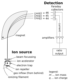
Schematic of an isotope-ratio mass spectrometer for measuring CO2
The isotope-ratio mass spectrometer (IRMS) allows the precise measurement of mixtures of naturally occurring isotopes.[4] Most instruments used for precise determination of isotope ratios are of the magnetic sector type. This type of analyzer is superior to the quadrupole type in this field of research for two reasons. First, it can be set up for multiple-collector analysis, and second, it gives high-quality 'peak shapes'. Both of these considerations are important for isotope-ratio analysis at very high precision and accuracy.[3]
The sector-type instrument designed by Alfred Nier was such an advance in mass spectrometer design that this type of instrument is often called the 'Nier type'. In the most general terms the instrument operates by ionizing the sample of interest, accelerating it over a potential in the kilo-volt range, and separating the resulting stream of ions according to their mass-to-charge ratio (m/z). Beams with lighter ions bend at a smaller radius than beams with heavier ions. The current of each ion beam is then measured using a 'Faraday cup' or multiplier detector.
Many radiogenic isotope measurements are made by ionization of a solid source, whereas stable isotope measurements of light elements (e.g. H, C, O) are usually made in an instrument with a gas source. In a "multicollector" instrument, the ion collector typically has an array of Faraday cups, which allows the simultaneous detection of multiple isotopes.[5]
Gas source mass spectrometry
Measurement of natural variations in the abundances of stable isotopes of the same element is normally referred to as stable isotope analysis. This field is of interest because the differences in mass between different isotopes leads to isotope fractionation, causing measurable effects on the isotopic composition of samples, characteristic of their biological or physical history.
As a specific example, the hydrogen isotope deuterium (heavy hydrogen) is almost double the mass of the common hydrogen isotope. Water molecules containing the common hydrogen isotope (and the common oxygen isotope, mass 16) have a mass of 18. Water incorporating a deuterium atom has a mass of 19, over 5% heavier. The energy to vaporise the heavy water molecule is higher than that to vaporize the normal water so isotope fractionation occurs during the process of evaporation. Thus a sample of sea water will exhibit a quite detectable isotopic-ratio difference when compared to Antarctic snowfall.
Samples must be introduced to the mass spectrometer as pure gases, achieved through combustion, gas chromatographic feeds,[6] or chemical trapping. By comparing the detected isotopic ratios to a measured standard, an accurate determination of the isotopic make up of the sample is obtained. For example, carbon isotope ratios are measured relative to the international standard for C. The C standard is produced from a fossil belemnite found in the Peedee Formation, which is a limestone formed in the Cretaceous period in South Carolina, U.S.A. The fossil is referred to as VPDB (Vienna Pee Dee Belemnite) and has 13C:12C ratio of 0.0112372. Oxygen isotope ratios are measured relative the standard, V-SMOW (Vienna Standard Mean Ocean Water).
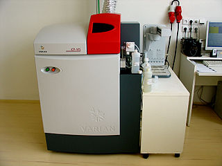
Isotope-ratio mass spectrometer used to measure stable isotope ratios, with gas bench in foreground
It is critical that the sample be processed before entering the mass spectrometer so that only a single chemical species enters at a given time. Generally, samples are combusted or pyrolyzed and the desired gas species (usually hydrogen (H2), nitrogen (N2), carbon dioxide (CO2), or sulfur dioxide (SO2)) is purified by means of traps, filters, catalysts and/or chromatography.
The two most common types of IRMS instruments are continuous flow[7] and dual inlet. In dual inlet IRMS, purified gas obtained from a sample is alternated rapidly with a standard gas (of known isotopic composition) by means of a system of valves, so that a number of comparison measurements are made of both gases. In continuous flow IRMS, sample preparation occurs immediately before introduction to the IRMS, and the purified gas produced from the sample is measured just once. The standard gas may be measured before and after the sample or after a series of sample measurements. While continuous-flow IRMS instruments can achieve higher sample throughput and are more convenient to use than dual inlet instruments, the yielded data is of approximately 10-fold lower precision.
Static gas mass spectrometry
A static gas mass spectrometer is one in which a gaseous sample for analysis is fed into the source of the instrument and then left in the source without further supply or pumping throughout the analysis. This method can be used for 'stable isotope' analysis of light gases (as above), but it is particularly used in the isotopic analysis of noble gases (rare or inert gases) for radiometric dating or isotope geochemistry. Important examples are argon–argon dating and helium isotope analysis.
When these isotope ratios are measured by TIMS, mass-dependent fractionation occurs as species are emitted by the hot filament. Fractionation occurs due to the excitation of the sample and therefore must be corrected for accurate measurement of the isotope ratio.[8]
There are several advantages of the TIMS method. It has a simple design, is less expensive than other mass spectrometers, and produces stable ion emissions. It requires a stable power supply, and is suitable for species with a low ionization potential, such as Strontium (Sr), and Lead (Pb).
The disadvantages of this method stem from the maximum temperature achieved in thermal ionization. The hot filament reaches a temperature of less than 2500 degrees Celsius, leading to the inability to create atomic ions of species with a high ionization potential, such as Osmium (Os), and Tungsten (Hf-W). Although the TIMS method can create molecular ions instead in this case, species with high ionization potential can be analyzed more effectively with MC-ICP-MS.
Secondary-ion mass spectrometry
Schematic diagram of a SHRIMP instrument illustrating the ion beam path. After Figure 4, Williams, 1998.
An alternative approach used to measure the relative abundance of radiogenic isotopes when working with a solid surface is secondary-ion mass spectrometry (SIMS). This type of ion-microprobe analysis normally works by focusing a primary (oxygen) ion beam on a sample in order to generate a series of secondary positive ions that can be focused and measured based on their mass/charge ratios.
SIMS is a common method used in U-Pb analysis, as the primary ion beam is used to bombard the surface of a single zircon grain in order to yield a secondary beam of Pb ions. The Pb ions are analyzed using a double focusing mass spectrometer that comprises both an electrostatic and magnetic analyzer. This assembly allows the secondary ions to be focused based on their kinetic energy and mass-charge ratio in order to be accurately collected using a series of Faraday cups.[10]
A major issue that arises in SIMS analysis is the generation of isobaric interference between sputtered molecular ions and the ions of interest. This issue occurs with U–Pb dating as Pb ions have essentially the same mass as HfO2+.[11] In order to overcome this problem, a sensitive high-resolution ion microprobe (SHRIMP) can be used. A SHRIMP is a double-focusing mass spectrometer that allows for a large spatial separation between different ion masses based on its relatively large size. For U-Pb analysis, the SHRIMP allows for the separation of Pb from other interfering molecular ions, such as HfO2+.
Multiple collector inductively coupled plasma mass spectrometry
An MC-ICP-MS instrument is a multiple collector mass spectrometer with a plasma source. MC-ICP-MS was developed to improve the precision achievable by ICP-MS during isotope-ratio measurements. Conventional ICP-MS analysis uses a quadrupole analyser, which only allows single-collector analysis. Due to the inherent instability of the plasma, this limits the precision of ICP-MS with a quadrupole analyzer to around 1%, which is insufficient for most radiogenic isotope systems.
Isotope-ratio analysis for radiometric dating has normally been determined by TIMS. However, some systems (e.g. Hf-W and Lu-Hf) are difficult or impossible to analyse by TIMS, due to the high ionization potential of the elements involved. Therefore, these methods can now be analysed using MC-ICP-MS.
The Ar-ICP produces an ion-beam with a large inherent kinetic energy distribution, which makes the design of the mass-spectrometer somewhat more complex than it is the case for conventional TIMS instruments. First, different from Quadrupole ICP-MS systems, magnetic sector instruments have to operate with a higher acceleration potential (several 1000 V) in order to minimize the energy distribution of the ion beam. Modern instruments operate at 6-10kV. The radius of deflection of an ion within a magnetic field depends on the kinetic energy and the mass/charge ratio of the ion (strictly, the magnet is a momentum analyzer not just a mass analyzer). Because of the large energy distribution, ions with similar mass/charge ratio can have very different kinetic energies and will thus experience different deflection for the same magnetic field. In practical terms one would see that ions with the same mass/charge ratio focus at different points in space. However, in a mass-spectrometer one wants ions with the same mass/charge ratio to focus at the same point, e.g. where the detector is located. In order to overcome these limitations, commercial MC-ICP-MS are double-focusing instruments. In a double-focusing mass-spectrometer ions are focused due to kinetic energy by the ESA (electro-static-analyzer) and kinetic energy + mass/charge (momentum) in the magnetic field. Magnet and ESA are carefully chosen to match the energy focusing properties of one another and are arranged so that the direction of energy focusing is in opposite directions. To simplify, two components have an energy focus term, when arranged properly, the energy term cancels out and ions with the same mass/charge ratio focus at the same point in space. It is important to note, double-focusing does not reduce the kinetic energy distribution and different kinetic energies are not filtered or homogenized. Double-focusing works for single as well as multi-collector instruments. In single collector instruments ESA and magnet can be arranged in either forward geometry (first ESA then magnet) or reversed geometry (magnet first then ESA), as only point-to-point focusing is required. In multi-collector instruments, only forward geometry (ESA then magnet) is possible due to the array of detectors and the requirements of a focal plane rather than a focal point.
Accelerator mass spectrometry
Accelerator mass spectrometry
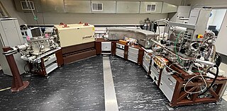
Accelerator mass spectrometer at Lawrence Livermore National Laboratory
For isotopes occurring at extremely low levels, accelerator mass spectrometry (AMS) can be used. For example, the decay rate of the radioisotope 14C is widely used to date organic materials, but this approach was once limited to relatively large samples no more than a few thousand years old. AMS extended the range of 14C dating to about 60,000 years BP, and is about 106 times more sensitive than conventional IRMS.
AMS works by accelerating negative ions through a large (mega-volt) potential, followed by charge exchange and acceleration back to ground. During charge exchange, interfering species can be effectively removed. In addition, the high energy of the beam allows the use of energy-loss detectors, that can distinguish between species with the same mass/charge ratio. Together, these processes allow the analysis of extreme isotope ratios above 1012.
Moving wire IRMS
Moving wire IRMS is useful for analyzing carbon-13 ratios of compounds in a solution, such as after purification by liquid chromatography. The solution (or outflow from the chromatography) is dried onto a nickel or stainless steel wire. After the residue is deposited on the wire, it enters a furnace where the sample is converted to CO2 and water by combustion. The gas stream finally enters a capillary, is dried, ionized, and analyzed.[12] This process allows a mixture of compounds to be purified and analyzed continuously, which can decrease the analysis time by a factor of four.[12] Moving wire IRMS is quite sensitive, and samples containing as little as 1 nanomole of carbon can yield precise (within 1‰) results.[13]
Inductively coupled plasma mass spectrometry (ICP-MS) is a type of mass spectrometry that uses an inductively coupled plasma to ionize the sample. It atomizes the sample and creates atomic and small polyatomic ions, which are then detected. It is known and used for its ability to detect metals and several non-metals in liquid samples at very low concentrations. It can detect different isotopes of the same element, which makes it a versatile tool in isotopic labeling.
Mass spectrometry (MS) is an analytical technique that is used to measure the mass-to-charge ratio of ions. The results are presented as a mass spectrum, a plot of intensity as a function of the mass-to-charge ratio. Mass spectrometry is used in many different fields and is applied to pure samples as well as complex mixtures.
An ion source is a device that creates atomic and molecular ions. Ion sources are used to form ions for mass spectrometers, optical emission spectrometers, particle accelerators, ion implanters and ion engines.
Electron ionization is an ionization method in which energetic electrons interact with solid or gas phase atoms or molecules to produce ions. EI was one of the first ionization techniques developed for mass spectrometry. However, this method is still a popular ionization technique. This technique is considered a hard ionization method, since it uses highly energetic electrons to produce ions. This leads to extensive fragmentation, which can be helpful for structure determination of unknown compounds. EI is the most useful for organic compounds which have a molecular weight below 600. Also, several other thermally stable and volatile compounds in solid, liquid and gas states can be detected with the use of this technique when coupled with various separation methods.
Secondary-ion mass spectrometry (SIMS) is a technique used to analyze the composition of solid surfaces and thin films by sputtering the surface of the specimen with a focused primary ion beam and collecting and analyzing ejected secondary ions. The mass/charge ratios of these secondary ions are measured with a mass spectrometer to determine the elemental, isotopic, or molecular composition of the surface to a depth of 1 to 2 nm. Due to the large variation in ionization probabilities among elements sputtered from different materials, comparison against well-calibrated standards is necessary to achieve accurate quantitative results. SIMS is the most sensitive surface analysis technique, with elemental detection limits ranging from parts per million to parts per billion.
Tandem mass spectrometry, also known as MS/MS or MS2, is a technique in instrumental analysis where two or more stages of analysis using one or more mass analyzer are performed with an additional reaction step in between these analyses to increase their abilities to analyse chemical samples. A common use of tandem MS is the analysis of biomolecules, such as proteins and peptides.
Gas chromatography–mass spectrometry (GC–MS) is an analytical method that combines the features of gas-chromatography and mass spectrometry to identify different substances within a test sample. Applications of GC–MS include drug detection, fire investigation, environmental analysis, explosives investigation, food and flavor analysis, and identification of unknown samples, including that of material samples obtained from planet Mars during probe missions as early as the 1970s. GC–MS can also be used in airport security to detect substances in luggage or on human beings. Additionally, it can identify trace elements in materials that were previously thought to have disintegrated beyond identification. Like liquid chromatography–mass spectrometry, it allows analysis and detection even of tiny amounts of a substance.
The sensitive high-resolution ion microprobe is a large-diameter, double-focusing secondary ion mass spectrometer (SIMS) sector instrument that was produced by Australian Scientific Instruments in Canberra, Australia and now has been taken over by Chinese company Dunyi Technology Development Co. (DTDC) in Beijing. Similar to the IMS 1270-1280-1300 large-geometry ion microprobes produced by CAMECA, Gennevilliers, France and like other SIMS instruments, the SHRIMP microprobe bombards a sample under vacuum with a beam of primary ions that sputters secondary ions that are focused, filtered, and measured according to their energy and mass.
In chemistry, isotopologues are molecules that differ only in their isotopic composition. They have the same chemical formula and bonding arrangement of atoms, but at least one atom has a different number of neutrons than the parent.
Fast atom bombardment (FAB) is an ionization technique used in mass spectrometry in which a beam of high energy atoms strikes a surface to create ions. It was developed by Michael Barber at the University of Manchester in 1980. When a beam of high energy ions is used instead of atoms, the method is known as liquid secondary ion mass spectrometry (LSIMS). In FAB and LSIMS, the material to be analyzed is mixed with a non-volatile chemical protection environment, called a matrix, and is bombarded under vacuum with a high energy beam of atoms. The atoms are typically from an inert gas such as argon or xenon. Common matrices include glycerol, thioglycerol, 3-nitrobenzyl alcohol (3-NBA), 18-crown-6 ether, 2-nitrophenyloctyl ether, sulfolane, diethanolamine, and triethanolamine. This technique is similar to secondary ion mass spectrometry and plasma desorption mass spectrometry.
Ion mobility spectrometry (IMS) It is a method of conducting analytical research that separates and identifies ionized molecules present in the gas phase based on the mobility of the molecules in a carrier buffer gas. Even though it is used extensively for military or security objectives, such as detecting drugs and explosives, the technology also has many applications in laboratory analysis, including studying small and big biomolecules. IMS instruments are extremely sensitive stand-alone devices, but are often coupled with mass spectrometry, gas chromatography or high-performance liquid chromatography in order to achieve a multi-dimensional separation. They come in various sizes, ranging from a few millimeters to several meters depending on the specific application, and are capable of operating under a broad range of conditions. IMS instruments such as microscale high-field asymmetric-waveform ion mobility spectrometry can be palm-portable for use in a range of applications including volatile organic compound (VOC) monitoring, biological sample analysis, medical diagnosis and food quality monitoring. Systems operated at higher pressure are often accompanied by elevated temperature, while lower pressure systems (1-20 hPa) do not require heating.
The history of mass spectrometry has its roots in physical and chemical studies regarding the nature of matter. The study of gas discharges in the mid 19th century led to the discovery of anode and cathode rays, which turned out to be positive ions and electrons. Improved capabilities in the separation of these positive ions enabled the discovery of stable isotopes of the elements. The first such discovery was with the element neon, which was shown by mass spectrometry to have at least two stable isotopes: 20Ne and 22Ne. Mass spectrometers were used in the Manhattan Project for the separation of isotopes of uranium necessary to create the atomic bomb.
Time-of-flight mass spectrometry (TOFMS) is a method of mass spectrometry in which an ion's mass-to-charge ratio is determined by a time of flight measurement. Ions are accelerated by an electric field of known strength. This acceleration results in an ion having the same kinetic energy as any other ion that has the same charge. The velocity of the ion depends on the mass-to-charge ratio. The time that it subsequently takes for the ion to reach a detector at a known distance is measured. This time will depend on the velocity of the ion, and therefore is a measure of its mass-to-charge ratio. From this ratio and known experimental parameters, one can identify the ion.
Ion mobility spectrometry–mass spectrometry (IMS-MS) is an analytical chemistry method that separates gas phase ions based on their interaction with a collision gas and their masses. In the first step, the ions are separated according to their mobility through a buffer gas on a millisecond timescale using an ion mobility spectrometer. The separated ions are then introduced into a mass analyzer in a second step where their mass-to-charge ratios can be determined on a microsecond timescale. The effective separation of analytes achieved with this method makes it widely applicable in the analysis of complex samples such as in proteomics and metabolomics.
Thermal ionization, also known as surface ionization or contact ionization, is a physical process whereby the atoms are desorbed from a hot surface, and in the process are ionized.
Thermal ionization mass spectrometry (TIMS) is also known as surface ionization and is a highly sensitive isotope mass spectrometry characterization technique. The isotopic ratios of radionuclides are used to get an accurate measurement for the elemental analysis of a sample. Singly charged ions of the sample are formed by the thermal ionization effect. A chemically purified liquid sample is placed on a metal filament which is then heated to evaporate the solvent. The removal of an electron from the purified sample is consequently achieved by heating the filament enough to release an electron, which then ionizes the atoms of the sample. TIMS utilizes a magnetic sector mass analyzer to separate the ions based on their mass to charge ratio. The ions gain velocity by an electrical potential gradient and are focused into a beam by electrostatic lenses. The ion beam then passes through the magnetic field of the electromagnet where it is partitioned into separate ion beams based on the ion's mass/charge ratio. These mass-resolved beams are directed into a detector where it is converted into voltage. The voltage detected is then used to calculate the isotopic ratio.
Aerosol mass spectrometry is the application of mass spectrometry to the analysis of the composition of aerosol particles. Aerosol particles are defined as solid and liquid particles suspended in a gas (air), with size range of 3 nm to 100 μm in diameter and are produced from natural and anthropogenic sources, through a variety of different processes that include wind-blown suspension and combustion of fossil fuels and biomass. Analysis of these particles is important owing to their major impacts on global climate change, visibility, regional air pollution and human health. Aerosols are very complex in structure, can contain thousands of different chemical compounds within a single particle, and need to be analysed for both size and chemical composition, in real-time or off-line applications.
A miniature mass spectrometer (MMS) is a type of mass spectrometer (MS) which has small size and weight and can be understood as a portable or handheld device. Current lab-scale mass spectrometers however, usually weigh hundreds of pounds and can cost on the range from thousands to millions of dollars. One purpose of producing MMS is for in situ analysis. This in situ analysis can lead to much simpler mass spectrometer operation such that non-technical personnel like physicians at the bedside, firefighters in a burning factory, food safety inspectors in a warehouse, or airport security at airport checkpoints, etc. can analyze samples themselves saving the time, effort, and cost of having the sample run by a trained MS technician offsite. Although, reducing the size of MS can lead to a poorer performance of the instrument versus current analytical laboratory standards, MMS is designed to maintain sufficient resolutions, detection limits, accuracy, and especially the capability of automatic operation. These features are necessary for the specific in-situ applications of MMS mentioned above.
Resonance ionization is a process in optical physics used to excite a specific atom beyond its ionization potential to form an ion using a beam of photons irradiated from a pulsed laser light. In resonance ionization, the absorption or emission properties of the emitted photons are not considered, rather only the resulting excited ions are mass-selected, detected and measured. Depending on the laser light source used, one electron can be removed from each atom so that resonance ionization produces an efficient selectivity in two ways: elemental selectivity in ionization and isotopic selectivity in measurement.
References
↑ Paul D, Skrzypek G, Fórizs I (2007). "Normalization of measured stable isotopic compositions to isotope reference scales - a review". Rapid Commun. Mass Spectrom. 21 (18): 3006–14. Bibcode:2007RCMS...21.3006P. doi:10.1002/rcm.3185. PMID17705258.
↑ Stellaard F, Elzinga H (2005). "Analytical techniques in biomedical stable isotope applications: (isotope ratio) mass spectrometry or infrared spectrometry?". Isotopes in Environmental and Health Studies. 41 (4): 345–61. doi:10.1080/10256010500384333. PMID16543190.
↑ Townsend, A., ed. (1995). Encyclopaedia of Analytical Science Encyclopaedia of Analytical Science. London: Academic Press Limited.
↑ C. B. Bouthitt; K. Garnett. "The Evolution of the Multicollector in Isotope Ratio Mass Spectromety". Proceedings of the 18th AMZSMS Conference: THO–07.
↑ Meier-Augenstein, W. (1999). "Applied gas chromatography coupled to isotope ratio mass spectrometry". J. Chromatogr. A. 842 (1–2): 351–371. doi:10.1016/S0021-9673(98)01057-7. PMID10377971.
↑ Dickin, A.P., 2005. Radiogenic Isotope Geology 2nd ed. (Cambridge: Cambridge University Press), pp. 21-22.
↑ Williams, I.S. (1998), "U-Th-Pb geochronology by ion microprobe", In: McKibben, M.A.; Shanks III, W.C.; Ridley, W.I.; (Editors), "Applications of microanalytical techniques to understanding mineralizing processes", Reviews in Economic Geology Special Publication 7: 1–35
↑ Dickin, A. P. (2005). Radiogenic Isotope Geology 2nd ed. Cambridge University Press.
↑ Hinton, R.W. and Long, J.V.P. (1979). High-resolution ion microprobe measurement of lead isotopes: variations within single zircons from Lac Seul, Northwestern Ontario. Earth Planet. Sci. lett. 45, 309-325.,
1 2 Caimi, R. J.; Brenna, J. T. (1996). "Direct analysis of carbon isotope variability in albumins by liquid flow-injection isotope ratio mass spectrometry". J. Am. Soc. Mass Spectrom. 7 (6): 605–610. doi:10.1016/1044-0305(96)00010-4. PMID24203433.
↑ Sessions, A.L.; Sylva, S.P.; Hayes, J.M. (2005). "Moving-wire device for carbon isotopic analyses of nanogram quantities of nonvolatile organic carbon". Analytical Chemistry. 77 (20): 6519–6527. doi:10.1021/ac051251z. PMID16223235.
Bibliography
Goetz, A.; Platzner, I. T. (Itzhak Thomas); Habfast, K.; Walder, A. J. (1997). Modern isotope ratio mass spectrometry. London: J. Wiley. ISBN978-0-471-97416-1. OCLC36461690.
This page is based on this Wikipedia article Text is available under the CC BY-SA 4.0 license; additional terms may apply. Images, videos and audio are available under their respective licenses.