Related Research Articles

Cell adhesion is the process by which cells interact and attach to neighbouring cells through specialised molecules of the cell surface. This process can occur either through direct contact between cell surfaces such as cell junctions or indirect interaction, where cells attach to surrounding extracellular matrix, a gel-like structure containing molecules released by cells into spaces between them. Cells adhesion occurs from the interactions between cell-adhesion molecules (CAMs), transmembrane proteins located on the cell surface. Cell adhesion links cells in different ways and can be involved in signal transduction for cells to detect and respond to changes in the surroundings. Other cellular processes regulated by cell adhesion include cell migration and tissue development in multicellular organisms. Alterations in cell adhesion can disrupt important cellular processes and lead to a variety of diseases, including cancer and arthritis. Cell adhesion is also essential for infectious organisms, such as bacteria or viruses, to cause diseases.

A retinal ganglion cell (RGC) is a type of neuron located near the inner surface of the retina of the eye. It receives visual information from photoreceptors via two intermediate neuron types: bipolar cells and retina amacrine cells. Retina amacrine cells, particularly narrow field cells, are important for creating functional subunits within the ganglion cell layer and making it so that ganglion cells can observe a small dot moving a small distance. Retinal ganglion cells collectively transmit image-forming and non-image forming visual information from the retina in the form of action potential to several regions in the thalamus, hypothalamus, and mesencephalon, or midbrain.
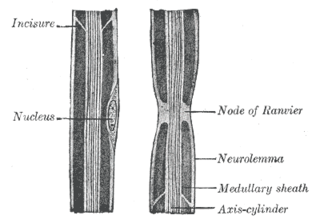
In neuroscience and anatomy, nodes of Ranvier, also known as myelin-sheath gaps, occur along a myelinated axon where the axolemma is exposed to the extracellular space. Nodes of Ranvier are uninsulated and highly enriched in ion channels, allowing them to participate in the exchange of ions required to regenerate the action potential. Nerve conduction in myelinated axons is referred to as saltatory conduction due to the manner in which the action potential seems to "jump" from one node to the next along the axon. This results in faster conduction of the action potential.

Cadherins (named for "calcium-dependent adhesion") are cell adhesion molecules important in forming adherens junctions that let cells adhere to each other. Cadherins are a class of type-1 transmembrane proteins, and they depend on calcium (Ca2+) ions to function, hence their name. Cell-cell adhesion is mediated by extracellular cadherin domains, whereas the intracellular cytoplasmic tail associates with numerous adaptors and signaling proteins, collectively referred to as the cadherin adhesome.
Cell adhesion molecules (CAMs) are a subset of cell surface proteins that are involved in the binding of cells with other cells or with the extracellular matrix (ECM), in a process called cell adhesion. In essence, CAMs help cells stick to each other and to their surroundings. CAMs are crucial components in maintaining tissue structure and function. In fully developed animals, these molecules play an integral role in generating force and movement and consequently ensuring that organs are able to execute their functions normally. In addition to serving as "molecular glue", CAMs play important roles in the cellular mechanisms of growth, contact inhibition, and apoptosis. Aberrant expression of CAMs may result in a wide range of pathologies, ranging from frostbite to cancer.

L1, also known as L1CAM, is a transmembrane protein member of the L1 protein family, encoded by the L1CAM gene. This protein, of 200-220 kDa, is a neuronal cell adhesion molecule with a strong implication in cell migration, adhesion, neurite outgrowth, myelination and neuronal differentiation. It also plays a key role in treatment-resistant cancers due to its function. It was first identified in 1984 by M. Schachner who found the protein in post-mitotic mice neurons.

Neural cell adhesion molecule (NCAM), also called CD56, is a homophilic binding glycoprotein expressed on the surface of neurons, glia and skeletal muscle. Although CD56 is often considered a marker of neural lineage commitment due to its discovery site, CD56 expression is also found in, among others, the hematopoietic system. Here, the expression of CD56 is mostly associated with, but not limited to, natural killer cells. CD56 has been detected on other lymphoid cells, including gamma delta (γδ) Τ cells and activated CD8+ T cells, as well as on dendritic cells. NCAM has been implicated as having a role in cell–cell adhesion, neurite outgrowth, synaptic plasticity, and learning and memory.

DSCAM and Dscam are both abbreviations for Down syndrome cell adhesion molecule. In humans, DSCAM refers to a gene that encodes one of several protein isoforms.
Axon guidance is a subfield of neural development concerning the process by which neurons send out axons to reach their correct targets. Axons often follow very precise paths in the nervous system, and how they manage to find their way so accurately is an area of ongoing research.

Netrins are a class of proteins involved in axon guidance. They are named after the Sanskrit word "netr", which means "one who guides". Netrins are genetically conserved across nematode worms, fruit flies, frogs, mice, and humans. Structurally, netrin resembles the extracellular matrix protein laminin.

Contactin-2 is a protein that in humans is encoded by the CNTN2 gene.

Neurofascin is a protein that in humans is encoded by the NFASC gene.

Receptor-type tyrosine-protein phosphatase mu is an enzyme that in humans is encoded by the PTPRM gene.

Neuronal cell adhesion molecule is a protein that in humans is encoded by the NRCAM gene.

Neurocan core protein is a protein that in humans is encoded by the NCAN gene.
Slit is a family of secreted extracellular matrix proteins which play an important signalling role in the neural development of most bilaterians. While lower animal species, including insects and nematode worms, possess a single Slit gene, humans, mice and other vertebrates possess three Slit homologs: Slit1, Slit2 and Slit3. Human Slits have been shown to be involved in certain pathological conditions, such as cancer and inflammation.
IgSF CAMs are cell adhesion molecules that belong to Immunoglobulin superfamily. It is regarded as the most diverse superfamily of CAMs. This family is characterized by their extracellular domains containing Ig-like domains. The Ig domains are then followed by Fibronectin type III domain repeats and IgSFs are anchored to the membrane by a GPI moiety. This family is involved in both homophilic or heterophilic binding and has the ability to bind integrins or different IgSF CAMs.

Cellular adhesions can be defined as proteins or protein aggregates that form mechanical and chemical linkages between the intracellular and extracellular space. Adhesions serve several critical processes including cell migration, signal transduction, tissue development and repair. Due to this functionality, adhesions and adhesion molecules have been a topic of study within the scientific community. Specifically, it has been found that adhesions are involved in tissue development, plasticity, and memory formation within the central nervous system (CNS), and may prove vital in the generation of CNS-specific therapeutics.
A follower neuron is a nerve cell that arises in the developmental stage of the brain and which growth and orientation is intrinsically related to pioneer neurons. These neurons can also be called later development neurons or follower cells. In the early stages of brain development, pioneer neurons define axonal trajectories that are later used as scaffolds by follower neurons, which project their growth cones and fasciculate with pioneer axons, forming a fiber tract and demonstrating a preference for axon-guided growth. It is thought that these neurons can read very accurate cues of direction and fasciculate or defasciculate in order to reach their target, even in a highly dense axon bundle.

Synaptic stabilization is crucial in the developing and adult nervous systems and is considered a result of the late phase of long-term potentiation (LTP). The mechanism involves strengthening and maintaining active synapses through increased expression of cytoskeletal and extracellular matrix elements and postsynaptic scaffold proteins, while pruning less active ones. For example, cell adhesion molecules (CAMs) play a large role in synaptic maintenance and stabilization. Gerald Edelman discovered CAMs and studied their function during development, which showed CAMs are required for cell migration and the formation of the entire nervous system. In the adult nervous system, CAMs play an integral role in synaptic plasticity relating to learning and memory.
References
- ↑ Hortsch M, Nagaraj K, Mualla R (2014). "The L1 Family of Cell Adhesion Molecules: A Sickening Number of Mutations and Protein Functions". Cell Adhesion Molecules. Advances in Neurobiology. Vol. 8. pp. 195–229. doi:10.1007/978-1-4614-8090-7_9. ISBN 978-1-4614-8089-1. PMID 25300138.
- ↑ Wei CH, Ryu SE (July 2012). "Homophilic interaction of the L1 family of cell adhesion molecules". Experimental & Molecular Medicine. 44 (7): 413–23. doi:10.3858/emm.2012.44.7.050. PMC 3406286 . PMID 22573111.
- ↑ Herron LR, Hill M, Davey F, Gunn-Moore FJ (May 2009). "The intracellular interactions of the L1 family of cell adhesion molecules". The Biochemical Journal. 419 (3): 519–31. doi:10.1042/BJ20082284. PMID 19356150.