Lesser occipital nerve

Last updated
Lesser occipital nerve
Gray1210.png
Side of neck, showing chief surface markings. (Lesser occip. nerve labeled at center right.)
Gray805.png
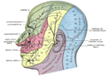
The nerves of the scalp, face, and side of neck. (Smaller occipital visible below and to the left of the ear.)
Details
From Cervical plexus (C2)
InnervatesCutaneous innervation of the posterior aspect of the auricle and mastoid region
Identifiers
Latin nervus occipitalis minor
TA98 A14.2.02.017
TA2 6384
FMA 6871
Anatomical terms of neuroanatomy

The lesser occipital nerve (or small occipital nerve [1] ) is a cutaneous spinal nerve of the cervical plexus. [2] It arises from second cervical (spinal) nerve (C2) (along with the greater occipital nerve). It innervates the skin of the back of the upper neck and of the scalp posterior to the ear.

Contents

Structure

Origin

It arises from the (lateral branch of the ventral ramus [3] [4] ) of cervical spinal nerve C2; [3] [4] [2] it (sources differ) receives [1] or may also receive fibres from cervical spinal nerve C3. [3] [4] It originates between the atlas, and axis. [4]

The lesser occipital nerve is one of the four cutaneous branches of the cervical plexus. [5]

Course and relations

It curves around the accessory nerve (CN XI) [4] [3] [6] to come to course anterior to it. [3] It then [3] curves around [4] and ascends along the posterior border of the sternocleidomastoid muscle; [3] [4] [6] rarely, it may pierce the muscle. [4] Near the cranium, it perforates the deep cervical fascia. It is continues upwards along the scalp posterior to the auricle. [3] [4] It divides into medial and lateral segments between the inion, and intermastoid line. [1]

Branches

It has an auricular, a mastoid, and an occipital branch. [1]

Its auricular branch supplies the skin of the upper and back part of the auricula, communicating with the mastoid branch of the great auricular. This branch is occasionally derived from the greater occipital nerve.[ citation needed ]

Distribution

The nerve provides sensory innervation to the upper part of the back of the neck and adjacent [6] scalp posterior to the auricle; [6] [1] it may also contribute to the sensory innervation of the auricle itself. [6]

Communications

It communicates with the greater occipital nerve, [4] [1] great auricular nerve, and the auricular branch of the facial nerve. [4]

Variation

Rarely, the lesser occipital nerve may be duplicated or triplicated. [4]

Clinical significance

Problems with the lesser occipital nerve cause occipital neuralgia. Nerve block is difficult due to variation in the course of the nerve. [4]

Additional images

Related Research Articles

<span class="mw-page-title-main">Axillary nerve</span> Nerve of the human body near the armpit

The axillary nerve or the circumflex nerve is a nerve of the human body, that originates from the brachial plexus at the level of the axilla (armpit) and carries nerve fibers from C5 and C6. The axillary nerve travels through the quadrangular space with the posterior circumflex humeral artery and vein to innervate the deltoid and teres minor.

<span class="mw-page-title-main">Greater occipital nerve</span> Nerve of the head

The greater occipital nerve is a nerve of the head. It is a spinal nerve, specifically the medial branch of the dorsal primary ramus of cervical spinal nerve 2. It arises from between the first and second cervical vertebrae, ascends, and then passes through the semispinalis muscle. It ascends further to supply the skin along the posterior part of the scalp to the vertex. It supplies sensation to the scalp at the top of the head, over the ear and over the parotid glands.

<span class="mw-page-title-main">Great auricular nerve</span> Cutaneous nerve of the head

The great auricular nerve is a cutaneous (sensory) nerve of the head. It originates from the second and third cervical (spinal) nerves (C2-C3) of the cervical plexus. It provides sensory innervation to the skin over the parotid gland and the mastoid process, parts of the outer ear, and to the parotid gland and its fascia.

<span class="mw-page-title-main">Scalp</span> Anatomical area bordered by the face at the front, and by the neck at the sides and back

The scalp is the area of the head where head hair grows. It is made up of skin, layers of connective and fibrous tissues, and the membrane of the skull. Anatomically, the scalp is part of the epicranium, a collection of structures covering the cranium. The scalp is bordered by the face at the front, and by the neck at the sides and back. The scientific study of hair and scalp is called trichology.

<span class="mw-page-title-main">Cervical plexus</span> Network of nerves in the neck

The cervical plexus is a nerve plexus of the anterior rami of the first four cervical spinal nerves C1-C4. The cervical plexus provides motor innervation to some muscles of the neck, and the diaphragm; it provides sensory innervation to parts of the head, neck, and chest.

<span class="mw-page-title-main">Obliquus capitis superior muscle</span>

The obliquus capitis superior muscle is a small muscle in the upper back part of the neck. It is one of the suboccipital muscles. It attaches inferiorly at the transverse process of the atlas ; it attaches superiorly at the external surface of the occipital bone. The muscle is innervated by the suboccipital nerve.

<span class="mw-page-title-main">Auriculotemporal nerve</span> Branch of the mandibular nerve

The auriculotemporal nerve is a sensory branch of the mandibular nerve (CN V3) that runs with the superficial temporal artery and vein, and provides sensory innervation to parts of the external ear, scalp, and temporomandibular joint. The nerve also conveys post-ganglionic parasympathetic fibres from the otic ganglion to the parotid gland.

<span class="mw-page-title-main">Ansa cervicalis</span> From cervical plexus

The ansa cervicalis is a loop formed by muscular branches of the cervical plexus formed by branches of cervical spinal nerves C1-C3. The ansa cervicalis has two roots - a superior root and an inferior root - that unite distally, forming a loop. It is situated within the carotid sheath.

<span class="mw-page-title-main">Rectus capitis posterior major muscle</span> Tendon

The rectus capitis posterior major is a muscle in the upper back part of the neck. It is one of the suboccipital muscles. Its inferior attachment is at the spinous process of the axis ; its superior attachment is onto the outer surface of the occipital bone on and around the side part of the inferior nuchal line. The muscle is innervated by the suboccipital nerve. The muscle acts to extend the head and rorate the head to its side.

<span class="mw-page-title-main">Occipital artery</span>

The occipital artery is a branch of the external carotid artery that provides arterial supply to the back of the scalp, sternocleidomastoid muscles, and deep muscles of the back and neck.

<span class="mw-page-title-main">Perforating cutaneous nerve</span>

The perforating cutaneous nerve is a cutaneous nerve of the sacral plexus that provides sensory innervation to the skin of the buttocks.

<span class="mw-page-title-main">Zygomaticotemporal nerve</span> Small nerve of the face

The zygomaticotemporal nerve (zygomaticotemporal branch, temporal branch) is a cutaneous (sensory) nerve of the head. It is a branch of the zygomatic nerve (itself a branch of the maxillary nerve (CN V2)). It arises in the orbit and exits the orbit through the zygomaticotemporal foramen in the zygomatic bone to enter the temporal fossa. It is distributed to the skin of the side of the forehead. It also contains a parasympathetic secretomotor component for the lacrimal gland which it confers to the lacrimal nerve (which then delivers it to the gland).

<span class="mw-page-title-main">Supraclavicular nerves</span>

The supraclavicular nerve is a cutaneous (sensory) nerve of the cervical plexus that arises from the third and fourth cervical (spinal) nerves. It emerges from beneath the posterior border of the sternocleidomastoid muscle, then split into multiple branches. Together, these innervate the skin over the shoulder.

<span class="mw-page-title-main">Transverse cervical nerve</span>

The transverse cervical nerve is a cutaneous (sensory) nerve of the cervical plexus that arises from the second and third cervical spinal nerves (C2-C3). It curves around the posterior border of the sternocleidomastoideus muscle, then pierces the fascia of the neck before dividing into two branches. It provides sensory innervation to the front of the neck.

<span class="mw-page-title-main">Superficial temporal vein</span>

The superficial temporal vein is a vein of the side of the head which collects venous blood from the region of the temple. It arises from an anastomosing venous plexus on the side and vertex of the head. The superficial temporal vein terminates within the substance of the parotid gland by uniting with the maxillary vein to form the retromandibular vein.

<span class="mw-page-title-main">Tympanic nerve</span> Branch of the glossopharyngeal nerve

The tympanic nerve is a branch of the glossopharyngeal nerve found near the ear. It gives sensation to the middle ear, the Eustachian tube, the parotid gland, and mastoid air cells. It gives parasympathetic to supply to the parotid gland via the otic ganglion and the auriculotemporal nerve.

<span class="mw-page-title-main">Posterior auricular nerve</span> Nerve of the head

The posterior auricular nerve is a nerve of the head. It is a branch of the facial nerve. It communicates with branches from the vagus nerve, the great auricular nerve, and the lesser occipital nerve. Its auricular branch supplies the posterior auricular muscle, the intrinsic muscles of the auricle, and gives sensation to the auricle. Its occipital branch supplies the occipitalis muscle.

<span class="mw-page-title-main">Mastoid cells</span> Air-filled cavities in the temporal bone

The mastoid cells are air-filled cavities within the mastoid process of the temporal bone of the cranium. The mastoid cells are a form of skeletal pneumaticity. Infection in these cells is called mastoiditis.

<span class="mw-page-title-main">Nerve to quadratus femoris</span>

The nerve to quadratus femoris is a nerve of the sacral plexus that provides motor innervation to the quadratus femoris muscle and gemellus inferior muscle, and an articular branch to the hip joint. The nerve leaves the pelvis through the greater sciatic foramen.

The parotid fascia is a tough fascia enclosing the parotid gland. It has a superficial layer and a deep layer.

References

  1. 1 2 3 4 5 6 Yu, Megan; Wang, Shu-Min (2023), "Anatomy, Head and Neck, Occipital Nerves", StatPearls, Treasure Island (FL): StatPearls Publishing, PMID   31194370 , retrieved 2023-06-22
  2. 1 2 Sinnatamby, Chummy S. (2011). Last's Anatomy (12th ed.). Elsevier Australia. p. 334. ISBN   978-0-7295-3752-0.
  3. 1 2 3 4 5 6 7 8 Standring, Susan (2020). Gray's Anatomy: The Anatomical Basis of Clinical Practice (42nd ed.). [New York]. ISBN   978-0-7020-7707-4. OCLC   1201341621.{{cite book}}: CS1 maint: location missing publisher (link)
  4. 1 2 3 4 5 6 7 8 9 10 11 12 13 Cesmebasi, Alper (2015). "31 - Anatomy of the Cervical Plexus and Its Branches". Nerves and Nerve Injuries. Vol. 1: History, Embryology, Anatomy, Imaging, and Diagnostics. Academic Press. pp. 441–449. doi:10.1016/B978-0-12-410390-0.00032-9. ISBN   978-0-12-410390-0.
  5. Rea, Paul (2016). "3 - Neck". Essential Clinically Applied Anatomy of the Peripheral Nervous System in the Head and Neck. Academic Press. pp. 131–183. doi:10.1016/B978-0-12-803633-4.00003-X. ISBN   978-0-12-803633-4.
  6. 1 2 3 4 5 Sinnatamby, Chummy S. (2011). Last's Anatomy (12th ed.). Elsevier Australia. p. 334. ISBN   978-0-7295-3752-0.