Mammillothalamic fasciculus

Last updated
Mammillothalamic tract
Details
Identifiers
Latin fasciculus mammillothalamicus
TA A14.1.08.671
A14.1.08.954
FMA 83849

Anatomical terminology

The mammillothalamic fasciculus (mammillothalamic tract, thalamomammillary fasciculus, bundle of Vicq d’Azyr) arises from cells in both the medial and lateral nuclei of the mammillary body and by fibers that are directly continued from the fornix. [1] [2]

Mammillary body pair of small round bodies, located on the undersurface of the brain that, as part of the diencephalon, form part of the limbic system

The mammillary bodies are a pair of small round bodies, located on the undersurface of the brain that, as part of the diencephalon, form part of the limbic system. They are located at the ends of the anterior arches of the fornix. They consist of two groups of nuclei, the medial mammillary nuclei and the lateral mammillary nuclei.

Contents

The mammillothalamic fasciculus then connects the mammillary body to the dorsal tegmental nuclei, the ventral tegmental nuclei, and the anterior thalamic nucleus. [1] [2] [3]

Tegmentum part of the brain stem

The tegmentum is a general area within the brainstem. The tegmentum is the ventral part of the midbrain and the tectum is the dorsal part of the midbrain. It is located between the ventricular system and distinctive basal or ventral structures at each level. It forms the floor of the midbrain (mesencephalon) whereas the tectum forms the ceiling. It is a multisynaptic network of neurons that is involved in many subconscious homeostatic and reflexive pathways. It is a motor center that relays inhibitory signals to the thalamus and basal nuclei preventing unwanted body movement. The tegmentum area includes various different structures, such as the "rostral (=frontal/cranial/oral) end of the reticular formation, several nuclei controlling eye movements, the periaqueductal gray matter, the red nucleus, the substantia nigra, and the ventral tegmental area".

Ventral tegmental area

The ventral tegmental area (VTA), also known as the ventral tegmental area of Tsai, or simply ventral tegmentum, is a group of neurons located close to the midline on the floor of the midbrain. The VTA is the origin of the dopaminergic cell bodies of the mesocorticolimbic dopamine system and other dopamine pathways; it is widely implicated in the drug and natural reward circuitry of the brain. The VTA plays an important role in a number of processes, including cognition, motivation, orgasm, and intense emotions relating to love, as well as several psychiatric disorders. Neurons in the VTA project to numerous areas of the brain, ranging from the prefrontal cortex to the caudal brainstem and several regions in between.

Structure

The mammillothalamic tract was first described by the French physician, Félix Vicq d'Azyr, from which it takes its alternate name (bundle of Vicq d'Azyr). There, axons divide within the gray matter; the coarser branches pass into the anterior nucleus of the thalamus as the bundle of Vicq d’Azyr. The finer branches pass downward as the mammillotegmental bundle of Gudden. [1] The bundle of Vicq d’Azyr spreads fan-like as it terminates in the medial dorsal nucleus. [1] Some fibers pass through the dorsal nucleus to the angular nucleus of the thalamus. ("The term 'angular thalamic nucleus' refers to a group of cells ventral to the lateral dorsal nucleus of thalamus." [4] ). The axons from these nuclei form part of the thalamocortical radiations.

The mammillotegmental fasciculus is a small bundle of efferent fibers from the hypothalamus running from the mammillary body to the tegmentum. Its functions are not well defined for humans, but based on animal studies it seems to be related to regulating visceral function and processing spatial information. The mammillotegmental fasciculus was first described by the German neuroanatomist, Bernhard von Gudden, from which it takes its alternate name, mammillo-tegmental bundle of Gudden.

Medial dorsal nucleus

The medial dorsal nucleus is a large nucleus in the thalamus.

Lateral dorsal nucleus of thalamus

The lateral dorsal nucleus is a nucleus of the thalamus. It is the most anterior of the dorsal lateral nuclei.

Function

The mammillothalamic tract carries signals from the mammillary bodies via the anterior thalamus to support spatial memory. [1] [2]

Spatial memory

In cognitive psychology and neuroscience, spatial memory is that part of the memory responsible for the recording of information about one's environment and spatial orientation. For example, a person's spatial memory is required in order to navigate around a familiar city, just as a rat's spatial memory is needed to learn the location of food at the end of a maze. It is often argued that in both humans and animals, spatial memories are summarized as a cognitive map. Spatial memory has representations within working, short-term memory and long-term memory. Research indicates that there are specific areas of the brain associated with spatial memory. Many methods are used for measuring spatial memory in children, adults, and animals.

Clinical significance

Infarction of the region including the mammillothalamic fasciculus has been associated with Korsakoff's syndrome. [5]

Infarction tissue death caused by a local lack of oxygen, due to an obstruction of the tissues blood supply

Infarction is tissue death (necrosis) due to inadequate blood supply to the affected area. It may be caused by artery blockages, rupture, mechanical compression, or vasoconstriction. The resulting lesion is referred to as an infarct (from the Latin infarctus, "stuffed into").

See also

Related Research Articles

Thalamus part of diencephalon, which is in turn part of prosencephalon (forebrain)

The thalamus is a large mass of gray matter in the dorsal part of the diencephalon of the brain with several functions such as relaying of sensory signals, including motor signals to the cerebral cortex, and the regulation of consciousness, sleep, and alertness.

Internal capsule

The internal capsule is a white matter structure situated in the inferomedial part of each cerebral hemisphere of the brain. It carries information past the basal ganglia, separating the caudate nucleus and the thalamus from the putamen and the globus pallidus. The internal capsule contains both ascending and descending axons, going to and coming from the cerebral cortex. It also separates the caudate nucleus and the putamen in the dorsal striatum, a brain region involved in motor and reward pathways.

Dorsal column–medial lemniscus pathway

The dorsal column–medial lemniscus pathway (DCML) is a sensory pathway of the central nervous system that conveys sensations of fine touch, vibration, two-point discrimination, and proprioception (position) from the skin and joints. It transmits information from the body to the primary somatosensory cortex in the postcentral gyrus of the parietal lobe of the brain. The pathway receives information from sensory receptors throughout the body, and carries this in nerve tracts in the white matter of the dorsal columns of the spinal cord, to the medulla where it is continued in the medial lemniscus, on to the thalamus and relayed from there through the internal capsule and transmitted to the somatosensory cortex. The name dorsal-column medial lemniscus comes from the two structures that carry the sensory information: the dorsal columns of the spinal cord, and the medial lemniscus in the brainstem.

Papez circuit

The Papez circuit, or medial limbic circuit, is a neural circuit for the control of emotional expression. In 1937, James Papez proposed that the circuit connecting the hypothalamus to the limbic lobe was the basis for emotional experiences. Paul D. MacLean reconceptualized Papez's proposal and coined the term limbic system. MacLean redefined the circuit as the "visceral brain" which consisted of the limbic lobe and its major connections in the forebrain – hypothalamus, amygdala, and septum. Over time, the concept of a forebrain circuit for the control of emotional expression has been modified to include the prefrontal cortex.

Septal nuclei

The septal nuclei are a set of structures that lie below the rostrum of the corpus callosum, anterior to the lamina terminalis. The septal nuclei are composed of medium-size neurons which are classified into medial, lateral, and posterior groups. The septal nuclei receive reciprocal connections from the olfactory bulb, hippocampus, amygdala, hypothalamus, midbrain, habenula, cingulate gyrus, and thalamus.

Anterior spinothalamic tract

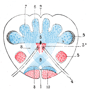
The ventral spinothalamic fasciculus situated in the marginal part of the anterior funiculus and intermingled more or less with the vestibulo-spinal fasciculus, is derived from cells in the posterior column or intermediate gray matter of the opposite side. Aβ fibres carry sensory information pertaining to crude touch from the skin. After entering the spinal cord the first order neurons synapse, and the second order neurons decussate via the anterior commissure. These second order neurons ascend synapsing in the VPL of the thalamus. It should also be noted incoming first order neurons can ascend or descend via the Lissauer tract.

The amygdalofugal pathway is one of the three major efferent pathways of the amygdala, meaning that it is one of the three principal pathways by which fibers leave the amygdala. It leads from the basolateral nucleus and central nucleus of the amygdala. The amygdala is a limbic structure in the medial temporal lobe of the brain. The other main efferent pathways from the amygdala are the stria terminalis and anterior commissure.

Dorsal column nuclei

In neuroanatomy, the dorsal column nuclei are a pair of nuclei in the dorsal columns in the brainstem. The name refers collectively to the cuneate nucleus and gracile nucleus, which are present at the junction between the spinal cord and the medulla oblongata. Both nuclei contain second-order neurons of the dorsal column-medial lemniscus pathway, which carries fine touch and proprioceptive information from the body to the brain. Each nucleus has an associated nerve tract, the gracile fasciculus and the cuneate fasciculus.

The isothalamus is a division used by some researchers in describing the thalamus.

Stria medullaris of thalamus

The stria medullaris is a part of the epithalamus. It is a fiber bundle containing afferent fibers from the septal nuclei, lateral preoptico-hypothalamic region, and anterior thalamic nuclei to the habenula. It forms a horizontal ridge on the medial surface of the thalamus, and is found on the border between dorsal and medial surfaces of thalamus. Superior and lateral to habenular trigone.

Anterior nuclei of thalamus

The anterior nuclei of thalamus are a collection of nuclei at the rostral end of the dorsal thalamus. They comprise the anteromedial, anterodorsal, and anteroventral nuclei.

Intralaminar nuclei of thalamus

The intralaminar nuclei are collections of neurons in the thalamus that are generally divided in two groups as follows:

Thalamic fasciculus

The thalamic fasciculus is a component of the subthalamus. It is synonymous with field H1 of Forel. Nerve fibres form a tract containing cerebellothalamic (crossed) and pallidothalamic (uncrossed) fibres, that is insinuated between the thalamus and the zona incerta.

Pallidothalamic tracts

The pallidothalamic tracts are a part of the basal ganglia. They provide connectivity between the internal globus pallidus (GPi) and the thalamus, primarily the ventral anterior nucleus and the ventral lateral nucleus.

The fields of Forel are areas in a deep part of the brain known as the diencephalon. They are below the thalamus and consist of three defined, white matter areas of the subthalamus. These three regions are also named "H fields":

The dorsal tegmental nucleus (DTN), also known as dorsal tegmental nucleus of Gudden (DTg), is a group of neurons located in the brain stem, which are involved in spatial navigation and orientation.

References

This article incorporates text in the public domain from page 869 of the 20th edition of Gray's Anatomy (1918)

The public domain consists of all the creative works to which no exclusive intellectual property rights apply. Those rights may have expired, been forfeited, expressly waived, or may be inapplicable.

<i>Grays Anatomy</i> English-language textbook of human anatomy

Gray's Anatomy is an English language textbook of human anatomy originally written by Henry Gray and illustrated by Henry Vandyke Carter. Earlier editions were called Anatomy: Descriptive and Surgical, Anatomy of the Human Body and Gray's Anatomy: Descriptive and Applied, but the book's name is commonly shortened to, and later editions are titled, Gray's Anatomy. The book is widely regarded as an extremely influential work on the subject, and has continued to be revised and republished from its initial publication in 1858 to the present day. The latest edition of the book, the 41st, was published in September 2015.

  1. 1 2 3 4 5 Dillingham, C. M; Frizzati, A; Nelson, A. J; Vann, S. D (2015). "How do mammillary body inputs contribute to anterior thalamic function?". Neuroscience & Biobehavioral Reviews. 54: 108–119. doi:10.1016/j.neubiorev.2014.07.025. PMC   4462591 Lock-green.svg.
  2. 1 2 3 Aggleton, J. P; O'Mara, S. M; Vann, S. D; Wright, N. F; Tsanov, M; Erichsen, J. T (2010). "Hippocampal–anterior thalamic pathways for memory: Uncovering a network of direct and indirect actions". European Journal of Neuroscience. 31 (12): 2292–2307. doi:10.1111/j.1460-9568.2010.07251.x. PMC   2936113 Lock-green.svg.
  3. Haines DE (2003). Neuroanatomy: Atlas of Structures, Sections, and Systems, 6th ed (page 148). Lippincott Williams & Wilkins. ISBN   0781746779.
  4. NeuroNames. Angular Thalamic Nucleus --> "What, Where and How Big is It?" BrainInfo. Accessed January 25, 2011.
  5. Yoneoka Y, Takeda N, Inoue A, et al. (2004). "Acute Korsakoff syndrome following mammillothalamic tract infarction". AJNR Am J Neuroradiol. 25 (6): 964–8. PMID   15205131.