Medical Renaissance

Last updated
The front cover illustration of De Humani Corporis Fabrica, On the Fabric of the Human Body, written by Andreas Vesalius, showing a public dissection being carried out by Vesalius himself. Vesalius01.jpg
The front cover illustration of De Humani Corporis Fabrica , On the Fabric of the Human Body, written by Andreas Vesalius, showing a public dissection being carried out by Vesalius himself.

The Medical Renaissance, from around 1400 to 1700 CE, was a period of progress in European medical knowledge, with renewed interest in the ideas of the ancient Greek, Roman civilizations and Islamic medicine, following the translation into Medieval Latin of many works from these societies. Medical discoveries during the Medical Renaissance are credited with paving the way for modern medicine.

Contents

Background

De Re Anatomica by Realdo Colombo, 1572 De re anatomica libri 15 V00044 00000004.tif
De Re Anatomica by Realdo Colombo, 1572

The Medical Renaissance began in the early 16th century. Medical researchers continued their Renaissance-evoked practices into the late 17th century. [1] [2] Progress made during the Medical Renaissance depended on several factors. [3] [4] Printed books based on movable type, adopted in Europe from the middle of the 15th century, allowed the diffusion of medical ideas and anatomical diagrams. Linacre, Erasmus, Leonicello and Sylvius are among the list of the first scholars most credited for the starting of the Medical Renaissance. [2] Following after is Andreas Vesalius's publication of De humani corporis fabrica (On the Fabric of the Human body) in 1543. Better knowledge of the original writings of Galen in particular, developed into the learned medicine tradition through the more open attitudes of Renaissance humanism. Religious control of the teachings of the medical profession and universities diminished, and dissection was more often possible.

Notable contributors

Leonardo da Vinci (1452–1519)

This drawing by Leonardo da Vinci of a foetus in the womb is one of many detailed anatomical drawings by the artist Leonardo da Vinci Studies of Embryos.jpg
This drawing by Leonardo da Vinci of a foetus in the womb is one of many detailed anatomical drawings by the artist

Leonardo da Vinci made many contributions in the fields of science and technology. His research centered around his desire to learn more about how the human brain processes visual and sensory information and how that connects to the soul. Though his artwork was widely observed before, some of his original research was not made public until the 20th century. Some of da Vinci's research involved studying vision. He believed that visual information entered the body through the eye, then continued by sending nerve impulses through the optic nerve, and eventually reaching the soul. Da Vinci subscribed to the ancient notion that the soul was housed in the brain.

He did research on the role of the spinal cord in humans by studying frogs. He noted that as soon as the frogs medulla of the spine is broken, the frog would die. This led him to believe that the spine is the basis for the sense of touch, cause of movement, and the origin of nerves. As a result of his studies on the spinal cord, he also came to the conclusion that all peripheral nerves begin from the spinal cord. Da Vinci also did some research on the sense of smell. He is credited with being the first to define the olfactory nerve as one of the cranial nerves. [5]

Leonardo da Vinci made his anatomical sketches based on observing and dissecting 30 cadavers. His sketches were very detailed and included organs, muscles of superior extremity, the hand, and the skull. Leonardo was well known for his three-dimensional drawings. His anatomical drawings were not found until 380 years after his death. [6]

Ambroise Paré (1510–1590)

Paré was a French surgeon, anatomist and an inventor of surgical instruments. He was a military surgeon during the French campaigns in Italy of 1533–36. It was here that, having run out of boiling oil (which was the accepted way of treating firearm wounds), Paré turned to an ancient Roman remedy: turpentine, egg yolk and oil of roses. He applied it to the wounds and found that it relieved pain and sealed the wound effectively. Paré also introduced the ligatures of arteries; silk threads would be used to tie up the arteries of amputated limbs to try to stop the bleeding. As antiseptics had not yet been invented this method led to an increased fatality rate and was abandoned by medical professionals of the time. [7]

Additionally, Paré's contributions extended to the practice of surgical amputation and on the design and development of limb prostheses. [8] [9] While caring for wounded soldiers, Paré recorded the pain endured by amputees, a phenomenon now recognized as phantom limb sensation. He believed that the phantom pain occurred in the brain and not in remnants of the limb. [10]

Illustration of the structure of the nervous system from De humani corporis fabrica by Vesalius Andreae Vesalii Bruxellensis Wellcome L0063854.jpg
Illustration of the structure of the nervous system from De humani corporis fabrica by Vesalius

Andreas Vesalius (1514–1564)

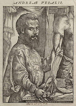
Andreas Vesalius was born in 1514 around midnight in Brussels, then part of Habsburg Netherlands, to a long lineage of doctors. [11] His father was also an apothecary to Charles V, Holy Roman Emperor. [11] He would be born into the period of The Revival of Learning. He gained an interest in anatomy at a young age and began dissecting mice, moles, cats, and dogs. [11] Vesalius picked up from the work of Galen (129–c. 200 CE) which was based on the dissection of animals from pigs to apes. [12]  The works of Galen would be accepted until Vesalius. He would challenge the medieval views of human anatomy made by Galen that had been taught for centuries. Vesalius paved he foundation of modern anatomy and most of his ideas are accepted today.

Vesalius went to school in Louvain and would enter the university there. [11] His education was majorly taught by Humanistic teachers. Vesalius spent many hours in cemeteries in the burial ground of the Church of the Innocents in Paris to study the bones that were misplaced from improper burial. [11] Eventually, Vesalius and his friend stole one complete skeleton from the gallows; this was the first body Vesalius was able to dissect completely. He would go on to befriend judges and doctors, to gain access to human bodies of those who had just died for dissection. This would start rumors that connected Vesalius to Vivisection. [13] Vesalius did want the most recent deceased cadavers and vivisection was frowned upon both then and currently. Vesalius eventually became a Doctor of Medicine at the age of 22. He noticed that many of his hands on experiences and findings with cadavers contradicted Galen's teachings, to the point he would discarded all them. [11] His students grew to be devoted followers and they would go grave robbing with Vesalius especially if a female was involved. [13]

A print portrait of Andreas Vesalius in his book De Humani Corporis Fabrica Libri Septem. Vesalius Fabrica portrait.jpg
A print portrait of Andreas Vesalius in his book De Humani Corporis Fabrica Libri Septem.
As mentioned, this is one of Vesalius' most known images used in the De Humani Corporis Fabrica that showed parts of the human body. Vesalius Fabrica p190.jpg
As mentioned, this is one of Vesalius' most known images used in the De Humani Corporis Fabrica that showed parts of the human body.

He wrote around 14 books on his findings in anatomy, including his best known book De humani corporis fabrica. [11] It was revolutionary because of the accuracy and precision of his descriptions and images of organs and would refute Galen's belief that human anatomy is closely related to apes. [14] The book was a dedication to Charles V, Holy Roman Emperor. Vesalius published De humani corporis fabrica at the age of 28. [14] With him being so young, it made his contributions harder to accept. De Fabrica was a milestone in medical science as it detailed many aspects of the human cadaver as well as presenting it in the form of art. Its imagery drew on new Renaissance methods in art. An example of his work is a picture of a dissected corpse hung by a rope through its eye sockets with the upper diaphragm on a wall behind the corpse. [14] The book gave clear identification of the organs in the human body while also removing the aspects that he found flawed with Galen's teachings.

Vesalius was an important part of the Medical Renaissance. He is remembered as a critic of the inaccurate teachings of Galen, and one of the founders of modern anatomy.

William Harvey (1578–1657)

William Harvey was an English medical doctor-physicist, known for his contributions in heart and blood movement. William Harvey fully believed all medical knowledge should be universal, and he made this his works goal. Accomplished historians credit him for his boldness in his experimental work and his everlasting eagerness to implement modern practice. [15] Although not the first to propose pulmonary circulation (Ibn al-Nafis, Michael Servetus and Realdo Colombo preceded him), he is credited as the first person in the Western world to give quantitative arguments for the circulation of blood around the body. [16] William Harvey's extensive work on the body's circulation can be found in the written work titles, "The Motu Cordis".This work opens up with clear definitions of anatomy as well as types of anatomy which clearly outlined a universal meaning of these words for various Renaissance physicians. Anatomy, as defined by William Harvey is, "the faculty that by ocular inspection and dissection [grasps] the uses and actions of the parts." [15] In other words, to be able to identify the actions or roles each part of the body plays in the overall function of the body by dissection, followed by visual identification. These were the foundation for the further research on the heart and blood vessels. [17]

Hieronymus Fabricius (1537–1619)

Hieronymus Fabricius is an anatomist and surgeon that prepared a human and animal anatomy atlas and these illustrations were used in his work, Tabulae Pictae. This work includes illustrations from many different artists and Fabricius is credited for providing a turning point in anatomical illustration. Fabricius' illustrations were of natural size and natural colors. After Fabricius' death, Tabulae Pictae disappeared and wasn't again discovered until 1909. [6] Fabricus focused on the human brain and the fissures that are inside of the brain. In Tabulae Pictae, he described the cerebral fissure that separates the temporal lobe from the frontal lobe. [18] He also studied veins and was the first to discover the valves inside of veins. [18]

See also

Related Research Articles

<span class="mw-page-title-main">Anatomy</span> Study of the structure of organisms and their parts

Anatomy is the branch of biology concerned with the study of the structure of organisms and their parts. Anatomy is a branch of natural science that deals with the structural organization of living things. It is an old science, having its beginnings in prehistoric times. Anatomy is inherently tied to developmental biology, embryology, comparative anatomy, evolutionary biology, and phylogeny, as these are the processes by which anatomy is generated, both over immediate and long-term timescales. Anatomy and physiology, which study the structure and function of organisms and their parts respectively, make a natural pair of related disciplines, and are often studied together. Human anatomy is one of the essential basic sciences that are applied in medicine.

<span class="mw-page-title-main">Galen</span> Greek physician, surgeon, and philosopher (129-c.216)

Aelius Galenus or Claudius Galenus, often anglicized as Galen or Galen of Pergamon, was a Roman and Greek physician, surgeon and philosopher. Considered to be one of the most accomplished of all medical researchers of antiquity, Galen influenced the development of various scientific disciplines, including anatomy, physiology, pathology, pharmacology, and neurology, as well as philosophy and logic.

<span class="mw-page-title-main">Andreas Vesalius</span> Anatomist, physician and author (1514–1564)

Andries van Wezel, latinised as Andreas Vesalius, was an anatomist and physician who wrote De Humani Corporis Fabrica Libri Septem, what is considered to be one of the most influential books on human anatomy and a major advance over the long-dominant work of Galen. Vesalius is often referred to as the founder of modern human anatomy. He was born in Brussels, which was then part of the Habsburg Netherlands. He was a professor at the University of Padua (1537–1542) and later became Imperial physician at the court of Emperor Charles V.

<span class="mw-page-title-main">History of anatomy</span> Aspect of history

The history of anatomy extends from the earliest examinations of sacrificial victims to the sophisticated analyses of the body performed by modern anatomists and scientists. Written descriptions of human organs and parts can be traced back thousands of years to ancient Egyptian papyri, where attention to the body was necessitated by their highly elaborate burial practices.

The human skeleton is the internal framework of the human body. It is composed of around 270 bones at birth – this total decreases to around 206 bones by adulthood after some bones get fused together. The bone mass in the skeleton makes up about 14% of the total body weight and reaches maximum mass between the ages of 25 and 30. The human skeleton can be divided into the axial skeleton and the appendicular skeleton. The axial skeleton is formed by the vertebral column, the rib cage, the skull and other associated bones. The appendicular skeleton, which is attached to the axial skeleton, is formed by the shoulder girdle, the pelvic girdle and the bones of the upper and lower limbs.

<span class="mw-page-title-main">Herophilos</span> Greek physician (335–280 BC)

Herophilos, sometimes Latinised Herophilus, was a Greek physician regarded as one of the earliest anatomists. Born in Chalcedon, he spent the majority of his life in Alexandria. He was the first scientist to systematically perform scientific dissections of human cadavers. He recorded his findings in over nine works, which are now all lost. The early Christian author Tertullian states that Herophilos vivisected at least 600 live prisoners; however, this account has been disputed by many historians. He is often seen as the father of anatomy.

<span class="mw-page-title-main">Bartolomeo Eustachi</span> Italian anatomist

Bartolomeo Eustachi, also known as Eustachio or by his Latin name of Bartholomaeus Eustachius, was an Italian anatomist and one of the founders of the science of human anatomy.

<span class="mw-page-title-main">Hieronymus Fabricius</span> Italian physician, anatomist and surgeon (1533–1619)

Girolamo Fabrici d'Acquapendente, also known as Girolamo Fabrizio or Hieronymus Fabricius, was a pioneering anatomist and surgeon known in medical science as "The Father of Embryology."

<span class="mw-page-title-main">Mondino de Luzzi</span> Italian physician and anatomist (c.1270–1326)

Mondino de Luzzi, or de Liuzzi or de Lucci,, also known as Mundinus, was an Italian physician, anatomist and professor of surgery, who lived and worked in Bologna. He is often credited as the restorer of anatomy because he made seminal contributions to the field by reintroducing the practice of public dissection of human cadavers and writing the first modern anatomical text.

<span class="mw-page-title-main">Realdo Colombo</span> Italian anatomy professor and surgery

Matteo Realdo Colombo was an Italian professor of anatomy and a surgeon at the University of Padua between 1544 and 1559.

<span class="mw-page-title-main">Moulage</span> Fake injuries for medical training

Moulage is the art of applying mock injuries for the purpose of training emergency response teams and other medical and military personnel. Moulage may be as simple as applying pre-made rubber or latex "wounds" to a healthy "patient's" limbs, chest, head, etc., or as complex as using makeup and theatre techniques to provide elements of realism to the training simulation. The practice dates to at least the Renaissance, when wax figures were used for this purpose.

<span class="mw-page-title-main">Dissection</span> Cutting procedure used in anatomy

Dissection is the dismembering of the body of a deceased animal or plant to study its anatomical structure. Autopsy is used in pathology and forensic medicine to determine the cause of death in humans. Less extensive dissection of plants and smaller animals preserved in a formaldehyde solution is typically carried out or demonstrated in biology and natural science classes in middle school and high school, while extensive dissections of cadavers of adults and children, both fresh and preserved are carried out by medical students in medical schools as a part of the teaching in subjects such as anatomy, pathology and forensic medicine. Consequently, dissection is typically conducted in a morgue or in an anatomy lab.

<i>De Humani Corporis Fabrica Libri Septem</i> 1543 Anatomy books written by Andreas Vesalius

De Humani Corporis Fabrica Libri Septem is a set of books on human anatomy written by Andreas Vesalius (1514–1564) and published in 1543. It was a major advance in the history of anatomy over the long-dominant work of Galen, and presented itself as such.

<i>The Anatomy Lesson of Dr. Nicolaes Tulp</i> Oil painting by Rembrandt

The Anatomy Lesson of Dr. Nicolaes Tulp is a 1632 oil painting on canvas by Rembrandt housed in the Mauritshuis museum in The Hague, the Netherlands. It was originally created to be displayed by the Surgeons Guild in their meeting room. The painting is regarded as one of Rembrandt's early masterpieces.

<span class="mw-page-title-main">Jacopo Berengario da Carpi</span> Italian physician

Jacopo Berengario da Carpi was an Italian physician. His book "Isagoge breves" published in 1522 made him the most important anatomist before Andreas Vesalius.

<span class="mw-page-title-main">Giambattista Canano</span> Italian anatomist and professor

Giambattista or Giovanni Battista Canano was a physician and anatomist, active mainly in his native Ferrara.

<i>Écorché</i> Drawing of a person that shows their muscles under the skin

An écorché is a figure drawn, painted, or sculpted showing the muscles of the body without skin, normally as a figure study for another work or as an exercise for a student artist. The Renaissance-era architect, theorist and all-around Renaissance man Leon Battista Alberti recommended that when painters intend to depict a nude, they should first arrange the muscles and bones, then depict the overlying skin.

<span class="mw-page-title-main">Cadaver</span> Dead body used for study or instruction

A cadaver or corpse is a dead human body. Cadavers are used by medical students, physicians and other scientists to study anatomy, identify disease sites, determine causes of death, and provide tissue to repair a defect in a living human being. Students in medical school study and dissect cadavers as a part of their education. Others who study cadavers include archaeologists and arts students. In addition, a cadaver may be used in the development and evaluation of surgical instruments.

An anatomy murder is a murder committed in order to use all or part of the cadaver for medical research or teaching. It is not a medicine murder because the body parts are not believed to have any medicinal use in themselves. The motive for the murder is created by the demand for cadavers for dissection, and the opportunity to learn anatomy and physiology as a result of the dissection. Rumors concerning the prevalence of anatomy murders are associated with the rise in demand for cadavers in research and teaching produced by the Scientific Revolution. During the 19th century, the sensational serial murders associated with Burke and Hare and the London Burkers led to legislation which provided scientists and medical schools with legal ways of obtaining cadavers. Rumors persist that anatomy murders are carried out wherever there is a high demand for cadavers. These rumors, like those concerning organ theft, are hard to substantiate, and may reflect continued, deep-held fears of the use of cadavers as commodities.

<span class="mw-page-title-main">Anatomical Theatre of Padua</span> Theatre in Padua, Italy

The Anatomical Theatre of Padua, Northern Italy, is the first permanent anatomical theatre in the world. Still preserved in the Palazzo del Bo, it was inaugurated in 1595 by Girolamo Fabrici of Acquapendente, according to the project of Paolo Sarpi and Dario Varotari. This theatre constituted the model for the anatomical theatres built during the seventeenth century in the main universities of Europe: all would have been based on the Paduan archetype. It is the symbol of a successful period in the University of Padua's history, and it is considered one of the most important achievements for the study of anatomy during the sixteenth century.

References

  1. Bradley, C. P. (2006-01-01). "Medical Renaissance in Florence". European Journal of General Practice. 12 (2): 51. doi: 10.1080/13814780600940767 . ISSN   1381-4788. PMID   16945876. S2CID   23988544.
  2. 1 2 Toledo-Pereyra, Luis H. (2015-05-04). "Medical Renaissance". Journal of Investigative Surgery. 28 (3): 127–130. doi:10.3109/08941939.2015.1054747. ISSN   0894-1939. PMID   26065591. S2CID   207482973.
  3. OCR GCSE: Medicine Through Time
  4. Parragon, World History Encyclopedia
  5. Pevsner, Jonathan (2002). "Leonardo da Vinci's contributions to neuroscience". Trends in Neurosciences. 25 (4): 217–220. doi:10.1016/S0166-2236(00)02121-4. PMID   11998691. S2CID   9833298.
  6. 1 2 Ghosh, Sanjib Kumar (2015-03-01). "Evolution of illustrations in anatomy: A study from the classical period in Europe to modern times". Anatomical Sciences Education. 8 (2): 175–188. doi:10.1002/ase.1479. ISSN   1935-9780. PMID   25053471. S2CID   15451344.
  7. Grendler, Paul F. (1999). Encyclopedia of the Renaissanc. New York: Scribner's. p. 399. ISBN   978-0-684-80511-5.
  8. "Prostheses by Ambroise Paré" drawings
  9. Thurston, Alan J. (2007) "Paré and prosthetics: the early history of artificial limbs" ANZ Journal of Surgery 77(12): pp. 1114–1119, doi : 10.1111/j.1445-2197.2007.04330.x
  10. News.nationalgeographic.com
  11. 1 2 3 4 5 6 7 Keeton, Morris (1936). "Andreas Vesalius: His Times, His Life, His Work". BIOS. 7 (2): 97–109. ISSN   0005-3155. JSTOR   4604123.
  12. Singer, P. N. (2021), "Galen", in Zalta, Edward N. (ed.), The Stanford Encyclopedia of Philosophy (Winter 2021 ed.), Metaphysics Research Lab, Stanford University, retrieved 2022-12-06
  13. 1 2 Park, Katharine (1994). "The Criminal and the Saintly Body: Autopsy and Dissection in Renaissance Italy*". Renaissance Quarterly. 47 (1): 1–33. doi: 10.2307/2863109 . ISSN   0034-4338. JSTOR   2863109. PMID   11639270. S2CID   31817948.
  14. 1 2 3 Gumpert, Martin (1948). "Vesalius". Scientific American. 178 (5): 24–31. doi:10.1038/scientificamerican0548-24. ISSN   0036-8733. JSTOR   24945814.
  15. 1 2 Distelzweig, Peter (2014-01-01). ""Meam de motu & usu cordis, & circuitu sanguinis sententiam": teleology in William Harvey's De motu cordis". Gesnerus. 71 (2): 258–270. doi: 10.1163/22977953-07102005 . ISSN   0016-9161. PMID   25707098. S2CID   35973541.
  16. Spotlight Science 9 (GCSE Science Text Book)
  17. Kids Work! > History of Medicine
  18. 1 2 Collice, Massimo; Collice, Rosa; Riva, Alessandro (2008-10-01). "Who Discovered the Sylvian Fissure?". Neurosurgery. 63 (4): 623–628. doi:10.1227/01.neu.0000327693.86093.3f. ISSN   0148-396X. PMID   18981875. S2CID   207140931.

Further reading