Meningeal lymphatic vessels

Last updated
Meningeal lymphatic vessels run parallel to the dural venous sinuses. Gray488.png
Meningeal lymphatic vessels run parallel to the dural venous sinuses.

The meningeal lymphatic vessels (or meningeal lymphatics) are a network of conventional lymphatic vessels located parallel to the dural venous sinuses and middle meningeal arteries of the mammalian central nervous system (CNS). As a part of the lymphatic system, the meningeal lymphatics are responsible for draining immune cells, small molecules, and excess fluid from the CNS into the deep cervical lymph nodes. [1] [2] Cerebrospinal fluid, and interstitial fluid are exchanged, and drained by the meningeal lymphatic vessels. [3]

Contents

While it was historically believed that both the brain and meninges were devoid of lymphatic vasculature, recent studies by Antoine Louveau and Jonathan Kipnis at the University of Virginia, submitted in October 2014, and by Aleksanteri Aspelund, Salli Antila and Kari Alitalo at the University of Helsinki submitted in December 2014, identified and described the basic biology of the meningeal lymphatics using a combination of histological, live-imaging, and genetic tools. [1] [2] In general, their work is thought to extend that of the Danish neuroscientist Maiken Nedergaard in identifying the pathway connecting the glymphatic system to the meningeal compartment.

The role that the meningeal lymphatics plays in neurological disease is yet to be explored. It is hypothesized that they may contribute to autoimmune and inflammatory diseases of the CNS due to their role in connecting the immune and nervous systems.

Background

In peripheral organs, lymphatic vessels are responsible for conducting lymph between different parts of the body. In general, lymphatic drainage is important for maintaining fluid homeostasis as well as providing a means for immune cells to traffic into draining lymph nodes from other parts of the body, allowing for immune surveillance of bodily tissues.[ citation needed ]

The first mention of meningeal lymphatic vessels can be attributed to Paolo Mascagni, whose anatomical work towards the end of the eighteenth century suggested their presence; however, this work received little attention or acceptance. [4] [5] In 1953, Italian scientist Lecco identified putative lymphatic vessels in post-mortem human dura. Further research in the 1960s described the existence of meningeal lymphatics, [6] but these findings were not accepted by the field due to their limited methodology. [7]

Prior to the discovery of true meningeal lymphatic vessels, it was generally believed that the mammalian CNS did not contain a lymphatic system and thus relied upon alternative routes of waste clearance such as the glymphatic system, [8] a cerebrospinal fluid (CSF) drainage pathway under the cribriform plate and into the lymphatics of the nasal mucosa, [9] and arachnoid granulations to clear itself of excess protein, fluid, and metabolic waste products. Furthermore, the presumed absence of CNS lymphatics was an important pillar in the long-held dogma that the CNS is an immune-privileged tissue to which immune cells have highly restricted access under normal physiological conditions.

Discovery

Although, several studies proposed the existence of lymphatic vessels in the dura mater, the presence of the meningeal lymphatic system was accepted in 2015, when two independent studies published by Louveau et al. [1] and Aspelund et al. [2] provided convincing data using novel methods. Louveau et al. noticed an unusual alignment of immune cells along the dural sinus using a meningeal whole-mount technique. Using lymphatic endothelial cell-specific markers and electron microscopy, the authors found that the immune cells were not inside blood vessels, but rather were organized inside lymphatic vessels within the meninges, a system of membranes that envelop the brain and spinal cord. [1]

Aspelund et al. had discovered that in the eye, another immune-privileged organ, the Schlemm's canal is a lymphatic-like vessel. [10] As Schlemm's canal was previously considered to be a venous sinus, the authors subsequently hypothesized that similar vessels may also be found in the brain due to its similarly immune-privileged status. [2] However, a recent study reported the absence of lymphatic vessels in the spinal cord dura of rat despite harboring numerous LYVE1+ cells. [11]

In an interview with Ira Flatow on NPR's Science Friday, Kipnis described the meningeal lymphatics as "well-hidden" when asked how, unlike the rest of the lymphatic system, they had remained unmapped into the 21st century. [12] While many scientists study the brain parenchyma proper, Kipnis explained, his lab is relatively unique in studying the meninges:

We are among the few labs who are interested in this very unique area of the brain: the coverings of the brain - the so-called 'meninges.' We've been looking into this area for a few years now," Kipnis said. "I was lucky to have a phenomenal post-doctoral fellow in my lab, Dr. Antoine Louveau, who developed a very unique technique of mounting this entire covering as a whole-mount. I think this is what allowed us to find those vessels. [12]

Visualization

Example of a meningeal whole-mount taken from an adult mouse. Laying the whole-mount on a glass slide allows for histological analysis of the entire dura, including the superior sagittal and transverse sinuses. Mouse whole-mount meninges.png
Example of a meningeal whole-mount taken from an adult mouse. Laying the whole-mount on a glass slide allows for histological analysis of the entire dura, including the superior sagittal and transverse sinuses.

To visualize the dura mater using immunohistochemistry, the dura must first be fixed within the skullcap. It is prepared by cutting around the base of the skull (inferior to the post-tympanic hook) and removing the lower portion of the skull and brain. Following fixation, the dura can be dissected out of the skullcap as a single piece of tissue that can be utilized for histological analysis. [13]

In transgenic mice containing Prox1-GFP or Vegfr3-LacZ reporter genes, the lymphatic vessels may be visualized by fluorescent microscopy or after X-gal staining, respectively. [2]

The meningeal lymphatics may also be visualized non-invasively by MRI, using MRI contrast agents such as gadobutrol and gadofosveset to reveal the presence of the vessels near the dura mater. [14]

Biology

Anatomy and route of drainage

The meningeal lymphatic system is composed of a network of vessels along the dural sinus in the dura which express lymphatic endothelial cell marker proteins, including PROX1, LYVE1, and PDPN. The vessels extend along the length of both the superior sagittal and transverse sinuses and directly connects to the deep cervical lymph nodes. [1] These meningeal lymphatic vessels drain down and exit the skull along the dural venous sinuses and meningeal arteries. Meningeal lymphatic vessels also drain out of the skull alongside cranial nerves and through the cribriform plate. Molecular profiling indicates that the vessels are conventional lymphatic vessels: they express high levels of PROX1, LYVE1, PDPN and VEGFR3, but low levels of PECAM1. Meningeal lymphatic vessels absorb cerebrospinal fluid and drain into the deep cervical lymph nodes. [2]

Several unique attributes differentiate meningeal lymphatic vessels from lymphatic vessels in peripheral organs. Compared to peripheral lymphatic vessels, the meningeal lymphatic network is markedly less complex, with far less tissue coverage and lymphatic branching. Furthermore, meningeal lymphatic vessels are generally smaller than those in the periphery and display a structural homogeneity along the dural sinuses, remaining thinner and mostly unbranched along the superior sagittal sinus while growing larger and more branched along the transverse sinuses. [1] The meningeal lymphatic vessels are also unique for their scarcity of valves, which prevent back-flow of lymph. While the vessels in the superior parts of the skull were mostly devoid of valves, the larger lymphatic vessels of the basal parts only contain scattered valves. [2]

Development

Development of the dural lymphatic system requires expression of vascular endothelial growth factor C (VEGFC) and its receptor, VEGFR3 (which is the major signaling pathway for lymphatic growth). [15] Meningeal lymphatic vessels increase in diameter when exposed to recombinant VEGFC [1] and completely fail to develop when VEGFC and VEGFD signaling is inhibited during embryogenesis, [2] indicating that meningeal lymphatics share developmental characteristics with peripheral lymphatics. In addition to its role in the development of the dural lymphatics, VEGFR3 signaling is required for lymphatic vessel maintenance in the adult meninges. [15] Mechanical forces and shear stress generated by lymph flow are also required for later stages of meningeal lymphatic vessel formation and maturation. [16]

Physiological functions

Confocal micrograph of meningeal lymphatic vessels and trafficking immune cells. LYVE1 (green), CD3e (red), and DAPI (blue) are shown. TJK Example lymphatics.jpg
Confocal micrograph of meningeal lymphatic vessels and trafficking immune cells. LYVE1 (green), CD3e (red), and DAPI (blue) are shown.

Like peripheral lymphatic vessels, the meningeal lymphatics serve both the tissue drainage and immune cell trafficking functions of the lymphatic system. Multiphoton live imaging experiments performed on anesthetized mice have demonstrated that the meningeal lymphatics are capable of draining fluorescent dyes injected intracisternally into the CSF, indicating that the meningeal lymphatics are capable of draining fluid from their surrounding environment. Histological analysis revealed that the meningeal lymphatics constitutively contain T cells, B cells, and MHC class II-expressing myeloid cells, demonstrating that meningeal lymphatic vessels are capable of carrying immune cells. [1]

Furthermore, tracing the outflow of compounds injected into the brain parenchyma have indicated that meningeal lymphatics function downstream of the glymphatic system. Genetically engineered mice which lack the meningeal lymphatic vessels demonstrated attenuated clearance of macromolecules from the brain. The uptake of tracers from the brain into deep cervical lymph nodes was completely abrogated. However, brain interstitial fluid pressure and water content were unaffected. These data suggested that meningeal lymphatic vessels are important for the clearance of macromolecules from the brain parenchyma, but in physiological settings the brain can compensate in solute clearance. [2]

Meningeal lymphatic ablation experiments performed on mice suggests other implications of dysfunctional meningeal lymphatic drainage; an impairment in fear memory and hippocampal-amygdala neuronal circuitry was observed in mice with impaired meningeal lymphatic vessel function. Similar deficits in spatial learning and memory were observed in mice with lymphatic ligation, indicating that the effect is a result of impaired meningeal lymphatic drainage. [17]

Role in disease

The role that the meningeal lymphatics play in diseases of the nervous system is an area of active research, particularly for neurological disorders in which immunity is a fundamental player, such as multiple sclerosis, Alzheimer's disease (AD), amyotrophic lateral sclerosis, Hennekam syndrome, and Prader-Willi syndrome. Impaired clearance of ISF waste has been associated with accelerated accumulation of toxic amyloid beta, the main component of amyloid plaques in AD. [7]

Significance

The paper by Jonathan Kipnis and his postdoctoral fellow Antoine Louveau was published in 2015 and by November 2022, this paper had been cited 3300 times. [1]

The discovery of meningeal lymphatic vessels has attracted attention from many sources, and was touted as a scientific breakthrough in lists such as Scientific American 's "Top 10 Science Stories of 2015", Science Magazine 's "Breakthrough of the Year", Huffington Post's "Eight Fascinating Things We Learned About the Mind in 2015" and the National Institutes of Health's director Francis Collins's year end review. [18] [19] In 2017, Business Insider highlighted this as the biggest discovery ever made in Virginia. [20] In 2019, the history of the brain lymphatic system was narrated by Stefano Sandrone et al in Nature Medicine . [5]

Related Research Articles

<span class="mw-page-title-main">Lymphatic system</span> Organ system in vertebrates

The lymphatic system, or lymphoid system, is an organ system in vertebrates that is part of the immune system, and complementary to the circulatory system. It consists of a large network of lymphatic vessels, lymph nodes, lymphoid organs, lymphoid tissues and lymph. Lymph is a clear fluid carried by the lymphatic vessels back to the heart for re-circulation..

<span class="mw-page-title-main">Lymph node</span> Organ of the lymphatic system

A lymph node, or lymph gland, is a kidney-shaped organ of the lymphatic system and the adaptive immune system. A large number of lymph nodes are linked throughout the body by the lymphatic vessels. They are major sites of lymphocytes that include B and T cells. Lymph nodes are important for the proper functioning of the immune system, acting as filters for foreign particles including cancer cells, but have no detoxification function.

<span class="mw-page-title-main">Meninges</span> Three membranes that envelop the brain and spinal cord

In anatomy, the meninges are the three membranes that envelop the brain and spinal cord. In mammals, the meninges are the dura mater, the arachnoid mater, and the pia mater. Cerebrospinal fluid is located in the subarachnoid space between the arachnoid mater and the pia mater. The primary function of the meninges is to protect the central nervous system.

<span class="mw-page-title-main">Pia mater</span> Delicate innermost layer of the meninges, the membranes surrounding the brain and spinal cord

Pia mater, often referred to as simply the pia, is the delicate innermost layer of the meninges, the membranes surrounding the brain and spinal cord. Pia mater is medieval Latin meaning "tender mother". The other two meningeal membranes are the dura mater and the arachnoid mater. Both the pia and arachnoid mater are derivatives of the neural crest while the dura is derived from embryonic mesoderm. The pia mater is a thin fibrous tissue that is permeable to water and small solutes. The pia mater allows blood vessels to pass through and nourish the brain. The perivascular space between blood vessels and pia mater is proposed to be part of a pseudolymphatic system for the brain. When the pia mater becomes irritated and inflamed the result is meningitis.

<span class="mw-page-title-main">Arachnoid granulation</span> Protrusions of the arachnoid mater for returning cerebrospinal fluid to circulation

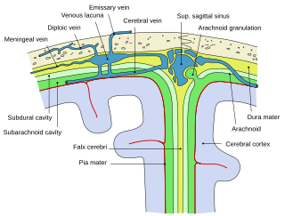
Arachnoid granulations are small protrusions of the arachnoid mater into the outer membrane of the dura mater. They protrude into the dural venous sinuses of the brain, and allow cerebrospinal fluid (CSF) to exit the subarachnoid space and enter the blood stream.

<span class="mw-page-title-main">Dura mater</span> Outermost layer of the protective tissues around the central nervous system (meninges)

In neuroanatomy, dura mater is a thick membrane made of dense irregular connective tissue that surrounds the brain and spinal cord. It is the outermost of the three layers of membrane called the meninges that protect the central nervous system. The other two meningeal layers are the arachnoid mater and the pia mater. It envelops the arachnoid mater, which is responsible for keeping in the cerebrospinal fluid. It is derived primarily from the neural crest cell population, with postnatal contributions of the paraxial mesoderm.

<span class="mw-page-title-main">Lymphatic vessel</span> Tubular vessels that are involved in the transport of lymph and lymphocytes

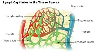
The lymphatic vessels are thin-walled vessels (tubes), structured like blood vessels, that carry lymph. As part of the lymphatic system, lymph vessels are complementary to the cardiovascular system. Lymph vessels are lined by endothelial cells, and have a thin layer of smooth muscle, and adventitia that binds the lymph vessels to the surrounding tissue. Lymph vessels are devoted to the propulsion of the lymph from the lymph capillaries, which are mainly concerned with the absorption of interstitial fluid from the tissues. Lymph capillaries are slightly bigger than their counterpart capillaries of the vascular system. Lymph vessels that carry lymph to a lymph node are called afferent lymph vessels, and those that carry it from a lymph node are called efferent lymph vessels, from where the lymph may travel to another lymph node, may be returned to a vein, or may travel to a larger lymph duct. Lymph ducts drain the lymph into one of the subclavian veins and thus return it to general circulation.

<span class="mw-page-title-main">Epidural space</span> Space between the dura mater and vertebrae

In anatomy, the epidural space is the potential space between the dura mater and vertebrae (spine).

<span class="mw-page-title-main">Falx cerebri</span> Anatomical structure of the brain

The falx cerebri is a large, crescent-shaped fold of dura mater that descends vertically into the longitudinal fissure between the cerebral hemispheres of the human brain, separating the two hemispheres and supporting dural sinuses that provide venous and CSF drainage to the brain. It is attached to the crista galli anteriorly, and blends with the tentorium cerebelli posteriorly.

The interstitium is a contiguous fluid-filled space existing between a structural barrier, such as a cell membrane or the skin, and internal structures, such as organs, including muscles and the circulatory system. The fluid in this space is called interstitial fluid, comprises water and solutes, and drains into the lymph system. The interstitial compartment is composed of connective and supporting tissues within the body – called the extracellular matrix – that are situated outside the blood and lymphatic vessels and the parenchyma of organs.

<span class="mw-page-title-main">Schlemm's canal</span> Lymphatic-like vessel in the eye

Schlemm's canal is a circular lymphatic-like vessel in the eye. It collects aqueous humor from the anterior chamber and delivers it into the episcleral blood vessels. Canaloplasty may be used to widen it.

<span class="mw-page-title-main">Dural venous sinuses</span> Venous channels in the dura mater

The dural venous sinuses are venous sinuses (channels) found between the endosteal and meningeal layers of dura mater in the brain. They receive blood from the cerebral veins, and cerebrospinal fluid (CSF) from the subarachnoid space via arachnoid granulations. They mainly empty into the internal jugular vein.

<span class="mw-page-title-main">Deep cervical lymph nodes</span> Group of cervical lymph nodes

The deep cervical lymph nodes are a group of cervical lymph nodes in the neck that form a chain along the internal jugular vein within the carotid sheath.

Certain sites of the mammalian body have immune privilege, meaning they are able to tolerate the introduction of antigens without eliciting an inflammatory immune response. Tissue grafts are normally recognised as foreign antigens by the body and attacked by the immune system. However, in immune privileged sites, tissue grafts can survive for extended periods of time without rejection occurring. Immunologically privileged sites include:

<span class="mw-page-title-main">LYVE1</span> Protein may function in lymphatic hyaluronan transport and have a role in tumor metastasis

Lymphatic vessel endothelial hyaluronan receptor 1 (LYVE1), also known as extracellular link domain containing 1 (XLKD1) is a Link domain-containing hyaladherin, a protein capable of binding to hyaluronic acid (HA), homologous to CD44, the main HA receptor. In humans it is encoded by the LYVE1 gene.

The Lymphatic Endothelium refers to a specialized subset of endothelial cells located in the sinus systems of draining lymph nodes. Specifically, these endothelial cells line the branched sinus systems formed by afferent lymphatic vessels, forming a single-cell layer which functions in a variety of critical physiological processes. These lymphatic endothelial cells contribute directly to immune function and response modulation, provide transport selectivity, and demonstrate orchestration of bidirectional signaling cascades. Additionally, lymphatic endothelial cells may be implicated in downstream immune cell development as well as lymphatic organogenesis. Until recently, lymphatic endothelial cells have not been characterized to their optimal potential. This system is very important in the function of continuous removal of interstitial fluid and proteins, while also having a significant function of entry for leukocytes and tumor cells. This leads to further research that is being developed on the relationship between lymphatic endothelium and metastasis of tumor cells . The lymphatic capillaries are described to be blind ended vessels, and they are made up of a single non-fenestrated layer of endothelial cells; The lymph capillaries function to aid in the uptake of fluids, macromolecules, and cells. Although they are generally similar to blood capillaries, the lymph capillaries have distinct structural differences. Lymph capillaries consist of a more wide and irregular lumen, and the endothelium in lymph capillaries is much thinner as well. Their origin has been speculated to vary based on them being dependent on specific tissue environments, and powered by organ-specific signals.(L. Gutierrez-Miranda, K. Yaniv, 2020). A lymph capillary endothelial cell is distinct from other endothelial cells in that collagen fibers are directly attached to its plasma membrane.

Lymph sacs are a part of the development of the lymphatic system, known as lymphangiogenesis. The lymph sacs are precursors of the lymph vessels. These sacs develop through the processes of vasculogenesis and angiogenesis. However, there is evidence of both of these processes in different organisms. In mice, it is thought that the lymphatic components form through an angiogenic process. But, there is evidence from bird embryos that gives rise to the idea that lymphatic vessels arise in the embryos through a vasculogenesis-like process from the lymphangioblastic endothelial precursor cells.

<span class="mw-page-title-main">Glymphatic system</span> System for waste clearance in the central nervous system of vertebrates

The glymphatic system is a system for waste clearance in the central nervous system (CNS) of vertebrates. According to this model, cerebrospinal fluid (CSF) flows into the paravascular space around cerebral arteries, combining with interstitial fluid (ISF) and parenchymal solutes, and exiting down venous paravascular spaces. The pathway consists of a para-arterial influx route for CSF to enter the brain parenchyma, coupled to a clearance mechanism for the removal of interstitial fluid (ISF) and extracellular solutes from the interstitial compartments of the brain and spinal cord. Exchange of solutes between CSF and ISF is driven primarily by arterial pulsation and regulated during sleep by the expansion and contraction of brain extracellular space. Clearance of soluble proteins, waste products, and excess extracellular fluid is accomplished through convective bulk flow of ISF, facilitated by astrocytic aquaporin 4 (AQP4) water channels.

<span class="mw-page-title-main">Jonathan Kipnis</span> Neuroscientist

Jonathan Kipnis is a neuroscientist, immunologist, and professor of pathology and immunology at the Washington University School of Medicine. His lab studies interactions between the immune system and nervous system. He is best known for his lab's discovery of meningeal lymphatic vessels in humans and mice, which has impacted research on neurodegenerative diseases such as Alzheimer's disease and multiple sclerosis, neuropsychiatric disorders, such as anxiety, and neurodevelopmental disorders such as autism and Rett syndrome.

The subarachnoid lymphatic-like membrane (SLYM) is a highly debated anatomical structure in the human brain that was proposed in 2023 as a possible fourth layer of the meninges.

References

  1. 1 2 3 4 5 6 7 8 9 Antoine Louveau; Igor Smirnov; Timothy J. Keyes; Jacob D. Eccles; Sherin J. Rouhani; J. David Peske; Noel C. Derecki; David Castle; James W. Mandell; Kevin S. Lee; Tajie H. Harris; Jonathan Kipnis. (2015). "Structural and functional features of central nervous system lymphatic vessels". Nature. 523 (7560): 337–41. Bibcode:2015Natur.523..337L. doi:10.1038/nature14432. PMC   4506234 . PMID   26030524.
  2. 1 2 3 4 5 6 7 8 9 Aleksanteri Aspelund; Salli Antila; Steven T. Proulx; Tine Veronica Karlsen; Sinem Karaman; Michael Detmar; Helge Wiig; Kari Alitalo. (2015). "A dural lymphatic vascular system that drains brain interstitial fluid and macromolecules". The Journal of Experimental Medicine. 212 (7): 991–9. doi:10.1084/jem.20142290. PMC   4493418 . PMID   26077718.
  3. Dupont, G; et al. (January 2019). "Our current understanding of the lymphatics of the brain and spinal cord". Clinical Anatomy. 32 (1): 117–121. doi: 10.1002/ca.23308 . PMID   30362622.
  4. Moreno-Zambrano, D; Santana, D; Avila, D; Santibáñez, Rocío (Apr 9, 2018). "Lymphatics of the Central Nervous System: Forgotten first descriptions. (S39.003)". Neurology. 90 (15).
  5. 1 2 Sandrone, S; Moreno-Zambrano, D; Kipnis, J; van Gijn, J (April 2019). "A (delayed) history of the brain lymphatic system". Nature Medicine. 25 (4): 538–540. doi:10.1038/s41591-019-0417-3. PMID   30948855. S2CID   96434900.
  6. Földi, M.; Gellért, A.; Kozma, M.; Poberai, M.; Zoltán, O. T.; Csanda, E. (1966). "New contributions to the anatomical connections of the brain and the lymphatic system". Acta Anatomica. 64 (4): 498–505. doi:10.1159/000142849. ISSN   0001-5180. PMID   5957959.
  7. 1 2 Da Mesquita S, Fu Z, Kipnis J (2018). "The Meningeal Lymphatic System: A New Player in Neurophysiology". Neuron. 100 (2): 375–388. doi:10.1016/j.neuron.2018.09.022. PMC   6268162 . PMID   30359603.
  8. Iliff JJ, Wang M, Liao Y, Plogg BA, Peng W, Gundersen GA, Benveniste H, Vates GE, Deane R, Goldman SA, Nagelhus EA, Nedergaard M (2012). "A Paravascular Pathway Facilitates CSF Flow Through the Brain Parenchyma and the Clearance of Interstitial Solutes, Including Amyloid β". Sci Transl Med. 4 (147): 147ra111. doi:10.1126/scitranslmed.3003748. PMC   3551275 . PMID   22896675.
  9. Cserr HF, Harling-Berg CJ, Knopf PM (1992). "Drainage of brain extracellular fluid into blood and deep cervical lymph and its immunological significance". Brain Pathol. 2 (4): 269–76. doi: 10.1111/j.1750-3639.1992.tb00703.x . PMID   1341962. S2CID   45444024.
  10. Aleksanteri Aspelund; Tuomas Tammela; Salli Antila; Harri Nurmi; Veli-Matti Leppänen; Georgia Zarkada; Lukas Stanczuk; Mathias Francois; Taija Mäkinen; Pipsa Saharinen; Ilkka Immonen; Kari Alitalo. (2014). "The Schlemm's canal is a VEGF-C/VEGFR-3-responsive lymphatic-like vessel". The Journal of Clinical Investigation. 124 (9): 3975–86. doi:10.1172/JCI75395. PMC   4153703 . PMID   25061878.
  11. Brezovakova, Veronika; Jadhav, Santosh (10 February 2020). "Identification of Lyve‐1 positive macrophages as resident cells in meninges of rats". Journal of Comparative Neurology. 528 (12): 2021–2032. doi:10.1002/cne.24870. PMID   32003471. S2CID   210984721.
  12. 1 2 Lim, Alexa (5 June 2015). "A potential "missing link" between the brain and immune system". National Public Radio . Retrieved 24 June 2015.
  13. 1 2 Antoine Louveau; Jonathan Kipnis. (2015). "Dissection and immunostaining of mouse whole-mount meninges". Protocol Exchange. doi: 10.1038/protex.2015.047 .
  14. Absinta M, Ha SK, Nair G, Sati P, Luciano NJ, Palisoc M, et al. (2017). "Human and nonhuman primate meninges harbor lymphatic vessels that can be visualized noninvasively by MRI". eLife. 6. doi: 10.7554/eLife.29738 . PMC   5626482 . PMID   28971799.
  15. 1 2 Antila, Salli; Karaman, Sinem; Nurmi, Harri; Airavaara, Mikko; Voutilainen, Merja H; Mathivet, Thomas; Chilov, Dmitri; Li, Zhilin; Koppinen, Tapani; Park, Jun-Hee; Fang, Shentong (2017-12-04). "Development and plasticity of meningeal lymphatic vessels". The Journal of Experimental Medicine. 214 (12): 3645–3667. doi:10.1084/jem.20170391. ISSN   1540-9538. PMC   5716035 . PMID   29141865.
  16. Bálint, László; Ocskay, Zsombor; Deák, Bálint András; Aradi, Petra; Jakus, Zoltán (14 January 2020). "Lymph Flow Induces the Postnatal Formation of Mature and Functional Meningeal Lymphatic Vessels". Frontiers in Immunology. 10: 3043. doi: 10.3389/fimmu.2019.03043 . ISSN   1664-3224. PMC   6970982 . PMID   31993056.
  17. Da Mesquita, Sandro; Louveau, Antoine; Vaccari, Andrea; Smirnov, Igor; Cornelison, R. Chase; Kingsmore, Kathryn M.; Contarino, Christian; Onengut-Gumuscu, Suna; Farber, Emily; Raper, Daniel; Viar, Kenneth E.; Powell, Romie D.; Baker, Wendy; Dabhi, Nisha; Bai, Robin (August 2018). "Functional aspects of meningeal lymphatics in ageing and Alzheimer's disease". Nature. 560 (7717): 185–191. Bibcode:2018Natur.560..185D. doi:10.1038/s41586-018-0368-8. ISSN   1476-4687. PMC   6085146 . PMID   30046111.
  18. "NIH, Scientific American, Science salute UVA brain discovery" . Retrieved 22 December 2016.
  19. "Media coverage of our discoveries" . Retrieved 22 December 2016.
  20. "The biggest scientific discoveries in every state". Business Insider . 2017-06-28.