
A rhabdomyoma is a benign tumor of striated muscle. Rhabdomyomas may be either "cardiac" or "extra cardiac". Extracardiac forms of rhabdomyoma are sub classified into three distinct types: adult type, fetal type, and genital type.

A leiomyoma, also known as a fibroid, is a benign smooth muscle tumor that very rarely becomes cancer (0.1%). They can occur in any organ, but the most common forms occur in the uterus, small bowel, and the esophagus. Polycythemia may occur due to increased erythropoietin production as part of a paraneoplastic syndrome.

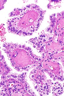
Surface epithelial-stromal tumors are a class of ovarian neoplasms that may be benign or malignant. Neoplasms in this group are thought to be derived from the ovarian surface epithelium or from ectopic endometrial or Fallopian tube (tubal) tissue. Tumors of this type are also called ovarian adenocarcinoma. This group of tumors accounts for 90% to 95% of all cases of ovarian cancer; however is mainly only found in postmenopausal women with the exception of the United States where 7% of cases occur in women under the age of 40. Serum CA-125 is often elevated but is only 50% accurate so it is not a useful tumor marker to assess the progress of treatment. 75% of women with epithelial ovarian cancer are found within the advanced-stages; however younger patients are more likely to have better prognoses than older patients.
Vaginal bleeding is any expulsion of blood from the vagina. This bleeding may originate from the uterus, vaginal wall, or cervix. Generally, it is either part of a normal menstrual cycle or is caused by hormonal or other problems of the reproductive system, such as abnormal uterine bleeding.

Uterine fibroids, also known as uterine leiomyomas or fibroids, are benign smooth muscle tumors of the uterus. Most women have no symptoms while others may have painful or heavy periods. If large enough, they may push on the bladder causing a frequent need to urinate. They may also cause pain during sex or lower back pain. A woman can have one uterine fibroid or many. Occasionally, fibroids may make it difficult to become pregnant, although this is uncommon.

Myomectomy, sometimes also called fibroidectomy, refers to the surgical removal of uterine leiomyomas, also known as fibroids. In contrast to a hysterectomy, the uterus remains preserved and the woman retains her reproductive potential.

Leiomyosarcoma is a malignant (cancerous) smooth muscle tumor. A benign tumor originating from the same tissue is termed leiomyoma. While leiomyosarcomas are not thought to arise from leiomyomas, some leiomyoma variants' classification is evolving.
Van der Woude syndrome (VDWS) is a genetic disorder characterized by the combination of lower lip pits, cleft lip with or without cleft palate (CL/P), and cleft palate only (CPO). The frequency of orofacial clefts ranges from 1:1000 to 1:500 births worldwide, and there are more than 400 syndromes that involve CL/P. VWS is distinct from other clefting syndromes due to the combination of cleft lip and palate (CLP) and CPO within the same family. Other features frequently associated with VWS include hypodontia in 10-81% of cases, narrow arched palate, congenital heart disease, heart murmur and cerebral abnormalities, syndactyly of the hands, polythelia, ankyloglossia, and adhesions between the upper and lower gum pads.

A neuromuscular disease is any disease affecting the peripheral nervous system (PNS), the neuromuscular junction, or skeletal muscle, all of which are components of the motor unit. Damage to any of these structures can cause muscle atrophy and weakness. Issues with sensation can also occur.

Ptosis, also known as blepharoptosis, is a drooping or falling of the upper eyelid. The drooping may be worse after being awake longer when the individual's muscles are tired. This condition is sometimes called "lazy eye", but that term normally refers to the condition amblyopia. If severe enough and left untreated, the drooping eyelid can cause other conditions, such as amblyopia or astigmatism. This is why it is especially important for this disorder to be treated in children at a young age, before it can interfere with vision development.

Intravenous leiomyomatosis is a rare condition seen exclusively in women in which leiomyomata, benign smooth muscle tumors, are found in veins. The masses are benign-appearing but can spread throughout the venous system leaving the uterus and even cause death when growing into the heart from the IVC. While the possibility that these arose de novo from the smooth muscle in the blood vessel wall was considered, chromosomal analysis suggests a uterine origin. Intravenous leiomyomata are usually but not always associated with uterine fibroids, and tend to recur.

Scrotalultrasound is a medical ultrasound examination of the scrotum. It is used in the evaluation of testicular pain, and can help identify solid masses.
Smooth muscle tumor of uncertain malignant potential, abbreviated STUMP, is an uncommon tumor of the uterine smooth muscle that may behave like a benign tumor or a cancerous tumor.
Reed’s syndrome is a rare inherited condition characterised by multiple cutaneous leiomyomas and, in women, uterine leiomyomas. It predisposes for renal cell cancer, an association denominated hereditary leiomyomatosis and renal cell cancer, and it is also associated with increased risk of uterine leiomyosarcoma. The syndrome is caused by a mutation in the fumarate hydratase gene, which leads to an accumulation of fumarate. The inheritance pattern is autosomal dominant.
Hereditary leiomyomatosis and renal cell carcinoma (HLRCC) is rare disorder associated with benign smooth muscle tumors and an increased risk of renal cell carcinoma.

The International Federation of Gynecology and Obstetrics is an international organization that links about 125 international professional societies of Obstetricians and Gynecologists. In 2011 FIGO recognized two systems designed to aid research, education, and clinical care of women with abnormal uterine bleeding (AUB) in the reproductive years. This page is a summary of the systems and their use in contemporary gynecology.
Myogel is a human-based extracellular matrix used mainly in the cancer research field to provide 3D cell culture environment for cancer cells. Unlike Matrigel, originated from mice sarcoma, and other synthesized matrices, myogel is extracted from human benign tumor tissue “leiomyoma”. Myogel was developed in Professor Tuula Salo's lab at the University of Oulu/Finland. The idea started by culturing cancer cells on myoma discs. Later on, these myoma tissues were processed to extract a gel form called Myogel . Myogel was compared with Matrigel and found to be superior in term of enhancing cancer cells proliferation, migration and invasion.
A bronchial leiomyoma is a relatively rare form of lung tumours. These tumours can form in the lower respiratory tract tissue of the bronchi, trachea and other lung tissue. They may also be derived from blood vessels. These tumors typically form from the smooth muscle tissue lining the bronchi. They grow as a solitary tumor attaching themselves to the sides of the bronchi.
Vulvar tumors are those neoplasms of the vulva. Vulvar and vaginal neoplasms make up a small percentage (3%) of female genital cancers. They can be benign or malignant. Vulvar neoplasms are divided into cystic or solid lesions and other mixed types. Vulvar cancers are those malignant neoplasms that originate from vulvar epithelium, while vulvar sarcomas develop from non-epithelial cells such as bone, cartilage, fat, muscle, blood vessels, or other connective or supportive tissue. Epithelial and mesenchymal tissue are the origin of vulvar tumors.
