Myxoma

Last updated
Myxoma
Atrial myxoma high mag.jpg
Micrograph of an atrial myxoma. H&E stain.
Specialty Oncology   OOjs UI icon edit-ltr-progressive.svg

A myxoma (New Latin from Greek muxa 'mucus') is a myxoid tumor of primitive connective tissue. [1] It is most commonly found in the heart (and is the most common primary tumor of the heart in adults) but can also occur in other locations.

Contents

Types

Table below: [2]

MyxomaMarginVascular patternCellularity Stroma Staining characteristicsRecurrence rateImage (see Histology)
Cutaneous myxoma or Superficial angiomyxomaPoor to moderately circumscribed, multilobularScattered thin-walled vesselsModerately cellular, bland spindled and stellate cells, variable inflammatory cell infiltrateAbundant mucin with clefts. Up to 30% have an associated epithelial component Vimentin; variable staining with CD34, factor XIIIA, SMA 1 , MSA 2 and S-100 20–30%
Cutaneous Myxoma CutaneousMyxoma.jpg
Cutaneous Myxoma
Intramuscular myxomaPoorly circumscribed merges with surrounding muscleHypovascular variant; hypervascular variantHypocellular variant; hypercellular variant; bland spindle cellsAbundant mucin with cystic spaces. Hypercellular variant has strands of collagenVimentin; variable staining with actin, desmin, CD34None
IntramuscularMyxoma.JPG
Juxta-articular myxomaPoorly circumscribed infiltrates surrounding tissueFocally vascularFocally hypercellular, peripheral spindle cells with occasional atypical cells and mitosesAbundant mucin, 89% of cases contain cystic spaces lined by fibrin or collagenVimentin; variable staining with actin, desmin, CD3434%
Aggressive angiomyxoma InfiltrativeUniformly distributed medium-sized blood vessels often with prominent hyalinizationLow to moderately cellular, evenly distributed round, spindled or stellate cellsLoose myxoid to focally collagenousVimentin, desmin, SMA 1 , MSA 2 , estrogen and progesterone receptor36–72%
AngiomyofibroblastomaWell circumscribedAbundant thin-walled blood vesselsAlternating hypercellular and hypocellular areas, perivascular condensations of spindled to epithelioid stromal cells Collagenous to edematous with minimal mucinVimentin, desmin, CD34, estrogen and progesterone receptor No recurrences reported, but rare cases of sarcomatous degeneration
Superficial acral fibromyxoma Pushing to infiltrativeMild to moderately accentuated vasculatureModerately cellular, spindle and stellate cells with a storiform to fascicular pattern, variable mast cellsMyxoid to collagenousCD34, EMA [ clarification needed ] 3 , CD99 Recurrence rare and primarily for incompletely excised lesions
Neurothekeoma (Nerve sheath myxoma)Well circumscribed, multilobularHypovascularModerately cellular, spindled cells in fascicles and whorlsNests of cells separated by collagenous bundlesS-100, EMA 3 47% if incompletely excised
Neurothekeoma2.JPG

1. ^ SMA, smooth muscle actin. 2. ^ MSA, muscle-specific actin. 3. ^ EMA, epithelial membrane antigen.

Symptoms and signs

Symptoms associated with cardiac myxomas are typically due to the effect of the mass of the tumor obstructing the normal flow of blood within the chambers of the heart. Because pedunculated myxomas are somewhat mobile, symptoms may only occur when the patient is in a particular position.

Some symptoms of myxoma may be associated with the release of interleukin 6 (IL-6) by the myxoma. [3] [4] High levels of IL-6 may be associated with a higher risk of embolism of the myxoma. [5]

Symptoms of a cardiac myxoma include: [6]

Location

Animated image of an MRI of the heart, showing a large myxoma plunging to and fro from atrium to ventricle across the mitral valve. Myxoma CMR.gif
Animated image of an MRI of the heart, showing a large myxoma plunging to and fro from atrium to ventricle across the mitral valve.

Ocular myxoma

Myxoma is a rare, benign stromal tumor of mesenchymal origin often confused with other conjunctival stromal tumors. Conjunctival myxomas are thought to originate in Tenon's capsule and can masquerade as conjunctival lymphoma, lymphangioma, ocular surface squamous neoplasia (OSSN), or amelanotic melanoma. [7]

A myxoma. A gelatinous tumor can be seen attached by a narrow pedicle to the atrial septum. The myxoma has an irregular surface and nearly fills the left atrium. Myxoma.jpg
A myxoma. A gelatinous tumor can be seen attached by a narrow pedicle to the atrial septum. The myxoma has an irregular surface and nearly fills the left atrium.

Atrial myxoma

Myxomas are usually located in either the left or right atrium of the heart; about 86 percent occur in the left atrium. [8]

Myxomas are typically pedunculated, with a stalk that is attached to the interatrial septum. The most common location for attachment of the stalk is the fossa ovalis region of the interatrial septum. [9]

An atrial myxoma may create an extra heart sound, audible to auscultation just after S2. It is most seen on echocardiography, as a pedunculated mass that is heterogeneous in appearance. A left atrial myxoma will cause an increase in pulmonary capillary wedge pressure.[ citation needed ]

The differential diagnosis include other cardiac tumors such as lipomas and rhabdomyomas (and rarely teratomas). These other tumors of the heart are typically not pedunculated, however, and are more likely to infiltrate the muscle of the heart. Cardiac magnetic resonance imaging (MRI) can help non-invasively diagnose cardiac tumors. However, diagnosis usually requires examination of a tissue sample by a pathologist.[ citation needed ]

Treatment

Myxomas are usually removed surgically. The surgeon removes the myxoma, along with at least 5 surrounding millimeters of atrial septum. The septum is then repaired, using material from the pericardium.[ citation needed ]

Epidemiology

Cardiac myxomas predominantly appear in females in their 30s to 40s. Myxomas are the most common primary cardiac tumor affecting adults, [9] accounting for one quarter to half of primary cardiac tumors seen in clinical practice. [10]

See also

Related Research Articles

<span class="mw-page-title-main">Atrioventricular node</span> Part of the electrical conduction system of the heart

The atrioventricular node or AV node electrically connects the heart's atria and ventricles to coordinate beating in the top of the heart; it is part of the electrical conduction system of the heart. The AV node lies at the lower back section of the interatrial septum near the opening of the coronary sinus, and conducts the normal electrical impulse from the atria to the ventricles. The AV node is quite compact.

Hypertrophic cardiomyopathy is a condition in which muscle tissues of the heart become thickened without an obvious cause. The parts of the heart most commonly affected are the interventricular septum and the ventricles. This results in the heart being less able to pump blood effectively and also may cause electrical conduction problems. Specifically, within the bundle branches that conduct impulses through the interventricular septum and into the Purkinje fibers, as these are responsible for the depolarization of contractile cells of both ventricles.

<span class="mw-page-title-main">Atrial septal defect</span> Human heart defect present at birth

Atrial septal defect (ASD) is a congenital heart defect in which blood flows between the atria of the heart. Some flow is a normal condition both pre-birth and immediately post-birth via the foramen ovale; however, when this does not naturally close after birth it is referred to as a patent (open) foramen ovale (PFO). It is common in patients with a congenital atrial septal aneurysm (ASA).

<span class="mw-page-title-main">Rhabdomyoma</span> Medical condition

A rhabdomyoma is a benign tumor of striated muscle. Rhabdomyomas may be either cardiac or extracardiac. Extracardiac forms of rhabdomyoma are sub-classified into three distinct types: adult type, fetal type, and genital type.

<span class="mw-page-title-main">Interatrial septum</span> Wall of tissue separating atria of human heart

The interatrial septum is the wall of tissue that separates the right and left atria of the heart.

<span class="mw-page-title-main">Carney complex</span> Medical condition

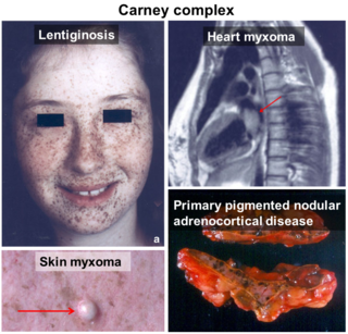
Carney complex and its subsets LAMB syndrome and NAME syndrome are autosomal dominant conditions comprising myxomas of the heart and skin, hyperpigmentation of the skin (lentiginosis), and endocrine overactivity. It is distinct from Carney's triad. Approximately 7% of all cardiac myxomas are associated with Carney complex.

<span class="mw-page-title-main">Bachmann's bundle</span> Anatomical cardiac structure

In the heart's conduction system, Bachmann's bundle is a branch of the anterior internodal tract that resides on the inner wall of the left atrium. It is a broad band of cardiac muscle that passes from the right atrium, between the superior vena cava and the ascending aorta. Bachmann's bundle is, during normal sinus rhythm, the preferential path for electrical activation of the left atrium. It is therefore considered to be part of the "atrial conduction system" of the heart.

Mitral valve replacement is a procedure whereby the diseased mitral valve of a patient's heart is replaced by either a mechanical or tissue (bioprosthetic) valve.

<span class="mw-page-title-main">Primary tumors of the heart</span> Medical condition

Primary tumors of the heart are extremely rare tumors that arise from the normal tissues that make up the heart. The incidence of primary cardiac tumors has been found to be approximately 0.02%. This is in contrast to secondary tumors of the heart, which are typically either metastatic from another part of the body, or infiltrate the heart via direct extension from the surrounding tissues. Metastatic tumors to the heart are about 20 times more common than primary cardiac tumors.

Heart cancer is an extremely rare form of cancer that is divided into primary tumors of the heart and secondary tumors of the heart.

<span class="mw-page-title-main">Cardiac myxoma</span> Medical condition

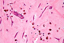
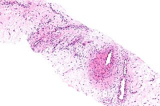
A myxoma is a rare benign tumor of the heart. Myxomata are the most common primary cardiac tumor in adults, and are most commonly found within the left atrium near the valve of the fossa ovalis. Myxomata may also develop in the other heart chambers. The tumor is derived from multipotent mesenchymal cells. Cardiac myxoma can affect adults between 30 and 60 years of age.

<span class="mw-page-title-main">Myocardial rupture</span> Medical condition

Myocardial rupture is a laceration of the ventricles or atria of the heart, of the interatrial or interventricular septum, or of the papillary muscles. It is most commonly seen as a serious sequela of an acute myocardial infarction.

<span class="mw-page-title-main">Aggressive angiomyxoma</span> Medical condition

Angiomyxoma is a myxoid tumor involving the blood vessels.

<span class="mw-page-title-main">Neurothekeoma</span>

A Neurothekeoma (NT) is a type of rare benign cutaneous tumor that usually develops on the head and neck. They often occur in the second and early third decades of life and tend to afflict women more frequently than men. First described by Gallager and Helwig, who proposed the term in order to reflect the presumed origin of the lesion from nerve sheath. Microscopically, the lesions described closely resembled the tumor, "nerve sheath myxoma (NSM)", an entity first described by Harkin and Reed. The latter had, through the years, been variously described as "bizarre cutaneous neurofibroma", "myxoma of nerve sheath", and "pacinian neurofibroma".

<span class="mw-page-title-main">Cutaneous myxoma</span>

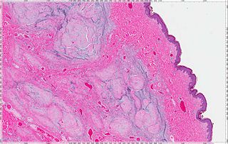
A cutaneous myxoma, or superficial angiomyxoma, consists of a multilobulated myxoid mass containing stellate or spindled fibroblasts with pools of mucin forming cleft-like spaces. There is often a proliferation of blood vessels and an inflammatory infiltrate. Staining is positive for vimentin, negative for cytokeratin and desmin, and variable for CD34, Factor VIIIa, SMA, MSA and S-100.

<span class="mw-page-title-main">Endometrial stromal sarcoma</span> Medical condition

Endometrial stromal sarcoma is a malignant subtype of endometrial stromal tumor arising from the stroma of the endometrium rather than the glands. There are three grades for endometrial stromal tumors, as follows. It was previously known as endolymphatic stromal myosis because of diffuse infiltration of myometrial tissue or the invasion of lymphatic channels.

Cardiac fibroma, also known as cardiac fibromatosis, cardiac fibrous hamartoma, fibroelastic hamartoma of heart and fibroma of heart is the second highest type of primary cardiac tumor seen in infants and children. This benign tumor made by connective tissue and fibroblast is largely observed in the ventricles of the heart. The left ventricle is the most common location of cardiac fibroma and accounts for approximately 57% of cardiac fibroma cases followed by the right ventricle with 27.5% of cases. Symptoms of the disease depend on the size of the tumor, its location relative to the conduction system, and whether it obstructs blood flow. Two-thirds of children with this tumor are asymptomatic, showing no signs and symptoms. Therefore the cause of cardiac fibroma is unexplained but has been associated with Gorlin Syndrome. Echocardiography is the primarily diagnostic method used to detect if an individual has cardiac fibroma. Resection of the tumor is recommended however heart transplantation is done if surgery is not possible. Overall prognosis of resection is favorable and the chance of recurrence is scarcely reported.

Tegumental angiomyxoma-neurothekeoma is a syndrome, an acronym, and eponym proposed by Malaysian ophthalmologist of Chinese Descent, Tan Aik Kah. Angiomyxomas are associated with LAMB syndrome, NAME syndrome and Carney syndrome.

<span class="mw-page-title-main">Myxoid tumor</span> Connective tissue tumor composed of clear, mucoid substance

A myxoid tumor is a connective tissue tumor with a "myxoid" background, composed of clear, mucoid substance.

Ectomesenchymal chondromyxoid tumor (ECT) is a benign intraoral tumor with presumed origin from undifferentiated (ecto)mesenchymal cells. There are some who think it is a myoepithelial tumor type.

References

  1. " Myxoma " at Dorland's Medical Dictionary
  2. Satter, Elizabeth K. (2009). "Solitary superficial angiomyxoma: an infrequent but distinct soft tissue tumor". Journal of Cutaneous Pathology. 36 (s1): 56–59. doi:10.1111/j.1600-0560.2008.01216.x. ISSN   1600-0560. PMID   19187115. S2CID   1528140.
  3. Seino Y, Ikeda U, Shimada K (1993). "Increased expression of interleukin 6 mRNA in cardiac myxomas". Br Heart J. 69 (6): 565–7. doi:10.1136/hrt.69.6.565. PMC   1025174 . PMID   8343326.
  4. Jourdan M, Bataille R, Seguin J, Zhang XG, Chaptal PA, Klein B (1990). "Constitutive production of interleukin-6 and immunologic features in cardiac myxomas". Arthritis Rheum. 33 (3): 398–402. doi:10.1002/art.1780330313. PMID   1690543.
  5. Wada A; Kanda t; Hayashi R; et al. (1993). "Cardiac myxoma metastasized to the brain: potential role of endogenous interleukin-6". Cardiology. 83 (3): 208–11. doi:10.1159/000015180. PMID   8281536. S2CID   22543670.
  6. Fisher J. (1983). "Cardiac myxoma". Cardiovasc Rev Rep (4): 1195–9.
  7. Jain, Puneet; Finger, Paul T; Iacob, Codrin E (2018-11-01). "Conjunctival myxoma: A case report with unique high frequency ultrasound (UBM) findings". Indian Journal of Ophthalmology. 66 (11): 1629–1631. doi: 10.4103/ijo.IJO_518_18 . PMC   6213696 . PMID   30355886.
  8. Knepper LE, Biller J, Adams HP, Bruno A (1988). "Neurologic manifestations of atrial myxoma. A 12-year experience and review". Stroke. 19 (11): 1435–40. doi: 10.1161/01.str.19.11.1435 . PMID   3188128.
  9. 1 2 Cheng, Davy C.H.; Martin, Janet; David, Tirone (2020). Evidence-Based Practice in Perioperative Cardiac Anesthesia and Surgery. Springer Nature. p. 210. ISBN   9783030478872.
  10. Takahashi, Ayaka; et al. (May 2016). "Multimodal Cardiovascular Imaging of Cardiac Tumors". Annals of Nuclear Cardiology. 2 (1): 61–67. doi: 10.17996/anc.2.1_61 .