Related Research Articles

A sarcoma is a malignant tumor, a type of cancer that arises from transformed cells of mesenchymal origin. Connective tissue is a broad term that includes bone, cartilage, fat, vascular, or hematopoietic tissues, and sarcomas can arise in any of these types of tissues. As a result, there are many subtypes of sarcoma, which are classified based on the specific tissue and type of cell from which the tumor originates. Sarcomas are primary connective tissue tumors, meaning that they arise in connective tissues. This is in contrast to secondary connective tissue tumors, which occur when a cancer from elsewhere in the body spreads to the connective tissue. The word sarcoma is derived from the Greek σάρκωμα sarkōma "fleshy excrescence or substance", itself from σάρξ sarx meaning "flesh".

Rhabdomyosarcoma, or RMS, is an aggressive and highly malignant form of cancer that develops from skeletal (striated) muscle cells that have failed to fully differentiate. It is generally considered to be a disease of childhood, as the vast majority of cases occur in those below the age of 18. It is commonly described as one of the "small, round, blue cell tumours of childhood" due to its appearance on an H&E stain. Despite being a relatively rare cancer, it accounts for approximately 40% of all recorded soft tissue sarcomas.

Brenner tumors are an uncommon subtype of the surface epithelial-stromal tumor group of ovarian neoplasms. The majority are benign, but some can be malignant.

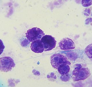
A mastocytoma or mast cell tumor is a type of round-cell tumor consisting of mast cells. It is found in humans and many animal species; it also can refer to an accumulation or nodule of mast cells that resembles a tumor.

Hemangioendotheliomas are a family of vascular neoplasms of intermediate malignancy.

Canalicular adenoma is a benign, epithelial salivary gland neoplasm arranged in interconnecting cords of columnar cells. This is a very rare benign neoplasm, that makes up about 1% of all salivary gland tumors, or about 4% of all benign salivary gland tumors.

A mammary tumor is a neoplasm originating in the mammary gland. It is a common finding in older female dogs and cats that are not spayed, but they are found in other animals as well. The mammary glands in dogs and cats are associated with their nipples and extend from the underside of the chest to the groin on both sides of the midline. There are many differences between mammary tumors in animals and breast cancer in humans, including tumor type, malignancy, and treatment options. The prevalence in dogs is about three times that of women. In dogs, mammary tumors are the second most common tumor over all and the most common tumor in female dogs with a reported incidence of 3.4%. Multiple studies have documented that spaying female dogs when young greatly decreases their risk of developing mammary neoplasia when aged. Compared with female dogs left intact, those spayed before puberty have 0.5% of the risk, those spayed after one estrous cycle have 8.0% of the risk, and dogs spayed after two estrous cycles have 26.0% of the risk of developing mammary neoplasia later in life. Overall, unspayed female dogs have a seven times greater risk of developing mammary neoplasia than do those that are spayed. While the benefit of spaying decreases with each estrous cycle, some benefit has been demonstrated in female dogs even up to 9 years of age. There is a much lower risk in male dogs and a risk in cats about half that of dogs.
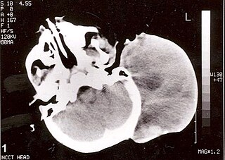
Sacrococcygeal teratoma (SCT) is a type of tumor known as a teratoma that develops at the base of the coccyx (tailbone) and is thought to be derived from the primitive streak. Sacrococcygeal teratomas are benign 75% of the time, malignant 12% of the time, and the remainder are considered "immature teratomas" that share benign and malignant features. Benign sacrococcygeal teratomas are more likely to develop in younger children who are less than 5 months old, and older children are more likely to develop malignant sacrococcygeal teratomas. The Currarino triad, due to an autosomal dominant mutation in the MNX1 gene, consists of a presacral mass, anorectal malformation and sacral dysgenesis.

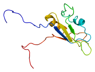
Neprilysin, also known as membrane metallo-endopeptidase (MME), neutral endopeptidase (NEP), cluster of differentiation 10 (CD10), and common acute lymphoblastic leukemia antigen (CALLA) is an enzyme that in humans is encoded by the MME gene. Neprilysin is a zinc-dependent metalloprotease that cleaves peptides at the amino side of hydrophobic residues and inactivates several peptide hormones including glucagon, enkephalins, substance P, neurotensin, oxytocin, and bradykinin. It also degrades the amyloid beta peptide whose abnormal folding and aggregation in neural tissue has been implicated as a cause of Alzheimer's disease. Synthesized as a membrane-bound protein, the neprilysin ectodomain is released into the extracellular domain after it has been transported from the Golgi apparatus to the cell surface.

RNA-binding protein EWS is a protein that in humans is encoded by the EWSR1 gene on human chromosome 22, specifically 22q12.2. This region of chromosome 22 encodes the N-terminal transactivation domain of the EWS protein and that region may become joined to one of several other chromosomes which encode various transcription factors, see The expression of a chimeric protein with the EWS transactivation domain fused to the DNA binding region of a transcription factor generates a powerful oncogenic protein causing Ewing sarcoma and other members of the Ewing family of tumors. These translocations can occur due to chromoplexy, a burst of complex chromosomal rearrangements seen in cancer cells. The normal EWS gene encodes an RNA binding protein closely related to FUS (gene) and TAF15, all of which have been associated to amyotrophic lateral sclerosis.

Angiomyxoma is a myxoid tumor involving the blood vessels.
Juxtaglomerular cell tumor is an extremely rare kidney tumour of the juxtaglomerular cells, with less than 100 cases reported in literature. This tumor typically secretes renin, hence the former name of reninoma. It often causes severe hypertension that is difficult to control, in adults and children, although among causes of secondary hypertension it is rare. It develops most commonly in young adults, but can be diagnosed much later in life. It is generally considered benign, but its malignant potential is uncertain.

Fetal adenocarcinoma (FA) of the lung is a rare subtype of pulmonary adenocarcinoma that exhibits tissue architecture and cell characteristics that resemble fetal lung tissue upon microscopic examination. It is currently considered a variant of solid adenocarcinoma with mucin production.
Large cell lung carcinoma with rhabdoid phenotype (LCLC-RP) is a rare histological form of lung cancer, currently classified as a variant of large cell lung carcinoma (LCLC). In order for a LCLC to be subclassified as the rhabdoid phenotype variant, at least 10% of the malignant tumor cells must contain distinctive structures composed of tangled intermediate filaments that displace the cell nucleus outward toward the cell membrane. The whorled eosinophilic inclusions in LCLC-RP cells give it a microscopic resemblance to malignant cells found in rhabdomyosarcoma (RMS), a rare neoplasm arising from transformed skeletal muscle. Despite their microscopic similarities, LCLC-RP is not associated with rhabdomyosarcoma.
Mucinous cystadenocarcinoma of the lung (MCACL) is a very rare malignant mucus-producing neoplasm arising from the uncontrolled growth of transformed epithelial cells originating in lung tissue.

Giant-cell carcinoma of the lung (GCCL) is a rare histological form of large-cell lung carcinoma, a subtype of undifferentiated lung cancer, traditionally classified within the non-small-cell lung carcinomas (NSCLC).
A sialoblastoma is a low-grade salivary gland neoplasm that recapitulates primitive salivary gland anlage. It has previously been referred to as congenital basal cell adenoma, embryoma, or basaloid adenocarcinoma. It is an extremely rare tumor, with less than 100 cases reported worldwide.
Rhabdomyoma is a benign mesenchymal tumor of skeletal muscle, separated into two major categories based on site: Cardiac and extracardiac. They are further separated by histology: fetal, juvenile (intermediate), and adult types. Genital types are recognized, but are often part of either the fetal or juvenile types. The fetal type is thought to recapitulate immature skeletal muscle at about week six to ten of gestational development.
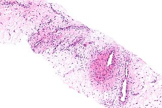
The Xanthogranulomatous Process (XP), is a form of acute and chronic inflammation characterized by an exuberant clustering of foamy macrophages among other inflammatory cells. Localization in the kidney and renal pelvis has been the most frequent and better known occurrence followed by that in the gallbladder but many others have been subsequently recorded. The pathological findings of the process and etiopathogenetic and clinical observations have been reviewed by Cozzutto and Carbone.
Ectomesenchymal chondromyxoid tumor (ECT) is a benign intraoral tumor with presumed origin from undifferentiated (ecto)mesenchymal cells. There are some who think it is a myoepithelial tumor type.
References
- ↑ Makhson AN, Bulycheva IV, Kuz'min IV, Denisov AK, Groshev IA (2006). "[Ossifying fibromyxoid tumor]". Arkh. Patol. (in Russian). 68 (2): 41–4. PMID 16752509.
- ↑ Seykora JT, Kutcher C, van de Rijn M, Dzubow L, Junkins-Hopkins J, Ioffreda M (2006). "Ossifying fibromyxoid tumor of soft parts presenting as a scalp cyst". J. Cutan. Pathol. 33 (8): 569–72. doi:10.1111/j.1600-0560.2006.00444.x. PMID 16919031.
- ↑ Mollaoglu N, Tokman B, Kahraman S, Cetiner S, Yucetas S, Uluoglu O (2006). "An unusual presentation of ossifying fibromyxoid tumor of the mandible: a case report". J Clin Pediatr Dent. 31 (2): 136–8. doi:10.17796/jcpd.31.2.f34037713m414l1u. PMID 17315811.
- ↑ Folpe AL, Weiss SW (2003). "Ossifying fibromyxoid tumor of soft parts: a clinicopathologic study of 70 cases with emphasis on atypical and malignant variants". Am. J. Surg. Pathol. 27 (4): 421–31. doi:10.1097/00000478-200304000-00001. PMID 12657926.