Plasmodium ovale

Last updated

Contents

Plasmodium ovale
Plasmodium ovale 01.png
Plasmodium ovale trophozoite, Giemsa stain.
Scientific classification OOjs UI icon edit-ltr.svg
Domain: Eukaryota
Clade: Diaphoretickes
Clade: SAR
Clade: Alveolata
Phylum: Apicomplexa
Class: Aconoidasida
Order: Haemospororida
Family: Plasmodiidae
Genus: Plasmodium
Species:
P. ovale
Binomial name
Plasmodium ovale
Stephens, 1922

Plasmodium ovale is a species of parasitic protozoon that causes tertian malaria in humans. It is one of several species of Plasmodium parasites that infect humans, including Plasmodium falciparum and Plasmodium vivax which are responsible for most cases of malaria in the world. P. ovale is rare compared to these two parasites, and substantially less dangerous than P. falciparum.

P. ovale has recently been shown by genetic methods to consist of what is considered to be two species (despite having been given subspecies names), namely P. ovale curtisi and P. ovale wallikeri . [1] The procedure necessary to rectify the nomenclature has been outlined. [2]

History

This species was first described in 1914 by Stephens in a blood sample taken in the autumn of 1913 from a patient in the sanitarium of Pachmari in central India and sent by Major W. H. Kenrick to Stephens (who was working in Liverpool). [3]

Epidemiology

Relative incidence of Plasmodium species by country of origin for imported cases to non-endemic countries, showing P. ovale in pink. Relative incidence of Plasmodium (malaria) species by country of origin for imported cases to non-endemic countries.png
Relative incidence of Plasmodium species by country of origin for imported cases to non-endemic countries, showing P. ovale in pink.

P. ovale is primarily concentrated in sub-Saharan Africa and islands in the western Pacific. [5] [6] However P. ovale has also been reported in the Philippines, eastern Indonesia, and Papua New Guinea, [7] as well as in Bangladesh, [8] Cambodia, [9] India, [10] Myanmar, [11] Thailand [12] and Vietnam. [13]

In several studies, the reported prevalence of P. ovale was low relative to other malaria parasites, with fewer than 5% of malaria cases being associated with P. ovale infection. Higher prevalences of P. ovale are possible under certain conditions, as at least one study in Cameroon found the prevalence of P. ovale infection to be greater than 10%. [5]

It has been estimated that there are about 15 million cases of infection each year with this parasite. [1]

While similar to P. vivax, P. ovale is able to infect individuals who are negative for the Duffy blood group, which is the case for many residents of sub-Saharan Africa. This has been said to explain the greater prevalence of P. ovale (versus P. vivax) in most of Africa. [14] However, low-parasitaemia or subpatent P. vivax cases might be more prevalent in Africa than has been thought. [15]

Clinical features

In humans, symptoms generally appear 12 to 20 days after the parasite has entered the blood. In the blood, the parasite's replication cycle lasts approximately 49 hours, causing tertian fever which spikes approximately every 49 hours as newly replicated parasites erupt out of red blood cells. Mean maximum parasite levels have been found to be 6,944/microl for sporozoite-induced infections and 7,310/microl for trophozoite-induced infections. [5]

In some cases, relapse may occur up to 4 years after infection. [5]

Diagnosis

Plasmodium ovale microgametocyte in Giemsa-stained thin blood film, with annotated Schuffner's dots and hemozoin pigment. Microphotograph of Plasmodium ovale microgametocyte in Giemsa-stained thin blood film, with Schuffner's dots and hemozoin pigment.png
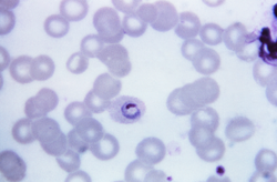
Plasmodium ovale microgametocyte in Giemsa-stained thin blood film, with annotated Schüffner's dots and hemozoin pigment.

The microscopic appearance of P. ovale is very similar to that of P. vivax and if there are only a small number of parasites seen, it may be impossible to distinguish the two species on morphological grounds alone. There is no difference between the medical treatment of P. ovale and P. vivax, and therefore some laboratory diagnoses report "P. vivax/ovale", which is perfectly acceptable as treatment for the two is very similar. Schüffner's dots are seen on the surface of the parasitised red blood cell, but these are larger and darker than in P. vivax and are sometimes called James' dots or James' stippling. About twenty percent of the parasitised cells are oval in shape (hence the species name) and some of the oval cells also have fimbriated edges (the so-called "comet cell"). The mature schizonts of P. ovale never have more than twelve nuclei within them and this is the only reliable way of distinguishing between the two species.

P. vivax and P. ovale that have been sitting in EDTA for more than half an hour before the blood film is made will look very similar in appearance to P. malariae , which is an important reason to warn the laboratory immediately when the blood sample is drawn so they can process the sample as soon as it arrives.

Molecular tests (tests that detect DNA in blood) must take into account the fact that there are two P. ovale sensu lato taxa. Tests designed for one will not necessarily detect the other. [16]

Treatment

Standard treatment is concurrent treatment with chloroquine and primaquine. The combination atovaquone-proguanil may be used in those patients who are unable to take chloroquine for whatever reason. [17] An overdose on Chloroquine can be very dangerous and can result in death.

Phylogenetics

Among the species infecting the great apes, Plasmodium schwetzi morphologically appears to be the closest relation to P. ovale. As of 2013 this had not been confirmed by DNA studies.

The original species has been shown to be two morphologically identical forms – Plasmodium ovale curtisi and Plasmodium ovale wallikeri – which can be differentiated only by genetic means. [1] Both species have been identified in Ghana, Myanmar, Nigeria, São Tomé, Sierra Leone and Uganda. The separation of the lineages is estimated to have occurred between 1.0 and 3.5 million years ago in hominid hosts. A second analysis suggests that these species separated 4.5  million years ago (95% confidence interval 0.5 – 7.7 Mya). [18] A third worked sequenced the whole genome of both species, confirmed the differences and dated the split at around million years. [19] Although dating is always difficult, the authors date that split to be 5 times older than the P. falciparum and P. reichenowi split.

These species appear to be more closely related to Plasmodium malariae than to Plasmodium vivax . [18]

The two species appear to differ biologically, with P. ovale wallikeri having a shorter latency period than P. ovale curtisi. [20]

Life cycle

Microphotographs of Plasmodium ovale in Giemsa-stained thin blood films. a-c ring stages, d, e young trophozoites, f trophozoite, g late trophozoite, h young schizont, i-k growing schizont, l late schizont, m ruptured schizont, n young gametocyte, o, p developing macrogametocytes, q macrogametocyte, r microgametocyte. Microphotographs of Plasmodium ovale in Giemsa-stained thin blood films.jpg
Microphotographs of Plasmodium ovale in Giemsa-stained thin blood films. a–c ring stages, d, e young trophozoites, f trophozoite, g late trophozoite, h young schizont, i–k growing schizont, l late schizont, m ruptured schizont, n young gametocyte, o, p developing macrogametocytes, q macrogametocyte, r microgametocyte.

P. ovale is introduced into the human host by the bite of an infected mosquito, in a motile form called a sporozoite. The sporozoites are carried by the blood to the liver, where they replicate asexually by merogony into non-motile merozoites. Several hundred merozoites are produced and released into the bloodstream where they infect erythrocytes. Inside the erythrocyte, the parasite's replication cycle takes approximately 49 hours, after which the erythrocyte ruptures and between 8 and 20 merozoites are released to infect other erythrocytes. Some of these merozoites will instead form gametocytes which remain in the blood and are ingested by a mosquito. [5]

When gametocytes are ingested by a mosquito, the gametocytes enter the mosquito gut where fertilisation occurs forming a zygote known as an ookinete. The ookinete moves to the outer wall of the mosquito midgut where it develops over the course of several weeks. This developing stage is called an oocyst. After the oocyst develops, it ruptures releasing several hundred sporozoites. The sporozoites are carried by the mosquito's circulation to the mosquito salivary glands. When the mosquito feeds again, the sporozoites enter through the salivary duct and are injected into a new host, starting the life cycle again. [5]

There are situations where some of the sporozoites hypothetically do not immediately start to grow and divide after entering the hepatocyte, but remain in a dormant, hypnozoite stage [22] for weeks or months. However, unlike the P. vivax situation, hypnozoites have yet to actually be seen in the life cycle of P. ovale. [23] The duration of latency is variable from one (assumed in the case of P. ovale) hypnozoite to another and the factors that will eventually trigger growth are not known; this could explain how a single infection can be responsible for a series of waves of parasitaemia or "relapses". [24]

Hosts

While humans appear to be the natural mammalian host of P. ovale, chimpanzees and Saimiri monkeys have also been experimentally infected. [5]

Anopheles gambiae and Anopheles funestus are likely the natural mosquito hosts of P. ovale. Experimentally, several other mosquito species have been shown to be capable of transmitting P. ovale to humans, including:

Genomes

The full genomes of the two P. ovale species can be seen on geneDB.org – P. ovali curtisi P. ovale wallikeri and plasmoDB.org, published 2017. [19]

See also

Related Research Articles

<span class="mw-page-title-main">Malaria</span> Mosquito-borne infectious disease

Malaria is a mosquito-borne infectious disease that affects humans and other vertebrates. Human malaria causes symptoms that typically include fever, fatigue, vomiting, and headaches. In severe cases, it can cause jaundice, seizures, coma, or death. Symptoms usually begin 10 to 15 days after being bitten by an infected Anopheles mosquito. If not properly treated, people may have recurrences of the disease months later. In those who have recently survived an infection, reinfection usually causes milder symptoms. This partial resistance disappears over months to years if the person has no continuing exposure to malaria.

<i>Plasmodium</i> Genus of parasitic protists that can cause malaria

Plasmodium is a genus of unicellular eukaryotes that are obligate parasites of vertebrates and insects. The life cycles of Plasmodium species involve development in a blood-feeding insect host which then injects parasites into a vertebrate host during a blood meal. Parasites grow within a vertebrate body tissue before entering the bloodstream to infect red blood cells. The ensuing destruction of host red blood cells can result in malaria. During this infection, some parasites are picked up by a blood-feeding insect, continuing the life cycle.

<i>Plasmodium falciparum</i> Protozoan species of malaria parasite

Plasmodium falciparum is a unicellular protozoan parasite of humans, and the deadliest species of Plasmodium that causes malaria in humans. The parasite is transmitted through the bite of a female Anopheles mosquito and causes the disease's most dangerous form, falciparum malaria. It is responsible for around 50% of all malaria cases. P. falciparum is therefore regarded as the deadliest parasite in humans. It is also associated with the development of blood cancer and is classified as a Group 2A (probable) carcinogen.

A trophozoite is the activated, feeding stage in the life cycle of certain protozoa such as malaria-causing Plasmodium falciparum and those of the Giardia group. The complementary form of the trophozoite state is the thick-walled cyst form. They are often different from the cyst stage, which is a protective, dormant form of the protozoa. Trophozoites are often found in the host's body fluids and tissues and in many cases, they are the form of the protozoan that causes disease in the host. In the protozoan, Entamoeba histolytica it invades the intestinal mucosa of its host, causing dysentery, which aid in the trophozoites traveling to the liver and leading to the production of hepatic abscesses.

<span class="mw-page-title-main">Gametocyte</span> Eukaryotic germ stem cell

A gametocyte is a eukaryotic germ cell that divides by mitosis into other gametocytes or by meiosis into gametids during gametogenesis. Male gametocytes are called spermatocytes, and female gametocytes are called oocytes.

<span class="mw-page-title-main">Giovanni Battista Grassi</span> Italian physician and zoologist (1854–1925)

Giovanni Battista Grassi was an Italian physician and zoologist, best known for his pioneering works on parasitology, especially on malariology. He was Professor of Comparative Zoology at the University of Catania from 1883, and Professor of Comparative Anatomy at Sapienza University of Rome from 1895 until his death. His first major research on the taxonomy and biology of termites earned him the Royal Society's Darwin Medal in 1896.

<span class="mw-page-title-main">Primaquine</span> Pharmaceutical drug

Primaquine is a medication used to treat and prevent malaria and to treat Pneumocystis pneumonia. Specifically it is used for malaria due to Plasmodium vivax and Plasmodium ovale along with other medications and for prevention if other options cannot be used. It is an alternative treatment for Pneumocystis pneumonia together with clindamycin. It is taken by mouth.

Recrudescence is the recurrence of an undesirable condition. In medicine, it is usually defined as the recurrence of symptoms after a period of remission or quiescence, in which sense it can sometimes be synonymous with relapse. In a narrower sense, it can also be such a recurrence with higher severity than before the remission. "Relapse" conventionally has a specific meaning when used in relation to malaria.

<i>Plasmodium vivax</i> Species of single-celled organism

Plasmodium vivax is a protozoal parasite and a human pathogen. This parasite is the most frequent and widely distributed cause of recurring malaria. Although it is less virulent than Plasmodium falciparum, the deadliest of the five human malaria parasites, P. vivax malaria infections can lead to severe disease and death, often due to splenomegaly. P. vivax is carried by the female Anopheles mosquito; the males do not bite.

<i>Plasmodium malariae</i> Species of single-celled organism

Plasmodium malariae is a parasitic protozoan that causes malaria in humans. It is one of several species of Plasmodium parasites that infect other organisms as pathogens, also including Plasmodium falciparum and Plasmodium vivax, responsible for most malarial infection. Found worldwide, it causes a so-called "benign malaria", not nearly as dangerous as that produced by P. falciparum or P. vivax. The signs include fevers that recur at approximately three-day intervals – a quartan fever or quartan malaria – longer than the two-day (tertian) intervals of the other malarial parasite.

<i>Plasmodium knowlesi</i> Species of single-celled organism

Plasmodium knowlesi is a parasite that causes malaria in humans and other primates. It is found throughout Southeast Asia, and is the most common cause of human malaria in Malaysia. Like other Plasmodium species, P. knowlesi has a life cycle that requires infection of both a mosquito and a warm-blooded host. While the natural warm-blooded hosts of P. knowlesi are likely various Old World monkeys, humans can be infected by P. knowlesi if they are fed upon by infected mosquitoes. P. knowlesi is a eukaryote in the phylum Apicomplexa, genus Plasmodium, and subgenus Plasmodium. It is most closely related to the human parasite Plasmodium vivax as well as other Plasmodium species that infect non-human primates.

<span class="mw-page-title-main">History of malaria</span> History of malaria infections

The history of malaria extends from its prehistoric origin as a zoonotic disease in the primates of Africa through to the 21st century. A widespread and potentially lethal human infectious disease, at its peak malaria infested every continent except Antarctica. Its prevention and treatment have been targeted in science and medicine for hundreds of years. Since the discovery of the Plasmodium parasites which cause it, research attention has focused on their biology as well as that of the mosquitoes which transmit the parasites.

Hematozoa is a subclass of blood parasites of the Apicomplexa clade. Well known examples include the Plasmodium spp. which cause malaria in humans and Theileria which causes theileriosis in cattle. A large number of species are known to infect birds and are transmitted by insect vectors. The pattern in which Haematozoa infect a host cell depends on the genera of the blood parasite. Plasmodium and Leucozytozoon displace the nucleus of the host cell so that the parasite can take control of the cell where as Hemoproteus completely envelops the nucleus in a host cell.

<span class="mw-page-title-main">Apicomplexan life cycle</span> Apicomplexa life cycle

Apicomplexans, a group of intracellular parasites, have life cycle stages that allow them to survive the wide variety of environments they are exposed to during their complex life cycle. Each stage in the life cycle of an apicomplexan organism is typified by a cellular variety with a distinct morphology and biochemistry.

Plasmodium coatneyi is a parasitic species that is an agent of malaria in nonhuman primates. P. coatneyi occurs in Southeast Asia. The natural host of this species is the rhesus macaque and crab-eating macaque, but there has been no evidence that zoonosis of P. coatneyi can occur through its vector, the female Anopheles mosquito.

<i>Plasmodium cynomolgi</i> Species of single-celled organism

Plasmodium cynomolgi is an apicomplexan parasite that infects mosquitoes and Asian Old World monkeys. In recent years, a number of natural infections of humans have also been documented. This species has been used as a model for human Plasmodium vivax because Plasmodium cynomolgi shares the same life cycle and some important biological features with P. vivax.

Plasmodium brasilianum is a parasite that infects many species of platyrrhine monkeys in South and Central America.

<span class="mw-page-title-main">Quartan fever</span> Medical condition

Quartan fever is one of the four types of malaria which can be contracted by humans.

References

  1. 1 2 3 Sutherland CJ, Tanomsing N, Nolder D, Oguike M, Jennison C, Pukrittayakamee S, et al. (May 2010). "Two nonrecombining sympatric forms of the human malaria parasite Plasmodium ovale occur globally". The Journal of Infectious Diseases. 201 (10): 1544–50. doi: 10.1086/652240 . PMID   20380562.
  2. Markus, M.B. (2024). "Type material of Plasmodium ovale sensu lato". Southern African Journal of Infectious Diseases. 39 (1): 615. doi:10.4102/sajid.v39i1.615.
  3. Stephens JWW (8 April 1914). "A new malaria parasite of man". Proc R Soc Lond B. 87 (596): 375–377. Bibcode:1914RSPSB..87..375S. doi:10.1098/rspb.1914.0024. S2CID   86121600.
  4. Tatem AJ, Jia P, Ordanovich D, Falkner M, Huang Z, Howes R; et al. (2017). "The geography of imported malaria to non-endemic countries: a meta-analysis of nationally reported statistics". Lancet Infect Dis. 17 (1): 98–107. doi:10.1016/S1473-3099(16)30326-7. PMC   5392593 . PMID   27777030.{{cite journal}}: CS1 maint: multiple names: authors list (link)
  5. 1 2 3 4 5 6 7 8 Collins WE, Jeffery GM (July 2005). "Plasmodium ovale: parasite and disease". Clinical Microbiology Reviews. 18 (3): 570–81. doi:10.1128/CMR.18.3.570-581.2005. PMC   1195966 . PMID   16020691.
  6. Faye FB, Konaté L, Rogier C, Trape JF (1998). "Plasmodium ovale in a highly malaria endemic area of Senegal". Transactions of the Royal Society of Tropical Medicine and Hygiene. 92 (5): 522–5. doi:10.1016/S0035-9203(98)90900-2. PMID   9861368.
  7. Baird JK, Hoffman SL (November 2004). "Primaquine therapy for malaria". Clinical Infectious Diseases. 39 (9): 1336–45. doi: 10.1086/424663 . PMID   15494911.
  8. Fuehrer HP, Starzengruber P, Swoboda P, Khan WA, Matt J, Ley B, et al. (July 2010). "Indigenous Plasmodium ovale malaria in Bangladesh". The American Journal of Tropical Medicine and Hygiene. 83 (1): 75–8. doi:10.4269/ajtmh.2010.09-0796. PMC   2912579 . PMID   20595481.
  9. Incardona S, Chy S, Chiv L, Nhem S, Sem R, Hewitt S, et al. (June 2005). "Large sequence heterogeneity of the small subunit ribosomal RNA gene of Plasmodium ovale in Cambodia". The American Journal of Tropical Medicine and Hygiene. 72 (6): 719–24. doi: 10.4269/ajtmh.2005.72.719 . PMID   15964956.
  10. Snounou G, Viriyakosol S, Jarra W, Thaithong S, Brown KN (April 1993). "Identification of the four human malaria parasite species in field samples by the polymerase chain reaction and detection of a high prevalence of mixed infections". Molecular and Biochemical Parasitology. 58 (2): 283–92. doi:10.1016/0166-6851(93)90050-8. PMID   8479452.
  11. Li, Nana; Parker, Daniel M.; Yang, Zhaoqing; Fan, Qi; Zhou, Guofa; Ai, Guoping; Duan, Jianhua; Lee, Ming-chieh; Yan, Guiyun; Matthews, Stephen A.; Cui, Liwang (2013-10-10). "Risk factors associated with slide positivity among febrile patients in a conflict zone of north-eastern Myanmar along the China-Myanmar border". Malaria Journal. 12 (1): 361. doi: 10.1186/1475-2875-12-361 . ISSN   1475-2875. PMC   3852943 . PMID   24112638.
  12. Cadigan FC, Desowitz RS (1969). "Two cases of Plasmodium ovale malaria from central Thailand". Transactions of the Royal Society of Tropical Medicine and Hygiene. 63 (5): 681–2. doi:10.1016/0035-9203(69)90194-1. PMID   5824291.
  13. Gleason NN, Fisher GU, Blumhardt R, Roth AE, Gaffney GW (1970). "Plasmodium ovale malaria acquired in Viet-Nam". Bulletin of the World Health Organization. 42 (3): 399–403. PMC   2427544 . PMID   4392940.
  14. "Biology: Malaria (CDC malaria)". Archived from the original on 2008-10-13.
  15. Markus, MB (2022). "Theoretical origin of genetically homologous Plasmodium vivax malarial recurrences". Southern African Journal of Infectious Diseases. 37 (1): 369. doi:10.4102/sajid.v37i1.369. PMC   8991251 . PMID   35399558.
  16. Fuehrer HP, Noedl H (February 2014). "Recent advances in detection of Plasmodium ovale: implications of separation into the two species Plasmodium ovale wallikeri and Plasmodium ovale curtisi". Journal of Clinical Microbiology. 52 (2): 387–91. doi:10.1128/JCM.02760-13. PMC   3911344 . PMID   24478466.
  17. Radloff PD, Philipps J, Hutchinson D, Kremsner PG (1996). "Atovaquone plus proguanil is an effective treatment for Plasmodium ovale and P. malariae malaria". Transactions of the Royal Society of Tropical Medicine and Hygiene. 90 (6): 682. doi:10.1016/S0035-9203(96)90435-6. PMID   9015517.
  18. 1 2 Putaporntip C, Hughes AL, Jongwutiwes S (2013). "Low level of sequence diversity at merozoite surface protein-1 locus of Plasmodium ovale curtisi and P. ovale wallikeri from Thai isolates". PLOS ONE. 8 (3): e58962. Bibcode:2013PLoSO...858962P. doi: 10.1371/journal.pone.0058962 . PMC   3594193 . PMID   23536840.
  19. 1 2 Rutledge GG, Böhme U, Sanders M, Reid AJ, Cotton JA, Maiga-Ascofare O, et al. (February 2017). "Plasmodium malariae and P. ovale genomes provide insights into malaria parasite evolution". Nature. 542 (7639): 101–104. Bibcode:2017Natur.542..101R. doi:10.1038/nature21038. PMC   5326575 . PMID   28117441.
  20. Nolder D, Oguike MC, Maxwell-Scott H, Niyazi HA, Smith V, Chiodini PL, Sutherland CJ (May 2013). "An observational study of malaria in British travellers: Plasmodium ovale wallikeri and Plasmodium ovale curtisi differ significantly in the duration of latency". BMJ Open. 3 (5): e002711. doi:10.1136/bmjopen-2013-002711. PMC   3657643 . PMID   23793668. Open Access logo PLoS transparent.svg
  21. Chavatte JM, Tan SB, Snounou G, Lin RT (2015). "Molecular characterization of misidentified Plasmodium ovale imported cases in Singapore". Malar J. 14: 454. doi: 10.1186/s12936-015-0985-8 . PMC   4650842 . PMID   26577930.{{cite journal}}: CS1 maint: multiple names: authors list (link)
  22. Markus MB (2011). "Malaria: origin of the term "hypnozoite"". Journal of the History of Biology. 44 (4): 781–6. doi:10.1007/s10739-010-9239-3. PMID   20665090. S2CID   1727294.
  23. Markus, MB (2021). "Safety and efficacy of tafenoquine for Plasmodium vivax malaria prophylaxis and radical cure: overview and perspectives". Therapeutics and Clinical Risk Management. 17: 989–999. doi: 10.2147/TCRM.S269336 . PMC   8435617 . PMID   34526770.
  24. "Malaria eModule – Exo-Erythrocytic Stages".