Ryanodine receptor 2

Last updated
RYR2
RYR2 protein.png
Available structures
PDB Ortholog search: PDBe RCSB
Identifiers
Aliases RYR2 , ARVC2, ARVD2, RYR-2, RyR, VTSIP, ryanodine receptor 2, VACRDS
External IDs OMIM: 180902 MGI: 99685 HomoloGene: 37423 GeneCards: RYR2
Orthologs
SpeciesHumanMouse
Entrez
Ensembl
UniProt
RefSeq (mRNA)

NM_001035

NM_023868

RefSeq (protein)

NP_001026

NP_076357

Location (UCSC) Chr 1: 237.04 – 237.83 Mb Chr 13: 11.55 – 12.11 Mb
PubMed search [3] [4]
Wikidata
View/Edit Human View/Edit Mouse

Ryanodine receptor 2 (RYR2) is one of a class of ryanodine receptors and a protein found primarily in cardiac muscle. In humans, it is encoded by the RYR2 gene. [5] [6] [7] In the process of cardiac calcium-induced calcium release, RYR2 is the major mediator for sarcoplasmic release of stored calcium ions.

Contents

Structure

The channel is composed of RYR2 homotetramers and  FK506-binding proteins  found in a 1:4 stoichiometric ratio. Calcium channel function is affected by the specific type of FK506 isomer interacting with the RYR2 protein, due to binding differences and other factors. [8]

Function

The RYR2 protein functions as the major component of a calcium channel located in the sarcoplasmic reticulum that supplies ions to the cardiac muscle during systole. To enable cardiac muscle contraction, calcium influx through voltage-gated  L-type calcium channels  in the plasma membrane allows calcium ions to bind to RYR2 located on the  sarcoplasmic reticulum. This binding causes the release of calcium through RYR2 from the sarcoplasmic reticulum into the cytosol, where it binds to the C domain of  troponin, which shifts tropomyosin and allows the myosin ATPase to bind to actin, enabling cardiac muscle contraction. [9] RYR2 channels are associated with many cellular functions, including mitochondrial metabolism, gene expression and cell survival, in addition to their role in cardiomyocyte contraction. [10]

Clinical significance

Deleterious mutations of the ryanodine receptor family, and especially the RYR2 receptor, lead to a constellation of pathologies leading to both acute and chronic heart failure collectively known as "Ryanopathies." [11]

Mutations in the RYR2 gene are associated with catecholaminergic polymorphic ventricular tachycardia and arrhythmogenic right ventricular dysplasia. [12]

Recently, sudden cardiac death in several young individuals in the Amish community (four of which were from the same family) was traced to homozygous duplication of a mutant RyR2 gene. [13] Normal (wild type) RyR2 functions primarily in the myocardium (heart muscle).

Mice with genetically reduced RYR2 exhibit a lower basal heart rate and fatal arrhythmias. [14]

Interactions

Ryanodine receptor 2 has been shown to interact with:

See also

Related Research Articles

<span class="mw-page-title-main">Sarcoplasmic reticulum</span> Menbrane-bound structure in muscle cells for storing calcium

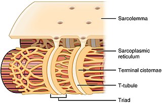
The sarcoplasmic reticulum (SR) is a membrane-bound structure found within muscle cells that is similar to the smooth endoplasmic reticulum in other cells. The main function of the SR is to store calcium ions (Ca2+). Calcium ion levels are kept relatively constant, with the concentration of calcium ions within a cell being 10,000 times smaller than the concentration of calcium ions outside the cell. This means that small increases in calcium ions within the cell are easily detected and can bring about important cellular changes (the calcium is said to be a second messenger). Calcium is used to make calcium carbonate (found in chalk) and calcium phosphate, two compounds that the body uses to make teeth and bones. This means that too much calcium within the cells can lead to hardening (calcification) of certain intracellular structures, including the mitochondria, leading to cell death. Therefore, it is vital that calcium ion levels are controlled tightly, and can be released into the cell when necessary and then removed from the cell.

<span class="mw-page-title-main">Flecainide</span> Antiarrhythmic medication

Flecainide is a medication used to prevent and treat abnormally fast heart rates. This includes ventricular and supraventricular tachycardias. Its use is only recommended in those with dangerous arrhythmias or when significant symptoms cannot be managed with other treatments. Its use does not decrease a person's risk of death. It is taken by mouth or injection into a vein.

Ryanodine receptors form a class of intracellular calcium channels in various forms of excitable animal tissue like muscles and neurons. There are three major isoforms of the ryanodine receptor, which are found in different tissues and participate in different signaling pathways involving calcium release from intracellular organelles. The RYR2 ryanodine receptor isoform is the major cellular mediator of calcium-induced calcium release (CICR) in animal cells.

<span class="mw-page-title-main">T-tubule</span> Extensions in cell membrane of muscle fibres

T-tubules are extensions of the cell membrane that penetrate into the center of skeletal and cardiac muscle cells. With membranes that contain large concentrations of ion channels, transporters, and pumps, T-tubules permit rapid transmission of the action potential into the cell, and also play an important role in regulating cellular calcium concentration.

<span class="mw-page-title-main">Desmoglein-2</span> Protein found in humans

Desmoglein-2 is a protein that in humans is encoded by the DSG2 gene. Desmoglein-2 is highly expressed in epithelial cells and cardiomyocytes. Desmoglein-2 is localized to desmosome structures at regions of cell-cell contact and functions to structurally adhere adjacent cells together. In cardiac muscle, these regions are specialized regions known as intercalated discs. Mutations in desmoglein-2 have been associated with arrhythmogenic right ventricular cardiomyopathy and familial dilated cardiomyopathy.

<span class="mw-page-title-main">Catecholaminergic polymorphic ventricular tachycardia</span> Medical condition

Catecholaminergic polymorphic ventricular tachycardia (CPVT) is an inherited genetic disorder that predisposes those affected to potentially life-threatening abnormal heart rhythms or arrhythmias. The arrhythmias seen in CPVT typically occur during exercise or at times of emotional stress, and classically take the form of bidirectional ventricular tachycardia or ventricular fibrillation. Those affected may be asymptomatic, but they may also experience blackouts or even sudden cardiac death.

<span class="mw-page-title-main">Calmodulin 1</span> Protein-coding gene in the species Homo sapiens

Calmodulin 1 is a protein in humans that is encoded by the CALM1 gene.

Ca<sub>v</sub>1.1 Mammalian protein found in Homo sapiens

Cav1.1 also known as the calcium channel, voltage-dependent, L type, alpha 1S subunit, (CACNA1S), is a protein which in humans is encoded by the CACNA1S gene. It is also known as CACNL1A3 and the dihydropyridine receptor.
<span class="mw-page-title-main">FKBP1B</span> Protein-coding gene in the species Homo sapiens

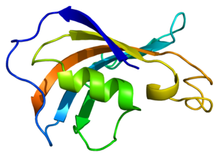
Peptidyl-prolyl cis-trans isomerase FKBP1B is an enzyme that in humans is encoded by the FKBP1B gene.

<span class="mw-page-title-main">Plakophilin-2</span> Protein-coding gene in the species Homo sapiens

Plakophilin-2 is a protein that in humans is encoded by the PKP2 gene. Plakophilin 2 is expressed in skin and cardiac muscle, where it functions to link cadherins to intermediate filaments in the cytoskeleton. In cardiac muscle, plakophilin-2 is found in desmosome structures located within intercalated discs. Mutations in PKP2 have been shown to be causal in arrhythmogenic right ventricular cardiomyopathy.

<span class="mw-page-title-main">Triadin</span> Protein-coding gene in humans

Triadin, also known as TRDN, is a human gene associated with the release of calcium ions from the sarcoplasmic reticulum triggering muscular contraction through calcium-induced calcium release. Triadin is a multiprotein family, arising from different processing of the TRDN gene on chromosome 6. It is a transmembrane protein on the sarcoplasmic reticulum due to a well defined hydrophobic section and it forms a quaternary complex with the cardiac ryanodine receptor (RYR2), calsequestrin (CASQ2) and junctin proteins. The luminal (inner compartment of the sarcoplasmic reticulum) section of Triadin has areas of highly charged amino acid residues that act as luminal Ca2+ receptors. Triadin is also able to sense luminal Ca2+ concentrations by mediating interactions between RYR2 and CASQ2. Triadin has several different forms; Trisk 95 and Trisk 51, which are expressed in skeletal muscle, and Trisk 32 (CT1), which is mainly expressed in cardiac muscle.

<span class="mw-page-title-main">AKAP6</span> Protein-coding gene in the species Homo sapiens

A-kinase anchor protein 6 is an enzyme that in humans is encoded by the AKAP6 gene.

<span class="mw-page-title-main">Ryanodine receptor 1</span> Protein and coding gene in humans

Ryanodine receptor 1 (RYR-1) also known as skeletal muscle calcium release channel or skeletal muscle-type ryanodine receptor is one of a class of ryanodine receptors and a protein found primarily in skeletal muscle. In humans, it is encoded by the RYR1 gene.

<span class="mw-page-title-main">MYL7</span> Protein-coding gene in the species Homo sapiens

Atrial Light Chain-2 (ALC-2) also known as Myosin regulatory light chain 2, atrial isoform (MLC2a) is a protein that in humans is encoded by the MYL7 gene. ALC-2 expression is restricted to cardiac muscle atria in healthy individuals, where it functions to modulate cardiac development and contractility. In human diseases, including hypertrophic cardiomyopathy, dilated cardiomyopathy, ischemic cardiomyopathy and others, ALC-2 expression is altered.

<span class="mw-page-title-main">Ankyrin-2</span> Protein-coding gene in the species Homo sapiens

Ankyrin-2, also known as Ankyrin-B, and Brain ankyrin, is a protein which in humans is encoded by the ANK2 gene. Ankyrin-2 is ubiquitously expressed, but shows high expression in cardiac muscle. Ankyrin-2 plays an essential role in the localization and membrane stabilization of ion transporters and ion channels in cardiomyocytes, as well as in costamere structures. Mutations in ANK2 cause a dominantly-inherited, cardiac arrhythmia syndrome known as long QT syndrome 4 as well as sick sinus syndrome; mutations have also been associated to a lesser degree with hypertrophic cardiomyopathy. Alterations in ankyrin-2 expression levels are observed in human heart failure.

<span class="mw-page-title-main">Ryanodine receptor 3</span> Transport protein and coding gene in humans

Ryanodine receptor 3 is one of a class of ryanodine receptors and a protein that in humans is encoded by the RYR3 gene. The protein encoded by this gene is both a calcium channel and a receptor for the plant alkaloid ryanodine. RYR3 and RYR1 control the resting calcium ion concentration in skeletal muscle.

JTV-519 (K201) is a 1,4-benzothiazepine derivative that interacts with many cellular targets. It has many structural similarities to diltiazem, a Ca2+ channel blocker used for treatment of hypertension, angina pectoris and some types of arrhythmias. JTV-519 acts in the sarcoplasmic reticulum (SR) of cardiac myocytes by binding to and stabilizing the ryanodine receptor (RyR2) in its closed state. It can be used in the treatment of cardiac arrhythmias, heart failure, catecholaminergic polymorphic ventricular tachycardia (CPVT) and store overload-induced Ca2+ release (SOICR). Currently, this drug has only been tested on animals and its side effects are still unknown. As research continues, some studies have also found a dose-dependent response; where there is no improvement seen in failing hearts at 0.3 μM and a decline in response at 1 μM.

CXL 1020 is an experimental drug that is being investigated as a treatment for acute decompensated heart failure. CXL 1020 functions as a nitroxyl donor; nitroxyl is the reduced, protonated version of nitric oxide. Nitroxyl is capable of enhancing left ventricular contractility without increasing heart rate by modifying normal Ca2+ cycling through the sarcoplasmic reticulum as well as increasing the sensitivity of cardiac myofilaments to Ca2+.

Calcium buffering describes the processes which help stabilise the concentration of free calcium ions within cells, in a similar manner to how pH buffers maintain a stable concentration of hydrogen ions. The majority of calcium ions within the cell are bound to intracellular proteins, leaving a minority freely dissociated. When calcium is added to or removed from the cytoplasm by transport across the cell membrane or sarcoplasmic reticulum, calcium buffers minimise the effect on changes in cytoplasmic free calcium concentration by binding calcium to or releasing calcium from intracellular proteins. As a result, 99% of the calcium added to the cytosol of a cardiomyocyte during each cardiac cycle becomes bound to calcium buffers, creating a relatively small change in free calcium.

The ryanodine-inositol 1,4,5-triphosphate receptor Ca2+ channel (RIR-CaC) family includes Ryanodine receptors and Inositol trisphosphate receptors. Members of this family are large proteins, some exceeding 5000 amino acyl residues in length. This family belongs to the Voltage-gated ion channel (VIC) superfamily. Ry receptors occur primarily in muscle cell sarcoplasmic reticular (SR) membranes, and IP3 receptors occur primarily in brain cell endoplasmic reticular (ER) membranes where they effect release of Ca2+ into the cytoplasm upon activation (opening) of the channel. They are redox sensors, possibly providing a partial explanation for how they control cytoplasmic Ca2+. Ry receptors have been identified in heart mitochondria where they provide the main pathway for Ca2+ entry. Sun et al. (2011) have demonstrated oxygen-coupled redox regulation of the skeletal muscle ryanodine receptor-Ca2+ release channel (RyR1;TC# 1.A.3.1.2) by NADPH oxidase 4.

References

  1. 1 2 3 GRCh38: Ensembl release 89: ENSG00000198626 Ensembl, May 2017
  2. 1 2 3 GRCm38: Ensembl release 89: ENSMUSG00000021313 Ensembl, May 2017
  3. "Human PubMed Reference:". National Center for Biotechnology Information, U.S. National Library of Medicine.
  4. "Mouse PubMed Reference:". National Center for Biotechnology Information, U.S. National Library of Medicine.
  5. Otsu K, Willard HF, Khanna VK, Zorzato F, Green NM, MacLennan DH (August 1990). "Molecular cloning of cDNA encoding the Ca2+ release channel (ryanodine receptor) of rabbit cardiac muscle sarcoplasmic reticulum". The Journal of Biological Chemistry. 265 (23): 13472–83. doi: 10.1016/S0021-9258(18)77371-7 . PMID   2380170.
  6. Otsu K, Fujii J, Periasamy M, Difilippantonio M, Uppender M, Ward DC, MacLennan DH (August 1993). "Chromosome mapping of five human cardiac and skeletal muscle sarcoplasmic reticulum protein genes". Genomics. 17 (2): 507–9. doi: 10.1006/geno.1993.1357 . PMID   8406504.
  7. Tiso N, Stephan DA, Nava A, Bagattin A, Devaney JM, Stanchi F, et al. (February 2001). "Identification of mutations in the cardiac ryanodine receptor gene in families affected with arrhythmogenic right ventricular cardiomyopathy type 2 (ARVD2)". Human Molecular Genetics. 10 (3): 189–94. doi: 10.1093/hmg/10.3.189 . PMID   11159936.
  8. Guo T, Cornea RL, Huke S, Camors E, Yang Y, Picht E, et al. (June 2010). "Kinetics of FKBP12.6 binding to ryanodine receptors in permeabilized cardiac myocytes and effects on Ca sparks". Circulation Research. 106 (11): 1743–52. doi:10.1161/CIRCRESAHA.110.219816. PMC   2895429 . PMID   20431056.
  9. "Q92736 - RYR2_HUMAN".
  10. Bround MJ, Wambolt R, Luciani DS, Kulpa JE, Rodrigues B, Brownsey RW, et al. (June 2013). "Cardiomyocyte ATP production, metabolic flexibility, and survival require calcium flux through cardiac ryanodine receptors in vivo". The Journal of Biological Chemistry. 288 (26): 18975–86. doi: 10.1074/jbc.M112.427062 . PMC   3696672 . PMID   23678000.
  11. Belevych AE, Radwański PB, Carnes CA, Györke S (May 2013). "'Ryanopathy': causes and manifestations of RyR2 dysfunction in heart failure". Cardiovascular Research. 98 (2): 240–7. doi:10.1093/cvr/cvt024. PMC   3633158 . PMID   23408344.
  12. "Entrez Gene: RYR2 ryanodine receptor 2 (cardiac)".
  13. Tester DJ, Bombei HM, Fitzgerald KK, Giudicessi JR, Pitel BA, Thorland EC, et al. (January 2020). "Identification of a Novel Homozygous Multi-Exon Duplication in RYR2 Among Children With Exertion-Related Unexplained Sudden Deaths in the Amish Community". JAMA Cardiology. 5 (3): 13–18. doi:10.1001/jamacardio.2019.5400. PMC   6990654 . PMID   31913406.
  14. Bround MJ, Asghari P, Wambolt RB, Bohunek L, Smits C, Philit M, et al. (December 2012). "Cardiac ryanodine receptors control heart rate and rhythmicity in adult mice". Cardiovascular Research. 96 (3): 372–80. doi:10.1093/cvr/cvs260. PMC   3500041 . PMID   22869620.
  15. 1 2 3 4 Marx SO, Reiken S, Hisamatsu Y, Jayaraman T, Burkhoff D, Rosemblit N, Marks AR (May 2000). "PKA phosphorylation dissociates FKBP12.6 from the calcium release channel (ryanodine receptor): defective regulation in failing hearts". Cell. 101 (4): 365–76. doi: 10.1016/S0092-8674(00)80847-8 . PMID   10830164. S2CID   6496567.
  16. Marx SO, Reiken S, Hisamatsu Y, Gaburjakova M, Gaburjakova J, Yang YM, et al. (May 2001). "Phosphorylation-dependent regulation of ryanodine receptors: a novel role for leucine/isoleucine zippers". The Journal of Cell Biology. 153 (4): 699–708. doi:10.1083/jcb.153.4.699. PMC   2192391 . PMID   11352932.
  17. Huke, Sabine; Bers, Donald M. (2008-11-07). "Ryanodine receptor phosphorylation at Serine 2030, 2808 and 2814 in rat cardiomyocytes". Biochemical and Biophysical Research Communications. 376 (1): 80–85. doi:10.1016/j.bbrc.2008.08.084. ISSN   1090-2104. PMC   2581610 . PMID   18755143.
  18. Witcher, D. R.; Kovacs, R. J.; Schulman, H.; Cefali, D. C.; Jones, L. R. (1991-06-15). "Unique phosphorylation site on the cardiac ryanodine receptor regulates calcium channel activity". The Journal of Biological Chemistry. 266 (17): 11144–11152. ISSN   0021-9258. PMID   1645727.
  19. Wehrens, Xander H.T.; Lehnart, Stephan E.; Reiken, Steven R.; Marks, Andrew R. (2004-04-02). "Ca 2+ /Calmodulin-Dependent Protein Kinase II Phosphorylation Regulates the Cardiac Ryanodine Receptor". Circulation Research. 94 (6). doi:10.1161/01.RES.0000125626.33738.E2. ISSN   0009-7330.
  20. Meyers MB, Pickel VM, Sheu SS, Sharma VK, Scotto KW, Fishman GI (November 1995). "Association of sorcin with the cardiac ryanodine receptor". The Journal of Biological Chemistry. 270 (44): 26411–8. doi: 10.1074/jbc.270.44.26411 . PMID   7592856.
  21. Huke, Sabine; Bers, Donald M. (2008-11-07). "Ryanodine receptor phosphorylation at Serine 2030, 2808 and 2814 in rat cardiomyocytes". Biochemical and Biophysical Research Communications. 376 (1): 80–85. doi:10.1016/j.bbrc.2008.08.084. ISSN   1090-2104. PMC   2581610 . PMID   18755143.
  22. Huke, Sabine; Bers, Donald M. (2008-11-07). "Ryanodine receptor phosphorylation at Serine 2030, 2808 and 2814 in rat cardiomyocytes". Biochemical and Biophysical Research Communications. 376 (1): 80–85. doi:10.1016/j.bbrc.2008.08.084. ISSN   1090-2104. PMC   2581610 . PMID   18755143.

Further reading