Cav1.2

Last updated
CACNA1C
Protein CACNA1C PDB 2be6.png
Available structures
PDB Ortholog search: PDBe RCSB
Identifiers
Aliases CACNA1C , CACH2, CACN2, CACNL1A1, CCHL1A1, CaV1.2, LQT8, TS, calcium voltage-gated channel subunit alpha1 C, TS. LQT8
External IDs OMIM: 114205 MGI: 103013 HomoloGene: 55484 GeneCards: CACNA1C
Orthologs
SpeciesHumanMouse
Entrez
Ensembl
UniProt
RefSeq (mRNA)
RefSeq (protein)
Location (UCSC) Chr 12: 1.97 – 2.7 Mb Chr 6: 118.56 – 119.17 Mb
PubMed search [3] [4]
Wikidata
View/Edit Human View/Edit Mouse

Calcium channel, voltage-dependent, L type, alpha 1C subunit (also known as Cav1.2) is a protein that in humans is encoded by the CACNA1C gene. [5] Cav1.2 is a subunit of L-type voltage-dependent calcium channel. [6]

Structure and function

This gene encodes an alpha-1 subunit of a voltage-dependent calcium channel. Calcium channels mediate the influx of calcium ions (Ca2+) into the cell upon membrane polarization (see membrane potential and calcium in biology). [7]

The alpha-1 subunit consists of 24 transmembrane segments and forms the pore through which ions pass into the cell. The calcium channel consists of a complex of alpha-1, alpha-2/delta and beta subunits in a 1:1:1 ratio. The S3-S4 linkers of Cav1.2 determine the gating phenotype and modulated gating kinetics of the channel. [8] Cav1.2 is widely expressed in the smooth muscle, pancreatic cells, fibroblasts, and neurons. [9] [10] However, it is particularly important and well known for its expression in the heart where it mediates L-type currents, which causes calcium-induced calcium release from the ER Stores via ryanodine receptors. It depolarizes at -30mV and helps define the shape of the action potential in cardiac and smooth muscle. [8] The protein encoded by this gene binds to and is inhibited by dihydropyridine. [11] In the arteries of the brain, high levels of calcium in mitochondria elevates activity of nuclear factor kappa B NF-κB and transcription of CACNA1c and functional Cav1.2 expression increases. [12] Cav1.2 also regulates levels of osteoprotegerin. [13]

CaV1.2 is inhibited by the action of STIM1. [14]

Regulation

The activity of CaV1.2 channels is tightly regulated by the Ca2+ signals they produce. An increase in intracellular Ca2+ concentration implicated in Cav1.2 facilitation, a form of positive feedback called Ca2+-dependent facilitation, that amplifies Ca2+ influx. In addition, increasing influx intracellular Ca2+ concentration has implicated to exert the opposite effect Ca2+ dependent inactivation. [15] These activation and inactivation mechanisms both involve Ca2+ binding to calmodulin (CaM) in the IQ domain in the C-terminal tail of these channels. [16] Cav1.2 channels are arranged in cluster of eight, on average, in the cell membrane. When calcium ions bind to calmodulin, which in turn binds to a Cav1.2 channel, it allows the Cav1.2 channels within a cluster to interact with each other. [17] This results in channels working cooperatively when they open at the same time to allow more calcium ions to enter and then close together to allow the cell to relax. [17]

Due to simplicity only two Calcium channels are shown to depict clustering. When depolarization occurs, calcium ions flow through the channel and some bind to Calmodulin. The Calcium/Calmodulin binding to the C-terminal pre-IQ domain of the Cav1.2 channel promotes interaction between channels that are beside each other. Ca1.2channelclutsering.png
Due to simplicity only two Calcium channels are shown to depict clustering. When depolarization occurs, calcium ions flow through the channel and some bind to Calmodulin. The Calcium/Calmodulin binding to the C-terminal pre-IQ domain of the Cav1.2 channel promotes interaction between channels that are beside each other.

Clinical significance

Mutation in the CACNA1C gene, the single-nucleotide polymorphism located in the third intron of the Cav1.2 gene, [18] are associated with a variant of Long QT syndrome called Timothy's syndrome [19] and more broadly with other CACNA1C-related disorders, [19] and also with Brugada syndrome. [20] Large-scale genetic analyses have shown the possibility that CACNA1C is associated with bipolar disorder [21] and subsequently also with schizophrenia. [22] [23] [24] Also, a CACNA1C risk allele has been associated to a disruption in brain connectivity in patients with bipolar disorder, while not or only to a minor degree, in their unaffected relatives or healthy controls. [25] .In a first study in Indian population, the Schizophrenia associated Genome-wide association study (GWAS) SNP was found not to be associated with the disease. Furthermore, the main effect of rs1006737 was found to be associated with spatial abilityefficiency scores. Subjects with genotypes carrying the risk allele of rs1006737 (G/A and A/A) were found to have higher spatial abilityefficiency scores as compared to those with the G/G genotype. While in healthy controls those with G/A and A/A genotypes were found to have higher spatial memoryprocessing speed scores than those with G/G genotypes, the former had lower scores than the latter in schizophrenia subjects. In the same study the genotypes with the risk allele of rs1006737 namely A/A was associated with a significantly lower Align rank transformed Abnormal and involuntary movement scale(AIMS) scores of Tardive dyskinesia(TD). [26]

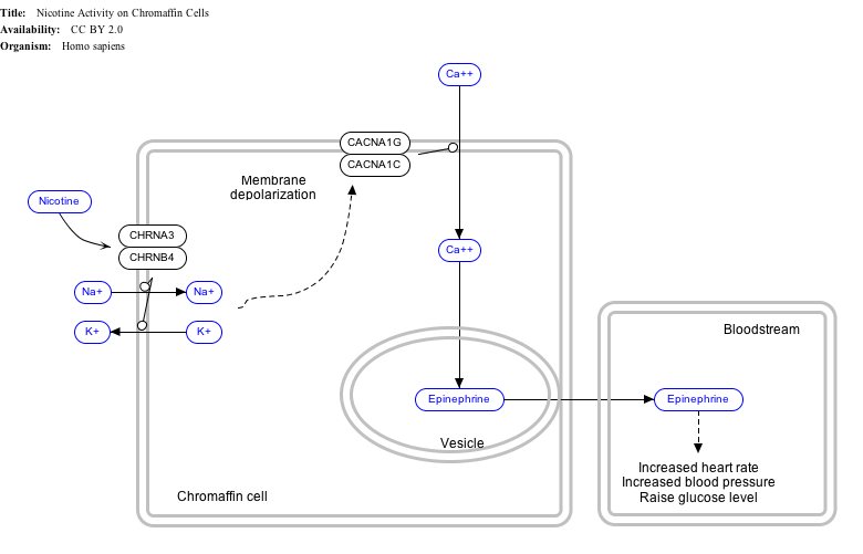
Interactive pathway map

Click on genes, proteins and metabolites below to link to respective Wikipedia articles. [§ 1]

[[File:
NicotineChromaffinCells WP1603.png Go to articlego to articleGo to articleGo to articleGo to articleGo to articlego to articlego to articleGo to articleGo to articlego to articleGo to articleGo to article
[[
]]
[[
]]
[[
]]
[[
]]
[[
]]
[[
]]
[[
]]
[[
]]
[[
]]
[[
]]
[[
]]
[[
]]
[[
]]
[[
]]
NicotineChromaffinCells WP1603.png Go to articlego to articleGo to articleGo to articleGo to articleGo to articlego to articlego to articleGo to articleGo to articlego to articleGo to articleGo to article
|alt=Nicotine Activity on Chromaffin Cells edit]]
Nicotine Activity on Chromaffin Cells edit
  1. The interactive pathway map can be edited at WikiPathways: "NicotineActivityonChromaffinCells_WP1603".

See also

Related Research Articles

Timothy syndrome is a rare autosomal-dominant disorder characterized by physical malformations, as well as neurological and developmental defects, including heart QT-prolongation, heart arrhythmias, structural heart defects, syndactyly, and autism spectrum disorders. Timothy syndrome represents one clinical manifestation of a range of disorders associated with mutations in CACNA1C, the gene encoding the calcium channel Cav1.2 α subunit.

Voltage-gated calcium channels (VGCCs), also known as voltage-dependent calcium channels (VDCCs), are a group of voltage-gated ion channels found in the membrane of excitable cells (e.g., muscle, glial cells, neurons, etc.) with a permeability to the calcium ion Ca2+. These channels are slightly permeable to sodium ions, so they are also called Ca2+–Na+ channels, but their permeability to calcium is about 1000-fold greater than to sodium under normal physiological conditions.

Sodium channels are integral membrane proteins that form ion channels, conducting sodium ions (Na+) through a cell's membrane. They belong to the superfamily of cation channels.

<span class="mw-page-title-main">Neuronal calcium sensor-1</span> Protein-coding gene in the species Homo sapiens

Neuronal calcium sensor-1 (NCS-1) also known as frequenin homolog (Drosophila) (freq) is a protein that is encoded by the FREQ gene in humans. NCS-1 is a member of the neuronal calcium sensor family, a class of EF hand containing calcium-myristoyl-switch proteins.

The R-type calcium channel is a type of voltage-dependent calcium channel. Like the others of this class, the α1 subunit forms the pore through which calcium enters the cell and determines most of the channel's properties. This α1 subunit is also known as the calcium channel, voltage-dependent, R type, alpha 1E subunit (CACNA1E) or Cav2.3 which in humans is encoded by the CACNA1E gene. They are strongly expressed in cortex, hippocampus, striatum, amygdala and interpeduncular nucleus.

<span class="mw-page-title-main">N-type calcium channel</span> Protein family

N-type calcium channels also called Cav2.2 channels are voltage gated calcium channels that are localized primarily on the nerve terminals and dendrites as well as neuroendocrine cells. The calcium N-channel consists of several subunits: the primary subunit α1B and the auxiliary subunits α2δ and β. The α1B subunit forms the pore through which the calcium enters and helps to determine most of the channel's properties. These channels play an important role in the neurotransmission during development. In the adult nervous system, N-type calcium channels are critically involved in the release of neurotransmitters, and in pain pathways. N-type calcium channels are the target of ziconotide, the drug prescribed to relieve intractable cancer pain. There are many known N-type calcium channel blockers that function to inhibit channel activity, although the most notable blockers are ω-conotoxins.

<span class="mw-page-title-main">L-type calcium channel</span> Family of transport proteins

The L-type calcium channel is part of the high-voltage activated family of voltage-dependent calcium channel. "L" stands for long-lasting referring to the length of activation. This channel has four isoforms: Cav1.1, Cav1.2, Cav1.3, and Cav1.4.

Ca<sub>v</sub>2.1 Protein-coding gene in the species Homo sapiens

Cav2.1, also called the P/Q voltage-dependent calcium channel, is a calcium channel found mainly in the brain. Specifically, it is found on the presynaptic terminals of neurons in the brain and cerebellum. Cav2.1 plays an important role in controlling the release of neurotransmitters between neurons. It is composed of multiple subunits, including alpha-1, beta, alpha-2/delta, and gamma subunits. The alpha-1 subunit is the pore-forming subunit, meaning that the calcium ions flow through it. Different kinds of calcium channels have different isoforms (versions) of the alpha-1 subunit. Cav2.1 has the alpha-1A subunit, which is encoded by the CACNA1A gene. Mutations in CACNA1A have been associated with various neurologic disorders, including familial hemiplegic migraine, episodic ataxia type 2, and spinocerebellar ataxia type 6.

Ca<sub>v</sub>1.4 Protein-coding gene in humans

Cav1.4 also known as the calcium channel, voltage-dependent, L type, alpha 1F subunit (CACNA1F), is a human gene.

Ca<sub>v</sub>1.1 Mammalian protein found in Homo sapiens

Cav1.1 also known as the calcium channel, voltage-dependent, L type, alpha 1S subunit, (CACNA1S), is a protein which in humans is encoded by the CACNA1S gene. It is also known as CACNL1A3 and the dihydropyridine receptor.

<span class="mw-page-title-main">CACNB2</span> Protein-coding gene in the species Homo sapiens

Voltage-dependent L-type calcium channel subunit beta-2 is a protein that in humans is encoded by the CACNB2 gene.

<span class="mw-page-title-main">CACNB4</span> Protein-coding gene in the species Homo sapiens

Voltage-dependent L-type calcium channel subunit beta-4 is a protein that in humans is encoded by the CACNB4 gene.

<span class="mw-page-title-main">CACNB1</span> Protein-coding gene in the species Homo sapiens

Voltage-dependent L-type calcium channel subunit beta-1 is a protein that in humans is encoded by the CACNB1 gene.

Ca<sub>v</sub>1.3 Protein-coding gene in the species Homo sapiens

Calcium channel, voltage-dependent, L type, alpha 1D subunit is a protein that in humans is encoded by the CACNA1D gene. Cav1.3 channels belong to the Cav1 family, which form L-type calcium currents and are sensitive to selective inhibition by dihydropyridines (DHP).

<span class="mw-page-title-main">Calcium-binding protein 1</span> Protein-coding gene in the species Homo sapiens

Calcium binding protein 1 is a protein that in humans is encoded by the CABP1 gene. Calcium-binding protein 1 is a calcium-binding protein discovered in 1999. It has two EF hand motifs and is expressed in neuronal cells in such areas as hippocampus, habenular nucleus of the epithalamus, Purkinje cell layer of the cerebellum, and the amacrine cells and cone bipolar cells of the retina.

<span class="mw-page-title-main">CACNB3</span> Protein-coding gene in humans

Voltage-dependent L-type calcium channel subunit beta-3 is a protein that in humans is encoded by the CACNB3 gene.

<span class="mw-page-title-main">CACNA2D1</span> Protein-coding gene in the species Homo sapiens

Voltage-dependent calcium channel subunit alpha-2/delta-1 is a protein that in humans is encoded by the CACNA2D1 gene.

<span class="mw-page-title-main">Calcium channel, voltage-dependent, T type, alpha 1H subunit</span> Protein-coding gene in the species Homo sapiens

Calcium channel, voltage-dependent, T type, alpha 1H subunit, also known as CACNA1H, is a protein which in humans is encoded by the CACNA1H gene.

<span class="mw-page-title-main">CACNA1B</span> Protein-coding gene in the species Homo sapiens

The voltage-dependent N-type calcium channel subunit alpha-1B is a protein that in humans is encoded by the CACNA1B gene. The α1B protein, together with β and α2δ subunits forms N-type calcium channel PMID 26386135. It is a R-type calcium channel.

<span class="mw-page-title-main">CACNA2D3</span> Protein-coding gene in the species Homo sapiens

Calcium channel, voltage-dependent, alpha 2/delta subunit 3 is a protein that in humans is encoded by the CACNA2D3 gene on chromosome 3 .

References

  1. 1 2 3 ENSG00000285479 GRCh38: Ensembl release 89: ENSG00000151067, ENSG00000285479 - Ensembl, May 2017
  2. 1 2 3 GRCm38: Ensembl release 89: ENSMUSG00000051331 - Ensembl, May 2017
  3. "Human PubMed Reference:". National Center for Biotechnology Information, U.S. National Library of Medicine.
  4. "Mouse PubMed Reference:". National Center for Biotechnology Information, U.S. National Library of Medicine.
  5. Lacerda AE, Kim HS, Ruth P, Perez-Reyes E, Flockerzi V, Hofmann F, Birnbaumer L, Brown AM (Aug 1991). "Normalization of current kinetics by interaction between the alpha 1 and beta subunits of the skeletal muscle dihydropyridine-sensitive Ca2+ channel". Nature. 352 (6335): 527–30. Bibcode:1991Natur.352..527L. doi:10.1038/352527a0. PMID   1650913. S2CID   4246540.
  6. Catterall WA, Perez-Reyes E, Snutch TP, Striessnig J (Dec 2005). "International Union of Pharmacology. XLVIII. Nomenclature and structure-function relationships of voltage-gated calcium channels". Pharmacological Reviews. 57 (4): 411–25. doi:10.1124/pr.57.4.5. PMID   16382099. S2CID   10386627.
  7. Shaw RM, Colecraft HM (May 2013). "L-type calcium channel targeting and local signalling in cardiac myocytes". Cardiovascular Research. 98 (2): 177–86. doi:10.1093/cvr/cvt021. PMC   3633156 . PMID   23417040.
  8. 1 2 Lipscombe D, Helton TD, Xu W (Nov 2004). "L-type calcium channels: the low down". Journal of Neurophysiology. 92 (5): 2633–41. doi:10.1152/jn.00486.2004. PMID   15486420. S2CID   52887174.
  9. Christel C, Lee A (Aug 2012). "Ca2+-dependent modulation of voltage-gated Ca2+ channels". Biochimica et Biophysica Acta (BBA) - General Subjects. 1820 (8): 1243–52. doi:10.1016/j.bbagen.2011.12.012. PMC   3345169 . PMID   22223119.
  10. Berger SM, Bartsch D (Aug 2014). "The role of L-type voltage-gated calcium channels Cav1.2 and Cav1.3 in normal and pathological brain function". Cell and Tissue Research. 357 (2): 463–76. doi:10.1007/s00441-014-1936-3. PMID   24996399. S2CID   15914718.
  11. "Entrez Gene: voltage-dependent, L type, alpha 1C subunit".
  12. Narayanan D, Xi Q, Pfeffer LM, Jaggar JH (Sep 2010). "Mitochondria control functional CaV1.2 expression in smooth muscle cells of cerebral arteries". Circulation Research. 107 (5): 631–41. doi:10.1161/CIRCRESAHA.110.224345. PMC   3050675 . PMID   20616314.
  13. Bergh JJ, Xu Y, Farach-Carson MC (Jan 2004). "Osteoprotegerin expression and secretion are regulated by calcium influx through the L-type voltage-sensitive calcium channel". Endocrinology. 145 (1): 426–36. doi: 10.1210/en.2003-0319 . PMID   14525906.
  14. Cahalan MD (Oct 2010). "Cell biology. How to STIMulate calcium channels". Science. 330 (6000): 43–4. doi:10.1126/science.1196348. PMC   3133971 . PMID   20929798.
  15. Isaev D, Solt K, Gurtovaya O, Reeves JP, Shirokov R (May 2004). "Modulation of the voltage sensor of L-type Ca2+ channels by intracellular Ca2+". The Journal of General Physiology. 123 (5): 555–71. doi:10.1085/jgp.200308876. PMC   2234499 . PMID   15111645.
  16. Kim EY, Rumpf CH, Van Petegem F, Arant RJ, Findeisen F, Cooley ES, Isacoff EY, Minor DL (Dec 2010). "Multiple C-terminal tail Ca(2+)/CaMs regulate Ca(V)1.2 function but do not mediate channel dimerization". The EMBO Journal. 29 (23): 3924–38. doi:10.1038/emboj.2010.260. PMC   3020648 . PMID   20953164.
  17. 1 2 Dixon RE, Moreno CM, Yuan C, Opitz-Araya X, Binder MD, Navedo MF, Santana LF (2015). "Graded Ca²⁺/calmodulin-dependent coupling of voltage-gated CaV1.2 channels". eLife. 4. doi: 10.7554/eLife.05608 . PMC   4360655 . PMID   25714924.
  18. Imbrici P, Camerino DC, Tricarico D (2013-05-07). "Major channels involved in neuropsychiatric disorders and therapeutic perspectives". Frontiers in Genetics. 4: 76. doi: 10.3389/fgene.2013.00076 . PMC   3646240 . PMID   23675382.
  19. 1 2 Napolitano C, Timothy KW, Bloise R, Priori SG (1993). Adam MP, Everman DB, Mirzaa GM, Pagon RA, Wallace SE, Bean LJ, Gripp KW, Amemiya A (eds.). CACNA1C-Related Disorders. Seattle (WA): University of Washington, Seattle. PMID   20301577 . Retrieved 2022-12-12.{{cite book}}: |work= ignored (help)
  20. Hedley PL, Jørgensen P, Schlamowitz S, Moolman-Smook J, Kanters JK, Corfield VA, Christiansen M (Sep 2009). "The genetic basis of Brugada syndrome: a mutation update". Human Mutation. 30 (9): 1256–66. doi:10.1002/humu.21066. PMID   19606473. S2CID   25207473.
  21. Ferreira MA, O'Donovan MC, Meng YA, Jones IR, Ruderfer DM, Jones L, et al. (Sep 2008). "Collaborative genome-wide association analysis supports a role for ANK3 and CACNA1C in bipolar disorder". Nature Genetics. 40 (9): 1056–8. doi:10.1038/ng.209. PMC   2703780 . PMID   18711365.
  22. Green EK, Grozeva D, Jones I, Jones L, Kirov G, Caesar S, Gordon-Smith K, Fraser C, Forty L, Russell E, Hamshere ML, Moskvina V, Nikolov I, Farmer A, McGuffin P, Holmans PA, Owen MJ, O'Donovan MC, Craddock N (Oct 2010). "The bipolar disorder risk allele at CACNA1C also confers risk of recurrent major depression and of schizophrenia". Molecular Psychiatry. 15 (10): 1016–22. doi:10.1038/mp.2009.49. PMC   3011210 . PMID   19621016.
  23. Curtis D, Vine AE, McQuillin A, Bass NJ, Pereira A, Kandaswamy R, Lawrence J, Anjorin A, Choudhury K, Datta SR, Puri V, Krasucki R, Pimm J, Thirumalai S, Quested D, Gurling HM (Feb 2011). "Case-case genome-wide association analysis shows markers differentially associated with schizophrenia and bipolar disorder and implicates calcium channel genes". Psychiatric Genetics. 21 (1): 1–4. doi:10.1097/YPG.0b013e3283413382. PMC   3024533 . PMID   21057379.
  24. Schizophrenia Working Group of the Psychiatric Genomics Consortium (2014-07-24). "Biological insights from 108 schizophrenia-associated genetic loci". Nature. 511 (7510): 421–427. Bibcode:2014Natur.511..421S. doi:10.1038/nature13595. ISSN   1476-4687. PMC   4112379 . PMID   25056061.
  25. Radua J, Surguladze SA, Marshall N, Walshe M, Bramon E, Collier DA, Prata DP, Murray RM, McDonald C (May 2013). "The impact of CACNA1C allelic variation on effective connectivity during emotional processing in bipolar disorder". Molecular Psychiatry. 18 (5): 526–7. doi: 10.1038/mp.2012.61 . PMID   22614292.
  26. Punchaichira TJ, Kukshal P, Bhatia T, Deshpande SN (2023). "Effect of rs1108580 of DBH and rs1006737 of CACNA1C on Cognition and Tardive Dyskinesia in a North Indian Schizophrenia Cohort". Molecular Neurobiology. 60 (12): 6826–6839. doi:10.1007/s12035-023-03496-4. PMID   37493923. S2CID   260162784.

Further reading

This article incorporates text from the United States National Library of Medicine, which is in the public domain.