Shigatoxigenic and verotoxigenic Escherichia coli

Last updated
Shigatoxigenic and verotoxigenic E. coli
Specialty Infectious disease

Shigatoxigenic Escherichia coli (STEC) and verotoxigenic E. coli (VTEC) are strains of the bacterium Escherichia coli that produce Shiga toxin (or verotoxin). [lower-alpha 1] Only a minority of the strains cause illness in humans. [2] [ failed verification ] The ones that do are collectively known as enterohemorrhagic E. coli (EHEC) and are major causes of foodborne illness. When infecting the large intestine of humans, they often cause gastroenteritis, enterocolitis, and bloody diarrhea (hence the name "enterohemorrhagic") and sometimes cause a severe complication called hemolytic-uremic syndrome (HUS). [3] [4] Cattle are an important natural reservoir for EHEC because the colonised adult ruminants are asymptomatic. This is because they lack vascular expression of the target receptor for Shiga toxins. [5] The group and its subgroups are known by various names. They are distinguished from other strains of intestinal pathogenic E. coli including enterotoxigenic E. coli (ETEC), enteropathogenic E. coli (EPEC), enteroinvasive E. coli (EIEC), enteroaggregative E. coli (EAEC), and diffusely adherent E. coli (DAEC). [6]

Contents

Background

The best known of these strains is O157:H7, but non-O157 strains cause an estimated 36,000[ citation needed ] illnesses, 1,000 hospitalizations and 30 deaths in the United States yearly. [7] Food safety specialists recognize "Big Six" strains: O26; O45; O103; O111; O121; and O145. [7] A 2011 outbreak in Germany was caused by another STEC, O104:H4. This strain has both enteroaggregative and enterohemorrhagic properties. Both the O145 and O104 strains can cause hemolytic-uremic syndrome (HUS); the former strain shown to account for 2% to 51% of known HUS cases; an estimated 56% of such cases are caused by O145 and 14% by other EHEC strains.[ citation needed ]

EHECs that induce bloody diarrhea lead to HUS in 10% of cases. The clinical manifestations of postdiarrheal HUS include acute renal failure, microangiopathic hemolytic anemia, and thrombocytopenia. The verocytotoxin (shiga-like toxin) can directly damage renal and endothelial cells. Thrombocytopenia occurs as platelets are consumed by clotting. Hemolytic anemia results from intravascular fibrin deposition, increased fragility of red blood cells, and fragmentation. [6]

Antibiotics are of questionable value and have not shown to be of clear clinical benefit. Antibiotics that interfere with DNA synthesis, such as fluoroquinolones, have been shown to induce the Stx-bearing bacteriophage and cause increased production of toxins. [8] Attempts to block toxin production with antibacterials which target the ribosomal protein synthesis are conceptually more attractive. Plasma exchange offers a controversial but possibly helpful treatment. The use of antimotility agents (medications that suppress diarrhea by slowing bowel transit) in children under 10 years of age or in elderly patients should be avoided, as they increase the risk of HUS with EHEC infections. [6]

The clinical presentation ranges from a mild and uncomplicated diarrhea to a hemorrhagic colitis with severe abdominal pain. Serotype O157:H7 may trigger an infectious dose with 100 bacterial cells or fewer; other strain such as 104:H4 has also caused an outbreak in Germany 2011. Infections are most common in warmer months and in children under five years of age and are usually acquired from uncooked beef and unpasteurized milk and juice. Initially a non-bloody diarrhea develops in patients after the bacterium attaches to the epithelium or the terminal ileum, cecum, and colon. The subsequent production of toxins mediates the bloody diarrhea. In children, a complication can be hemolytic uremic syndrome which then uses cytotoxins to attack the cells in the gut, so that bacteria can leak out into the blood and cause endothelial injury in locations such as the kidney by binding to globotriaosylceramide (Gb3).[ citation needed ]

Names

Names of the group and its subgroups include the following. [9] There is some polysemy involved. Invariable synonymity is indicated by having the same color. Beyond that there is also some wider but variable synonymity. The first two (purple) in their narrowest sense are generally treated as hypernyms of the others (red and blue), although in less precise usage the red and blue have often been treated as synonyms of the purple. At least one reference holds "EHEC" to be mutually exclusive of "VTEC" and "STEC", [3] but this does not match common usage, as many more publications lump all of the latter in with the former.

The current microbiology-based view on "Shiga-like toxin" (SLT) or "verotoxin" is that they should all be referred to as (versions of) Shiga toxin, as the difference is negligible. Following this view, all "VTEC" (blue) should be called "STEC" (red). [1] [10] :2–3 Historically, a different name was sometimes used because the toxins are not exactly the same as the one found in Shigella dysenteriae , down to every last amino acid residue, although by this logic every "STEC" would be a "VTEC". The line can also be drawn to use "STEC" for Stx1-producing strains and "VTEC" for Stx2-producing strains, since Stx1 is closer to the Shiga toxin. Practically, the choice of words and categories is not as important as the understanding of clinical relevance.

NameShort form
enterohemorrhagic E. coliEHEC
hemolytic uremic syndrome–associated enterohemorrhagic E. coliHUSEC
shiga toxin–producing E. coliSTEC
shigatoxigenic E. coliSTEC
shiga-like toxin–producing E. coliSLTEC
verotoxin-producing E. coliVTEC
verotoxigenic E. coliVTEC
verocytotoxin-producing E. coliVTEC
verocytotoxigenic E. coliVTEC

Infectivity and virulence

The infectivity or the virulence of an EHEC strain depends on several factors, including the presence of fucose in the medium, the sensing of this sugar and the activation of EHEC pathogenicity island.[ citation needed ]

Scheme of the signalling cascade in EHEC where we can observe the effect of the sensing of some signalling molecules in the virulence of eneterohaemorragic E. coli QseCE signalling cascade for EHEC.jpg
Scheme of the signalling cascade in EHEC where we can observe the effect of the sensing of some signalling molecules in the virulence of eneterohaemorragic E. coli

Regulation of the pathogenicity island

EHEC becomes pathogenic through the expression of the locus of enterocyte effacement (LEE) encoded on its pathogenicity island. However, when EHEC is not in a host this expression is a waste of energy and resources, so it is only activated if some molecules are sensed in the environment. [ citation needed ]

When QseC or QseE bind with one of their interacting signalling molecule, they autophosphorylate and transfer its phosphate to the response regulator. QseC senses adrenaline, noradrenaline, and an Endonuclease I-SceIII, encoded by a mobile group I intron within the mitochondrial COX1 gene (AI3); whereas QseE senses adrenaline, noradrenaline, SO4 and PO4. These signals are a clear indication to the bacteria that they are no longer free in the environment, but in the gut.[ citation needed ]

As a result, QseC phosphorylates QseB (which activates flagella), KpdE (which activates the LEE) and QseF. QseE phosphorylates QseF. The products QseBC and QseEF repress the expression of FusK and FusR. FusK and FusR are the two components of a system to repress the transcription of the LEE genes. FusK is a sensor kinase which is able to sense many sugars among which fucose. When fucose is present in the medium FusK phosphorylates FusR which represses LEE expression. [ citation needed ]

Thus when EHEC enters the gut there is a competition between the signals coming from QseC and QseF, and the signal coming from FusK. The first two would like to activate virulence, but Fusk stops it because the mucous layer, which is a source of fucose, isolates enterocytes from bacteria making the synthesis of the virulence factors useless. However, when fucose concentration decreases because bacterial cells find an unprotected area of the epithelium, then the expression of LEE genes will not be repressed by FusR, and KpdE will strongly activate them. In summary, the combined effect of the QseC/QseF and FusKR provide a fine-tuning system of LEE expression which saves energy and allow the mechanisms of virulence to be expressed only when the chances of success are higher.[ citation needed ]

Shiga toxins

Shiga toxins are a major virulence factor of EHEC. The toxins interact with intestinal epithelium and can cause systematic complications in humans like HUS and cerebral dysfunction if they enter the circulation. [11] In EHEC, Shiga toxins are encoded lysogenic bacteriophages. The toxins bind to cell-surface glycolipid receptor Gb3, which causes the cell to take the toxin in via endocytosis. The Shiga toxins target ribosomal RNA, which inhibits protein synthesis and causes apoptosis. [12] The reason EHEC are symptomless in cattle is because the cattle do not have vascular expression of Gb3 unlike humans. Thus, the Shiga toxins cannot pass through the intestinal epithelium into circulation. [5]

FusKR complex

This complex, formed by two components (FusK and FusR) has the function in EHEC to detect the presence of fucose in the environment and regulate the activation of LEE genes.[ citation needed ]

Inactivation of LEE genes ( | [fucose] ) INACTIVATION OF LEE GENES.jpg
Inactivation of LEE genes ( ↑ [fucose] )

Fucose increases the activation of the FusKR system, which inhibits the z0461 gene, which controls the metabolism of fucose. This is a mechanisms that is useful to avoid the competition for fucose with other strains of E. coli which are usually more efficient at using fucose as a carbon source. High concentrations of fucose in the medium also increases the repression of the LEE genes.

Activation of LEE genes ( | [fucose] ) ACTIVATION OF LEE GENES.jpg
Activation of LEE genes ( ↓ [fucose] )

With low levels of fucose in the environment, the FusKR system is inactive, and this means that z0461 gene is transcribed, thus increasing the metabolism of fucose. Furthermore, a low concentration of fucose is an indication of unprotected epithelium, thus the repression of ler genes will disappear and the expression of the LEE genes will allow to attack the adjacent cells.[ citation needed ]

See also

Notes

  1. Current classifications consider the two identical, and only use the "Shiga toxin" name. See § Names. [1]

Related Research Articles

<i>Escherichia coli</i> Enteric, rod-shaped, gram-negative bacterium

Escherichia coli ( ESH-ə-RIK-ee-ə KOH-ly) is a gram-negative, facultative anaerobic, rod-shaped, coliform bacterium of the genus Escherichia that is commonly found in the lower intestine of warm-blooded organisms. Most E. coli strains are harmless, but some serotypes such as EPEC, and ETEC are pathogenic and can cause serious food poisoning in their hosts, and are occasionally responsible for food contamination incidents that prompt product recalls. Most strains are part of the normal microbiota of the gut and are harmless or even beneficial to humans (although these strains tend to be less studied than the pathogenic ones). For example, some strains of E. coli benefit their hosts by producing vitamin K2 or by preventing the colonization of the intestine by pathogenic bacteria. These mutually beneficial relationships between E. coli and humans are a type of mutualistic biological relationship — where both the humans and the E. coli are benefitting each other. E. coli is expelled into the environment within fecal matter. The bacterium grows massively in fresh fecal matter under aerobic conditions for three days, but its numbers decline slowly afterwards.

<i>Escherichia coli</i> O157:H7 Serotype of the bacteria Escherichia coli

Escherichia coli O157:H7 is a serotype of the bacterial species Escherichia coli and is one of the Shiga-like toxin–producing types of E. coli. It is a cause of disease, typically foodborne illness, through consumption of contaminated and raw food, including raw milk and undercooked ground beef. Infection with this type of pathogenic bacteria may lead to hemorrhagic diarrhea, and to kidney failure; these have been reported to cause the deaths of children younger than five years of age, of elderly patients, and of patients whose immune systems are otherwise compromised.

<span class="mw-page-title-main">Shiga toxin</span> Family of related toxins

Shiga toxins are a family of related toxins with two major groups, Stx1 and Stx2, expressed by genes considered to be part of the genome of lambdoid prophages. The toxins are named after Kiyoshi Shiga, who first described the bacterial origin of dysentery caused by Shigella dysenteriae. Shiga-like toxin (SLT) is a historical term for similar or identical toxins produced by Escherichia coli. The most common sources for Shiga toxin are the bacteria S. dysenteriae and some serotypes of Escherichia coli (STEC), which includes serotypes O157:H7, and O104:H4.

<span class="mw-page-title-main">Hemolytic–uremic syndrome</span> Group of blood disorders related to bacterial infection

Hemolytic–uremic syndrome (HUS) is a group of blood disorders characterized by low red blood cells, acute kidney injury, and low platelets. Initial symptoms typically include bloody diarrhea, fever, vomiting, and weakness. Kidney problems and low platelets then occur as the diarrhea progresses. Children are more commonly affected, but most children recover without permanent damage to their health, although some children may have serious and sometimes life-threatening complications. Adults, especially the elderly, may present a more complicated presentation. Complications may include neurological problems and heart failure.

<span class="mw-page-title-main">Coliform bacteria</span> Group of bacterial species

Coliform bacteria are defined as either motile or non-motile Gram-negative non-spore forming bacilli that possess β-galactosidase to produce acids and gases under their optimal growth temperature of 35–37 °C. They can be aerobes or facultative aerobes, and are a commonly used indicator of low sanitary quality of foods, milk, and water. Coliforms can be found in the aquatic environment, in soil and on vegetation; they are universally present in large numbers in the feces of warm-blooded animals as they are known to inhabit the gastrointestinal system. While coliform bacteria are not normally causes of serious illness, they are easy to culture, and their presence is used to infer that other pathogenic organisms of fecal origin may be present in a sample, or that said sample is not safe to consume. Such pathogens include disease-causing bacteria, viruses, or protozoa and many multicellular parasites.

The AB5 toxins are six-component protein complexes secreted by certain pathogenic bacteria known to cause human diseases such as cholera, dysentery, and hemolytic–uremic syndrome. One component is known as the A subunit, and the remaining five components are B subunits. All of these toxins share a similar structure and mechanism for entering targeted host cells. The B subunit is responsible for binding to receptors to open up a pathway for the A subunit to enter the cell. The A subunit is then able to use its catalytic machinery to take over the host cell's regular functions.

<span class="mw-page-title-main">Intimin</span>

Intimin is a virulence factor (adhesin) of EPEC and EHEC E. coli strains. It is an attaching and effacing (A/E) protein, which with other virulence factors is necessary and responsible for enteropathogenic and enterohaemorrhagic diarrhoea.

Escherichia coli O121 is a pathogenic serotype of Escherichia coli, associated with Shiga toxin, intestinal bleeding, and hemolytic-uremic syndrome (HUS). HUS, if left untreated, can lead to kidney failure.

Escherichia coli O104:H21 is a rare serotype of Escherichia coli, a species of bacteria that lives in the lower intestines of mammals. Although there are many serotypes of E. coli, when in animals, there are benefits or do not cause disease. Some serotypes of E. coli have been recognized as pathogenic to humans, e.g. E. coli O157:H7, E. coli O121 and E. coli O104:H21.

Microbial toxins are toxins produced by micro-organisms, including bacteria, fungi, protozoa, dinoflagellates, and viruses. Many microbial toxins promote infection and disease by directly damaging host tissues and by disabling the immune system. Endotoxins most commonly refer to the lipopolysaccharide (LPS) or lipooligosaccharide (LOS) that are in the outer plasma membrane of Gram-negative bacteria. The botulinum toxin, which is primarily produced by Clostridium botulinum and less frequently by other Clostridium species, is the most toxic substance known in the world. However, microbial toxins also have important uses in medical science and research. Currently, new methods of detecting bacterial toxins are being developed to better isolate and understand these toxins. Potential applications of toxin research include combating microbial virulence, the development of novel anticancer drugs and other medicines, and the use of toxins as tools in neurobiology and cellular biology.

Enteroinvasive Escherichia coli (EIEC) is a type of pathogenic bacteria whose infection causes a syndrome that is identical to shigellosis, with profuse diarrhea and high fever. EIEC are highly invasive, and they use adhesin proteins to bind to and enter intestinal cells. They produce no toxins, but severely damage the intestinal wall through mechanical cell destruction.

<span class="mw-page-title-main">Vibrio regulatory RNA of OmpA</span>

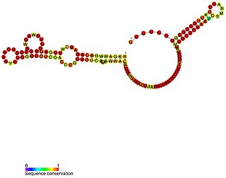
VrrA is a non-coding RNA that is conserved across all Vibrio species of bacteria and acts as a repressor for the synthesis of the outer membrane protein OmpA. This non-coding RNA was initially identified from Tn5 transposon mutant libraries of Vibrio cholerae and its location within the bacterial genome was mapped to the intergenic region between genes VC1741 and VC1743 by RACE analysis.

The RTX toxin superfamily is a group of cytolysins and cytotoxins produced by bacteria. There are over 1000 known members with a variety of functions. The RTX family is defined by two common features: characteristic repeats in the toxin protein sequences, and extracellular secretion by the type I secretion systems (T1SS). The name RTX refers to the glycine and aspartate-rich repeats located at the C-terminus of the toxin proteins, which facilitate export by a dedicated T1SS encoded within the rtx operon.

2011 Germany <i>E. coli</i> O104:H4 outbreak 2011 foodborne illness outbreak in northern Germany

A novel strain of Escherichia coli O104:H4 bacteria caused a serious outbreak of foodborne illness focused in northern Germany in May through June 2011. The illness was characterized by bloody diarrhea, with a high frequency of serious complications, including hemolytic–uremic syndrome (HUS), a condition that requires urgent treatment. The outbreak was originally thought to have been caused by an enterohemorrhagic (EHEC) strain of E. coli, but it was later shown to have been caused by an enteroaggregative E. coli (EAEC) strain that had acquired the genes to produce Shiga toxins, present in organic fenugreek sprouts.

Escherichia coli O104:H4 is an enteroaggregative Escherichia coli strain of the bacterium Escherichia coli, and the cause of the 2011 Escherichia coli O104:H4 outbreak. The "O" in the serological classification identifies the cell wall lipopolysaccharide antigen, and the "H" identifies the flagella antigen.

Antimotility agents are drugs used to alleviate the symptoms of diarrhea. These include loperamide (Imodium), diphenoxylate with atropine (Lomotil), and opiates such as paregoric, tincture of opium, codeine, and morphine. In diarrhea caused by invasive pathogens such as Salmonella, Shigella, and Campylobacter, the use of such agents has generally been strongly discouraged, though evidence is lacking that they are harmful when administered in combination with antibiotics in Clostridium difficile cases. Use of antimotility agents in children and the elderly has also been discouraged in treatment of EHEC due to an increased rate of hemolytic uremic syndrome.

Pathogenic <i>Escherichia coli</i> Strains of E. coli that can cause disease

Escherichia coli is a gram-negative, rod-shaped bacterium that is commonly found in the lower intestine of warm-blooded organisms (endotherms). Most E. coli strains are harmless, but pathogenic varieties cause serious food poisoning, septic shock, meningitis, or urinary tract infections in humans. Unlike normal flora E. coli, the pathogenic varieties produce toxins and other virulence factors that enable them to reside in parts of the body normally not inhabited by E. coli, and to damage host cells. These pathogenic traits are encoded by virulence genes carried only by the pathogens.

Enteroaggregative Escherichia coli are a pathotype of Escherichia coli which cause acute and chronic diarrhea in both the developed and developing world. They may also cause urinary tract infections. EAEC are defined by their "stacked-brick" pattern of adhesion to the human laryngeal epithelial cell line HEp-2. The pathogenesis of EAEC involves the aggregation of and adherence of the bacteria to the intestinal mucosa, where they elaborate enterotoxins and cytotoxins that damage host cells and induce inflammation that results in diarrhea.

Proteobiotics are natural metabolites which are produced by fermentation process of specific probiotic strains. These small oligopeptides were originally discovered in and isolated from culture media used to grow probiotic bacteria and may account for some of the health benefits of probiotics.

References

  1. 1 2 Scheutz F, Teel LD, Beutin L, Piérard D, Buvens G, Karch H, Mellmann A, Caprioli A, Tozzoli R, Morabito S, Strockbine NA, Melton-Celsa AR, Sanchez M, Persson S, O'Brien AD (September 2012). "Multicenter evaluation of a sequence-based protocol for subtyping Shiga toxins and standardizing Stx nomenclature". Journal of Clinical Microbiology. 50 (9): 2951–63. doi:10.1128/JCM.00860-12. PMC   3421821 . PMID   22760050.
  2. Croxen MA, Law RJ, Scholz R, Keeney KM, Wlodarska M, Finlay BB (2013). "Recent advances in understanding enteric pathogenic Escherichia coli". Clinical Microbiology Reviews. 26 (4): 822–80. doi:10.1128/CMR.00022-13. PMC   3811233 . PMID   24092857.
  3. 1 2 Mainil, J (1999), "Shiga/verocytotoxins and Shiga/verotoxigenic Escherichia coli in animals", Vet Res, 30 (2–3): 235–57, PMID   10367357.
  4. Phillips, A; Navabpour, S; Hicks, S; Dougan, G; Wallis, T; Frankel, G (2000). "Enterohaemorrhagic Escherichia coli O157:H7 target Peyer's patches in humans and cause attaching/effacing lesions in both human and bovine intestine". Gut. 47 (3): 377–381. doi:10.1136/gut.47.3.377. PMC   1728033 . PMID   10940275.
  5. 1 2 Pruimboom-Brees, I; Morgan, T; Ackermann, M; Nystrom, E; Samuel, J; Cornick, N; Moon, H (2000). "Cattle lack vascular receptors for Escherichia coli O157:H7 Shiga toxins". Proceedings of the National Academy of Sciences. 97 (19): 10325–10329. Bibcode:2000PNAS...9710325P. doi: 10.1073/pnas.190329997 . ISSN   0027-8424. PMC   27023 . PMID   10973498.
  6. 1 2 3 Bae, Woo Kyun; Lee, Youn Kyoung; Cho, Min Seok; Ma, Seong Kwon; Kim, Soo Wan; Kim, Nam Ho; Choi, Ki Chul (2006-06-30). "A Case of Hemolytic Uremic Syndrome Caused by Escherichia coli O104:H4". Yonsei Med J. 47 (3): 437–439. doi:10.3349/ymj.2006.47.3.437. PMC   2688167 . PMID   16807997. Two sentences were taken from this source verbatim.
  7. 1 2 Mallove, Zach (26 April 2010). "Lawyer Battles FSIS on Non-O157 E. coli". Food Safety News . Retrieved 2 June 2011.
  8. Zhang, X; McDaniel, AD; Wolf, LE; Keusch, GT; Waldor, MK; Acheson, DW (2000). "Quinolone antibiotics induce Shiga toxin-encoding bacteriophages, toxin production, and death in mice". The Journal of Infectious Diseases. 181 (2): 664–70. doi: 10.1086/315239 . PMID   10669353.
  9. Karch, Helge; Tarr, Phillip I.; Bielaszewska, Martina (2005). "Enterohaemorrhagic Escherichia coli in human medicine". International Journal of Medical Microbiology. 295 (6–7): 405–18. doi:10.1016/j.ijmm.2005.06.009. PMID   16238016.
  10. Silva, Christopher J.; Brandon, David L.; Skinner, Craig B.; He, Xiaohua; et al. (2017), "Structure of Shiga Toxins and Other AB5 Toxins", Shiga toxins: A Review of Structure, Mechanism, and Detection, Springer, pp. 21–45, doi:10.1007/978-3-319-50580-0_3, ISBN   978-3319505800.
  11. Detzner, J; Pohlentz, G; Müthing, J (2020). "Valid Presumption of Shiga Toxin-Mediated Damage of Developing Erythrocytes in EHEC-Associated Hemolytic Uremic Syndrome". Toxins. 12 (6): 373. doi: 10.3390/toxins12060373 . ISSN   2072-6651. PMC   7354503 . PMID   32512916.
  12. Smith, D; Naylor, S; Gally, D (2002). "Consequences of EHEC colonisation in humans and cattle". International Journal of Medical Microbiology. 292 (3): 169–183. doi:10.1078/1438-4221-00202. ISSN   1438-4221. PMID   12398208.

Further reading

  1. Bardiau, M.; M. Szalo & J.G. Mainil (2010). "Initial adherence of EPEC, EHEC and VTEC to host cells". Vet Res. 41 (5): 57. doi:10.1051/vetres/2010029. PMC   2881418 . PMID   20423697.
  2. Wong, A.R.; et al. (2011). "Enteropathogenic and enterohaemorrhagic Escherichia coli: even more subversive elements". Mol Microbiol. 80 (6): 1420–38. doi: 10.1111/j.1365-2958.2011.07661.x . PMID   21488979. S2CID   24606261.
  3. Tatsuno, I. (2007). "[Adherence of enterohemorrhagic Escherichia coli O157:H7 to human epithelial cells]". Nihon Saikingaku Zasshi. 62 (2): 247–53. doi: 10.3412/jsb.62.247 . PMID   17575791.
  4. Kaper, J.B.; J.P. Nataro & H.L. Mobley (2004). "Pathogenic Escherichia coli". Nat Rev Microbiol. 2 (2): 123–40. doi:10.1038/nrmicro818. PMID   15040260. S2CID   3343088.
  5. Garcia, A.; J.G. Fox & T.E. Besser (2010). "Zoonotic enterohemorrhagic Escherichia coli: A One Health perspective". ILAR J. 51 (3): 221–32. doi: 10.1093/ilar.51.3.221 . PMID   21131723.
  6. Shimizu, T. (2010). "[Expression and extracellular release of Shiga toxin in enterohemorrahgic Escherichia coli]". Nihon Saikingaku Zasshi. 65 (2–4): 297–308. doi: 10.3412/jsb.65.297 . PMID   20505269.