Sinodonty and Sundadonty

Last updated

In anthropology, Sinodonty and Sundadonty are two patterns of features widely found in the dentitions of different East Asians. These patterns were identified by anthropologist Christy G. Turner II as being within the greater "Mongoloid dental complex". [1]

Contents

The combining forms Sino- and Sunda- refer to China and Sundaland, respectively, while -dont refers to teeth.

Proto-sundadonty hypothesis

Tsunehiko Hanihara (1993) believed that the dental features of Aboriginal Australians have the characteristic of high frequencies of "evolutionarily conservative characteristics," which he called the "proto- sundadont" pattern, as he believed that the dental pattern of Aboriginal Australians was ancestral to that of Southeast Asians. [2]

C.G Turner II shows with his analysis of 2016 that sundadonty is the proto-East Eurasian dental morphology and is not connected to the Australian dental morphology, rendering the term "proto-sundadont" inaccurate for the Australian dental morphology. He also shows that sinodonty is predominant in Native Americans. [3]

Super-Sinodont

Analysis on the Sinodonty and Sundadonty of New world groups by G.R. Sott et al. (2016) shows the distinction between East Asians is not nearly as dramatic as the difference between all Asians and all New World groups. Other researchers like Stojanowski et al., 2013; Stojanowski and Johnson, (2015) suggest New World groups may be neither Sinodont nor Sundadont and in most regards, could be viewed as super-Sinodont. A clear dental morphology not only ties New World groups to Asians, particularly northeast Asians, but it also exhibits a pattern largely consistent with the Beringian Standstill model (BSM) based on a Sinodont source population. [4]

Mongoloid dental complex

Turner defined the Sinodont and Sundadont dental complexes in contrast to a broader Mongoloid dental complex. [5] Hanihara defined the Mongoloid dental complex in 1966. In 1984, Turner separated the Mongoloid dental complex into the Sinodont and Sundadont dental complexes. [6]

Ryuta Hamada, Shintaro Kondo and Eizo Wakatsuki (1997) said, on the basis of dental traits, that Mongoloids are separated into sinodonts and sundadonts, which is supported by Christy G. Turner II (1989). [7] [8]

Sundadont

Turner found the Sundadont pattern in the skeletal remains of Jōmon people of Japan, and in living populations of Taiwanese indigenous peoples, Filipinos, Indonesians, Borneans, and Malays.

In 1996, Rebecca Haydenblit of the Hominid Evolutionary Biology Research Group at Cambridge University did a study on the dentition of four Pre-Columbian era Mesoamerican populations and compared their data to other Eastern Eurasian populations. [9] She found that "Tlatilco", "Cuicuilco", "Monte Albán" and "Cholula" populations followed an overall Sundadont dental pattern "characteristic of Southeast Asia" rather than a Sinodont dental pattern "characteristic of Northeast Asia". [9]

Sinodont

Turner found the Sinodont pattern in the Han Chinese, in the inhabitants of Mongolia and eastern Siberia, in the Native Americans, and in the Yayoi people of Japan.

Sinodonty is a particular pattern of teeth characterized by the following features:

Associated traits

The EDAR gene causes the Sinodont tooth pattern, and also affects hair texture, [11] jaw morphology, [12] and perhaps the nutritional profile of breast milk. [13]

Applicability

In the 1990s, Turner's dental morphological traits were frequently mentioned as one of three new tools for studying origins and migrations of human populations. The other two were linguistic methods such as Joseph Greenberg's mass comparison of vocabulary or Johanna Nichols's statistical study of language typology and its evolution, and genetic studies pioneered by Cavalli-Sforza.[ original research? ]

Today, the largest number of references to Turner's work are from discussions of the origin of Paleo-Amerindians and modern Native Americans, including the Kennewick Man controversy. Turner found that the dental remains of both ancient and modern Amerindians are more similar to each other than they are to dental complexes from other continents, but that the Sinodont patterns of the Paleo-Amerindians identify their ancestral homeland as north-east Asia. Some later studies[which?] have questioned this and found Sundadont features in some American peoples.

A study done by Stojonowski et al in 2015 found a "significant interobserver error" in the earlier studies and their statistical analysis of matched wear and morphology scores suggests trait downgrading for some traits. [14]

See also

Related Research Articles

<span class="mw-page-title-main">Dentition</span> Development and arrangement of teeth

Dentition pertains to the development of teeth and their arrangement in the mouth. In particular, it is the characteristic arrangement, kind, and number of teeth in a given species at a given age. That is, the number, type, and morpho-physiology of the teeth of an animal.

<span class="mw-page-title-main">Forensic dentistry</span> Aspect of criminal investigation

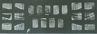
Forensic dentistry or forensic odontology involves the handling, examination, and evaluation of dental evidence in a criminal justice context. Forensic dentistry is used in both criminal and civil law. Forensic dentists assist investigative agencies in identifying human remains, particularly in cases when identifying information is otherwise scarce or nonexistent—for instance, identifying burn victims by consulting the victim's dental records. Forensic dentists may also be asked to assist in determining the age, race, occupation, previous dental history, and socioeconomic status of unidentified human beings.

<span class="mw-page-title-main">Hyperdontia</span> Condition of having extra teeth beyond the regular number of teeth

Hyperdontia is the condition of having supernumerary teeth, or teeth that appear in addition to the regular number of teeth. They can appear in any area of the dental arch and can affect any dental organ. The opposite of hyperdontia is hypodontia, where there is a congenital lack of teeth, which is a condition seen more commonly than hyperdontia. The scientific definition of hyperdontia is "any tooth or odontogenic structure that is formed from tooth germ in excess of usual number for any given region of the dental arch." The additional teeth, which may be few or many, can occur on any place in the dental arch. Their arrangement may be symmetrical or non-symmetrical.

<span class="mw-page-title-main">Premolar</span> Transitional teeth located between the canine and molar teeth

The premolars, also called premolar teeth, or bicuspids, are transitional teeth located between the canine and molar teeth. In humans, there are two premolars per quadrant in the permanent set of teeth, making eight premolars total in the mouth. They have at least two cusps. Premolars can be considered transitional teeth during chewing, or mastication. They have properties of both the canines, that lie anterior and molars that lie posterior, and so food can be transferred from the canines to the premolars and finally to the molars for grinding, instead of directly from the canines to the molars.

Hypodontia is defined as the developmental absence of one or more teeth excluding the third molars. It is one of the most common dental anomalies, and can have a negative impact on function, and also appearance. It rarely occurs in primary teeth and the most commonly affected are the adult second premolars and the upper lateral incisors. It usually occurs as part of a syndrome that involves other abnormalities and requires multidisciplinary treatment.

<span class="mw-page-title-main">Black hair</span> Human hair color

Black hair is the darkest and most common of all human hair colors globally, due to large populations with this trait. This hair type contains a much more dense quantity of eumelanin pigmentation in comparison to other hair colors, such as brown, blonde and red. In English, various types of black hair are sometimes described as soft-black, raven black, or jet-black. The range of skin colors associated with black hair is vast, ranging from the palest of light skin tones to dark skin. Black-haired humans can have dark or light eyes.

<span class="mw-page-title-main">Shovel-shaped incisors</span> Possible shape of human incisor teeth

Shovel-shaped incisors are incisors whose lingual surfaces are scooped as a consequence of lingual marginal ridges, crown curvature, or basal tubercles, either alone or in combination.

<span class="mw-page-title-main">Talon cusp</span> Rare dental anomaly resulting in teeth having more than one cusp

Talon cusp is a rare dental anomaly resulting in an extra cusp or cusp-like projection on an anterior tooth, located on the inside surface of the affected tooth. Sometimes it can also be found on the facial surface of the anterior tooth.

<span class="mw-page-title-main">Overjet</span> Medical condition

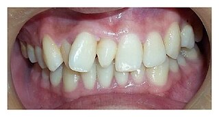
Overjet is the extent of horizontal (anterior-posterior) overlap of the maxillary central incisors over the mandibular central incisors. In class II malocclusion the overjet is increased as the maxillary central incisors are protruded.

Ectodysplasin A receptor (EDAR) is a protein that in humans is encoded by the EDAR gene. EDAR is a cell surface receptor for ectodysplasin A which plays an important role in the development of ectodermal tissues such as the skin. It is structurally related to members of the TNF receptor superfamily.

<span class="mw-page-title-main">Toothcomb</span> Dental structure found in some mammals

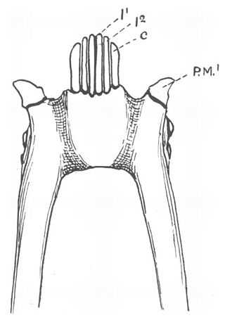
A toothcomb is a dental structure found in some mammals, comprising a group of front teeth arranged in a manner that facilitates grooming, similar to a hair comb. The toothcomb occurs in lemuriform primates, treeshrews, colugos, hyraxes, and some African antelopes. The structures evolved independently in different types of mammals through convergent evolution and varies both in dental composition and structure. In most mammals the comb is formed by a group of teeth with fine spaces between them. The toothcombs in most mammals include incisors only, while in lemuriform primates they include incisors and canine teeth that tilt forward at the front of the lower jaw, followed by a canine-shaped first premolar. The toothcombs of colugos and hyraxes take a different form with the individual incisors being serrated, providing multiple tines per tooth.

<span class="mw-page-title-main">Enamel hypoplasia</span> Medical condition

Enamel hypoplasia is a defect of the teeth in which the enamel is deficient in quantity, caused by defective enamel matrix formation during enamel development, as a result of inherited and acquired systemic condition(s). It can be identified as missing tooth structure and may manifest as pits or grooves in the crown of the affected teeth, and in extreme cases, some portions of the crown of the tooth may have no enamel, exposing the dentin. It may be generalized across the dentition or localized to a few teeth. Defects are categorized by shape or location. Common categories are pit-form, plane-form, linear-form, and localised enamel hypoplasia. Hypoplastic lesions are found in areas of the teeth where the enamel was being actively formed during a systemic or local disturbance. Since the formation of enamel extends over a long period of time, defects may be confined to one well-defined area of the affected teeth. Knowledge of chronological development of deciduous and permanent teeth makes it possible to determine the approximate time at which the developmental disturbance occurred. Enamel hypoplasia varies substantially among populations and can be used to infer health and behavioural impacts from the past. Defects have also been found in a variety of non-human animals.

<span class="mw-page-title-main">Tooth</span> Hard, calcified structure found in the mouths of many vertebrates

A tooth is a hard, calcified structure found in the jaws of many vertebrates and used to break down food. Some animals, particularly carnivores and omnivores, also use teeth to help with capturing or wounding prey, tearing food, for defensive purposes, to intimidate other animals often including their own, or to carry prey or their young. The roots of teeth are covered by gums. Teeth are not made of bone, but rather of multiple tissues of varying density and hardness that originate from the outermost embryonic germ layer, the ectoderm.

<span class="mw-page-title-main">Odontometrics</span> Measurement and study of tooth size

Odontometrics is the measurement and study of tooth size. It is used in biological anthropology and bioarchaeology to study human phenotypic variation. The rationale for use is similar to that of the study of dentition, the structure and arrangement of teeth. There are a number of features that can be observed in human teeth through the use of odontometrics.

<span class="mw-page-title-main">Mammal tooth</span> Details of teeth found in many warm-blooded vertebrate animals

Teeth are common to most vertebrates, but mammalian teeth are distinctive in having a variety of shapes and functions. This feature first arose among early therapsids during the Permian, and has continued to the present day. All therapsid groups with the exception of the mammals are now extinct, but each of these groups possessed different tooth patterns, which aids with the classification of fossils.

<span class="mw-page-title-main">Maxillary lateral incisor agenesis</span>

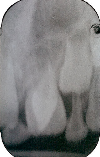
Maxillary lateral incisor agenesis (MLIA) is lack of development (agenesis) of one or both of the maxillary lateral incisor teeth. In normal human dentition, this would be the second tooth on either side from the center of the top row of teeth. The condition is bilateral if the incisor is absent on both sides or unilateral if only one is missing. It appears to have a genetic component.

Changes to the dental morphology and jaw are major elements of hominid evolution. These changes were driven by the types and processing of food eaten. The evolution of the jaw is thought to have facilitated encephalization, speech, and the formation of the uniquely human chin.

<span class="mw-page-title-main">ASUDAS</span> References system for human tooth morphology

The ASUDAS is a reference system for collecting data on human tooth morphology and variation created by Christy G. Turner II, Christian R. Nichol, and G. Richard Scott. The ASUDAS gives detailed descriptions for common crown and root shape variants and their different degrees of expression. It also comprises a set of reference plaques illustrating dental variants as well as showing their expression levels in 3D. The ASUDAS was designed to ensure a standardized scoring procedure with minimum error in order to warrant comparability between data collected by different observers.

<span class="mw-page-title-main">Near Eastern bioarchaeology</span> Archaeological sub-discipline

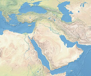
Near Eastern bioarchaeology covers the study of human skeletal remains from archaeological sites in Cyprus, Egypt, Levantine coast, Jordan, Turkey, Iran, Saudi Arabia, Qatar, Kuwait, Bahrain, United Arab Emirates, Oman, and Yemen.

The analysis of dental remains is a valuable tool to archaeologists. Teeth are hard, highly mineralised and chemically stable, so therefore preserve well and are one of the most commonly found animals remains. Analysis of these remains also yields a wealth of information. It can not only be used to determine the sex and age of the individual whose mandibular or dental remains have been found, but can also shed light on their diet, pathology, and even their geographic origins through isotope analysis.

References

  1. G. Richard Scott, Christy G. Turner, (2000). The Anthropology of Modern Human Teeth: Dental Morphology and Its Variation in Recent Human Populations . Cambridge University Press. ISBN   0521784530
  2. Hanihara, Tsunehiko. (1993). Craniofacial Features of Southeast Asians and Jomonese: A Reconsideration of Their Microevolution Since the Late Pleistocene. Anthropological Science, 101(1). Page 26. Retrieved March 8, 2018, from link to the PDF document.
  3. Pilloud, Marin; Heim, Kelly; Schmitz, Kirk; Paul, Kathleen (2018). "Sinodonty, Sundadonty, and the Beringian Standstill model: Issues of timing and migrations into the New World". Quaternary International. 466: 233. Bibcode:2018QuInt.466..233S. doi:10.1016/j.quaint.2016.04.027.
  4. Pilloud, Marin; Heim, Kelly; Schmitz, Kirk; Paul, Kathleen (2018). "Sinodonty, Sundadonty, and the Beringian Standstill model: Issues of timing and migrations into the New World". Quaternary International. 466: 233. Bibcode:2018QuInt.466..233S. doi:10.1016/j.quaint.2016.04.027.
  5. 1 2 Scott, R.G. (1997). Encyclopedia of Human Biology. Second Edition. Volume 3. Pages 175-190. Retrieved December 14, 2016, from link.
  6. Díaz, E. et al. (2014). Frequency and variability of dental morphology in deciduous and permanent dentition of a Nasa indigenous group in the municipality of Morales, Cauca, Colombia. In Colombia Médica, 45(1). Pages 15–24. Retrieved December 14, 2016, from link.
  7. Hamada, Ryuta, Kondo, Shintaro & Wakatsuki, Eizo. (1997). Odontometrical Analysis of Filipino Dentition. The Journal of Showa University Dental Society, 17. Page 197. Retrieved March 8, 2018, from link to the PDF document.
  8. SAO/NASA Astrophysics Data System. Teeth and Prehistory in Asia. Retrieved March 9, 2018, from link to the web page.
  9. 1 2 Haydenblit, Rebeca (June 1996). "Dental variation among four prehispanic Mexican populations". American Journal of Physical Anthropology. 100 (2): 225–246. doi:10.1002/(SICI)1096-8644(199606)100:2<225::AID-AJPA5>3.0.CO;2-W.
  10. 1 2 Kimura, R. et al. (2009). A Common Variation in EDAR Is a Genetic Determinant of Shovel-Shaped Incisors. In American Journal of Human Genetics, 85(4). Page 528. Retrieved December 24, 2016, from link.
  11. Kamberov YG, Wang S, Tan J, Gerbault P, Wark A, Tan L, Yang Y, Li S, Tang K, Chen H, Powell A, Itan Y, Fuller D, Lohmueller J, Mao J, Schachar A, Paymer M, Hostetter E, Byrne E, Burnett M, McMahon AP, Thomas MG, Lieberman DE, Jin L, Tabin CJ, Morgan BA, Sabeti PC (Feb 2013). "Modeling recent human evolution in mice by expression of a selected EDAR variant". Cell. 152 (4): 691–702. doi:10.1016/j.cell.2013.01.016. PMC   3575602 . PMID   23415220.
  12. Adhikari, K.; Fuentes-Guajardo, M.; Quinto-Sánchez, M.; Mendoza-Revilla, J.; Chacón-Duque, J. C.; Acuña-Alonzo, V.; Gómez-Valdés, J. (2016). "A genome-wide association scan implicates DCHS2, RUNX2, GLI3, PAX1 and EDAR in human facial variation". Nature Communications. 7: 11616. Bibcode:2016NatCo...711616A. doi:10.1038/ncomms11616. PMC   4874031 . PMID   27193062.
  13. Lozovschi, Alexandra (24 April 2018). "Ancient Teeth Reveal Breastfeeding-Related Gene Helped Early Americans Survive The Ice Age [Study]". Inquisitr. Retrieved 25 April 2018.
  14. Stojanowski, Christopher M.; Johnson, Kent M. (March 2015). "Observer error, dental wear, and the inference of new world sundadonty". American Journal of Physical Anthropology. 156 (3): 349–362. doi:10.1002/ajpa.22653. ISSN   1096-8644. PMID   25363296.