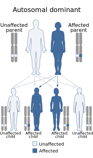
Spinocerebellar ataxia type 13 (SCA13) is a rare autosomal dominant disorder, which, like other types of SCA, is characterized by dysarthria, nystagmus, and ataxia of gait, stance and the limbs due to cerebellar dysfunction. Patients with SCA13 also tend to present with epilepsy, an inability to run, and increased reflexes. This cerebellar dysfunction is permanent and progressive. SCA13 is caused by mutations in KCNC3, a gene encoding a voltage-gated potassium channel KV3.3. There are two known mutations in this gene causative for SCA13. Unlike many other types of SCA, these are not polyglutamine expansions but, rather, point mutations resulting in channels with no current or altered kinetics.
SCA13 is typified by early onset, mildly progressive cerebellar ataxia with accompanying dysarthria, mental retardation, and nystagmus. Symptoms and age of onset can vary slightly according to the causative mutation.[1][2]
Pathophysiology
Mutations in KCNC3 are responsible for SCA13. This gene is expressed heavily in Purkinje cells, as is the case for some other SCA subtypes, where it is believed to play an important role in facilitating high-frequency action potential firing. There are two known mutations in this gene associated with SCA13. The first mutation, R420H, is located in the voltage-sensing S4 segment of the channel. As this mutation neutralizes a site important for voltage sensing, it is not surprising that it results in non-conducting channels. Neurons expressing such channels are unable to follow high-frequency input with adequate fidelity.[citation needed]
The second SCA13 associated mutation, F448L, results in functional channels that have altered kinetics. The voltage for half activation of these channels (V½) is shifted 13mV hyperpolarized compared to wild-type. Deactivation of these channels is also slowed drastically compared to wild-type. This results in neurons with longer after-hyperpolarizations and thus, a decreased maximal firing rate.[3]
Diagnosis
This section is empty.You can help by adding to it.(October 2017)
Treatment
This section is empty.You can help by adding to it.(October 2017)
Prognosis
There is no known prevention of spinocerebellar ataxia. Those who are believed to be at risk can have genetic sequencing of known SCA loci performed to confirm inheritance of the disorder.
↑ Waters M, Fee D, Figueroa K, Nolte D, Müller U, Advincula J, Coon H, Evidente V, Pulst S (2005). "An autosomal dominant ataxia maps to 19q13: Allelic heterogeneity of SCA13 or novel locus?". Neurology. 65 (7): 1111–3. doi:10.1212/01.wnl.0000177490.05162.41. PMID16135769. S2CID38318745.
↑ Waters M, Minassian N, Stevanin G, Figueroa K, Bannister J, Nolte D, Mock A, Evidente V, Fee D, Müller U, Dürr A, Brice A, Papazian D, Pulst S (2006). "Mutations in voltage-gated potassium channel KCNC3 cause degenerative and developmental central nervous system phenotypes". Nat Genet. 38 (4): 447–51. doi:10.1038/ng1758. PMID16501573. S2CID16790821.
Spinocerebellar ataxia (SCA) is a progressive, degenerative, genetic disease with multiple types, each of which could be considered a neurological condition in its own right. An estimated 150,000 people in the United States have a diagnosis of spinocerebellar ataxia at any given time. SCA is hereditary, progressive, degenerative, and often fatal. There is no known effective treatment or cure. SCA can affect anyone of any age. The disease is caused by either a recessive or dominant gene. In many cases people are not aware that they carry a relevant gene until they have children who begin to show signs of having the disorder.
Machado–Joseph disease (MJD), also known as Machado–Joseph Azorean disease, Machado's disease, Joseph's disease or spinocerebellar ataxia type 3 (SCA3), is a rare autosomal dominantly inherited neurodegenerative disease that causes progressive cerebellar ataxia, which results in a lack of muscle control and coordination of the upper and lower extremities. The symptoms are caused by a genetic mutation that results in an expansion of abnormal "CAG" trinucleotide repeats in the ATXN3 gene that results in an abnormal form of the protein ataxin which causes degeneration of cells in the hindbrain. Some symptoms, such as clumsiness and rigidity, make MJD commonly mistaken for drunkenness or Parkinson's disease.
Behr syndrome is characterized by the association of early-onset optic atrophy with spinocerebellar degeneration resulting in ataxia, pyramidal signs, peripheral neuropathy and developmental delay.
Familial hemiplegic migraine (FHM) is an autosomal dominant type of hemiplegic migraine that typically includes weakness of half the body which can last for hours, days, or weeks. It can be accompanied by other symptoms, such as ataxia, coma, and paralysis. Migraine attacks may be provoked by minor head trauma. Some cases of minor head trauma in patients with hemiplegic migraine can develop into delayed cerebral edema, a life-threatening medical emergency. Clinical overlap occurs in some FHM patients with episodic ataxia type 2 and spinocerebellar ataxia type 6, benign familial infantile epilepsy, and alternating hemiplegia of childhood.
Spinocerebellar ataxia type 6 (SCA6) is a rare, late-onset, autosomal dominant disorder, which, like other types of SCA, is characterized by dysarthria, oculomotor disorders, peripheral neuropathy, and ataxia of the gait, stance, and limbs due to cerebellar dysfunction. Unlike other types, SCA 6 is not fatal. This cerebellar function is permanent and progressive, differentiating it from episodic ataxia type 2 (EA2) where said dysfunction is episodic. In some SCA6 families, some members show these classic signs of SCA6 while others show signs more similar to EA2, suggesting that there is some phenotypic overlap between the two disorders. SCA6 is caused by mutations in CACNA1A, a gene encoding a calcium channel α subunit. These mutations tend to be trinucleotide repeats of CAG, leading to the production of mutant proteins containing stretches of 20 or more consecutive glutamine residues; these proteins have an increased tendency to form intracellular agglomerations. Unlike many other polyglutamine expansion disorders expansion length is not a determining factor for the age that symptoms present.
Episodic ataxia (EA) is an autosomal dominant disorder characterized by sporadic bouts of ataxia with or without myokymia. There are seven types recognized but the majority are due to two recognized entities. Ataxia can be provoked by psychological stress or startle, or heavy exertion, including exercise. Symptoms can first appear in infancy. There are at least six loci for EA, of which 4 are known genes. Some patients with EA also have migraine or progressive cerebellar degenerative disorders, symptomatic of either familial hemiplegic migraine or spinocerebellar ataxia. Some patients respond to acetazolamide though others do not.
Ataxin-2 is a protein that in humans is encoded by the ATXN2 gene. Mutations in ATXN2 cause spinocerebellar ataxia type 2 (SCA2).
The Cav2.1 P/Q voltage-dependent calcium channel is encoded by the CACNA1A gene.
Ataxin-10 is a protein that in humans is encoded by the ATXN10 gene.
Fibroblast growth factor 14 is a protein that in humans is encoded by the FGF14 gene.
Puratrophin-1 is a protein that in humans is encoded by the PLEKHG4 gene.
Tau tubulin kinase 2 is a protein in humans that is encoded by the TTBK2 gene. This gene encodes a serine-threonine kinase that putatively phosphorylates tau and tubulin proteins. Mutations in this gene cause spinocerebellar ataxia type 11 (SCA11); a neurodegenerative disease characterized by progressive ataxia and atrophy of the cerebellum and brainstem.
Potassium voltage-gated channel, Shaw-related subfamily, member 3 also known as KCNC3 or Kv3.3 is a protein that in humans is encoded by the KCNC3.
Gillespie syndrome, also called aniridia, cerebellar ataxia and mental deficiency. is a rare genetic disorder. The disorder is characterized by partial aniridia, ataxia, and, in most cases, intellectual disability. It is heterogeneous, inherited in either an autosomal dominant or autosomal recessive manner. Gillespie syndrome was first described by American ophthalmologist Fredrick Gillespie in 1965.
AFG3 ATPase family gene 3-like 2 is a protein that in humans is encoded by the AFG3L2 gene.
Autosomal recessive cerebellar ataxia type 1 (ARCA1) is a condition characterized by progressive problems with movement. Signs and symptoms of the disorder first appear in early to mid-adulthood. People with this condition initially experience impaired speech (dysarthria), problems with coordination and balance (ataxia), or both. They may also have difficulty with movements that involve judging distance or scale (dysmetria). Other features of ARCA1 include abnormal eye movements (nystagmus) and problems following the movements of objects with their eyes. The movement problems are slowly progressive, often resulting in the need for a cane, walker, or wheelchair.
Oculomotor apraxia (OMA), is the absence or defect of controlled, voluntary, and purposeful eye movement. It was first described in 1952 by the American ophthalmologist David Glendenning Cogan. People with this condition have difficulty moving their eyes horizontally and moving them quickly. The main difficulty is in saccade initiation, but there is also impaired cancellation of the vestibulo-ocular reflex. Patients have to turn their head in order to compensate for the lack of eye movement initiation in order to follow an object or see objects in their peripheral vision, but they often exceed their target. There is controversy regarding whether OMA should be considered an apraxia, since apraxia is the inability to perform a learned or skilled motor action to command, and saccade initiation is neither a learned nor a skilled action.
Autosomal dominant cerebellar ataxia (ADCA) is a form of spinocerebellar ataxia inherited in an autosomal dominant manner. ADCA is a genetically inherited condition that causes deterioration of the nervous system leading to disorder and a decrease or loss of function to regions of the body.
Spinocerebellar ataxia type 1 (SCA1) is a rare autosomal dominant disorder, which, like other spinocerebellar ataxias, is characterized by neurological symptoms including dysarthria, hypermetric saccades, and ataxia of gait and stance. This cerebellar dysfunction is progressive and permanent. First onset of symptoms is normally between 30 and 40 years of age, though juvenile onset can occur. Death typically occurs within 10 to 30 years from onset.
Christianson syndrome is an X linked syndrome associated with intellectual disability, microcephaly, seizures, ataxia and absent speech.
This page is based on this Wikipedia article Text is available under the CC BY-SA 4.0 license; additional terms may apply. Images, videos and audio are available under their respective licenses.