Subcutaneous fat necrosis of the newborn

Last updated
Subcutaneous fat necrosis of the newborn
Other namesSCFN or SFN
Specialty Pediatrics   OOjs UI icon edit-ltr-progressive.svg

Subcutaneous fat necrosis of the newborn is a rare form of lobular panniculitis occurring in newborns that is usually self-remitting and non-recurring. Proposed causes include perinatal stress, local trauma, hypoxia and hypothermia, though the exact cause is unknown. It has been suggested that the brown fat seen in newborns is more sensitive to hypoxic injury than fat seen in adults, and that such hypoxia, usually in the context of a complicated birth, leads to the fat necrosis. Complications can include hypercalcemia, hyperlipidemia, dehydration, hypoglycemia, seizures, vomiting, constipation, and thrombocytopenia, and can present months after the onset of SCFN symptoms. [1] [2] :490 [3]

Contents

Signs and symptoms

Subcutaneous fat necrosis of the newborn (SCFN) occurs in full-term newborns usually in the first six weeks of life as indurated plaques or erythematous to violaceous nodules on the cheeks, back, buttocks, or proximal extremities. Usually, the abdomen and chest are unaffected. [4] There has been a case report of SCFN with localized scalp alopecia covering an erythematous nodule. [5] Tenderness is common in lesions. [6]

About 50% of newborns with subcutaneous fat necrosis of the newborn (SCFN) experience hypercalcemia, a potentially fatal consequence. [7] Some infants with SCFN experience anemia, hypoglycemia, thrombocytopenia, and elevated triglycerides. [8] [9] Rarely, thrombocytosis is seen in infants with SCFN. [10]

Causes

Although the exact cause of the newborn's subcutaneous fat necrosis is unknown, several systemic maternal diseases, including preeclampsia and diabetes, as well as asphyxia, hypothermia, meconium aspiration, and obstetric trauma, may act as precipitating factors in the development of SCFN. [7]

Diagnosis

A skin biopsy and the clinical presentation must both be taken into account for the most accurate diagnosis. [11] But in order to prevent skin biopsy, ultrasonography examination has been shown to be useful in identifying the newborn's subcutaneous fat necrosis when combined with Doppler blood flow analysis; this usually indicates a subcutaneous high echo signal, either with or without calcifications. [12]

See also

Related Research Articles

<span class="mw-page-title-main">Panniculitis</span> Medical condition

Panniculitis is a group of diseases whose hallmark is inflammation of subcutaneous adipose tissue. Symptoms include tender skin nodules, and systemic signs such as weight loss and fatigue.

Bromoderma is a skin condition characterized by an eruption of papules and pustules on the skin. It is caused by hypersensitivity to bromides, such as those found in certain drugs. There is at least one reported case of bromoderma caused by excessive consumption of a soft drink containing brominated vegetable oil.

<span class="mw-page-title-main">Pyogenic granuloma</span> Vascular tumor on both mucosa and skin

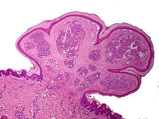
A pyogenic granuloma or lobular capillary hemangioma is a vascular tumor that occurs on both mucosa and skin, and appears as an overgrowth of tissue due to irritation, physical trauma, or hormonal factors. It is often found to involve the gums, skin, or nasal septum, and has also been found far from the head, such as in the thigh.

Atrophia Maculosa Varioliformis Cutis (AMVC) is an idiopathic noninflammatory macular atrophy subtype that affects young people. Clinically, it is distinguished by a variety of shaped, shallow, sharply demaracated depressions.

<span class="mw-page-title-main">Elejalde syndrome</span> Medical condition

Elejalde syndrome or neuroectodermal melanolysosomal disease is an extremely rare autosomal recessive syndrome consisting of moderate pigment dilution, profound central nervous system dysfunction, no immune defects, and hair with a metallic silvery sheen. The changes to hair and skin pigmentation are associated with altered melanosome trafficking.

Lupus erythematosus panniculitis presents with subcutaneous nodules that are commonly firm, sharply defined and nontender.

Anetoderma is a benign but uncommon disorder that causes localized areas of flaccid or herniated sac-like skin due to a focal reduction of dermal elastic tissue. Anetoderma is subclassified as primary anetoderma, secondary anetoderma, iatrogenic anetoderma of prematurity, congenital anetoderma, familial anetoderma, and drug-induced anetoderma.

Palisaded neutrophilic and granulomatous dermaititis (PNGS) is usually associated with a well-defined connective tissue disease, lupus erythematosus or rheumatoid arthritis most commonly, and often presents with eroded or ulcerated symmetrically distributed umbilicated papules or nodules on the elbows.

Eccrine angiomatous hamartoma (EAH), first described by Lotzbeck in 1859, is a rare benign vascular hamartoma characterized histologically by a proliferation of eccrine and vascular components. EAH exists on a spectrum of cutaneous tumors that include eccrine nevus, mucinous eccrine nevus and EAH. Each diagnostic subtype is characterized by an increase in the number as well as size of mature eccrine glands or ducts, with EAH being distinguished by the added vascular component.

Congenital smooth muscle hamartoma is typically a skin colored or lightly pigmented patch or plaque with hypertrichosis. Congenital smooth muscle hamartoma was originally reported in 1969 by Sourreil et al.

<span class="mw-page-title-main">Annular erythema of infancy</span> Medical condition

Annular erythema of infancy(AEI) consists of self-limited eruptions of erythematous, annular to polycyclic patches and plaques. It is an idiopathic figurate erythema. Over several days, a single lesion disappears without leaving behind any scale or hyperpigmentation. Mostly affecting the trunk, face, and extremities, this rash has no symptoms. The diagnosis of AEI is made through a combination of histopathologic and clinical examinations. The disease first manifests in infancy, and if treatment is not received, the periodic eruptions usually stop after the first year of life.

Sclerema neonatorum is a rare and severe skin condition that is characterized by diffuse hardening of the subcutaneous tissue with minimal inflammation.

Self-healing juvenile cutaneous mucinosis is a skin condition caused by fibroblasts producing abnormally large amounts of mucopolysaccharides, and is characterized by the sudden onset of skin lesions and polyarthritis.

Reticular erythematous mucinosis (REM) is a skin condition caused by fibroblasts producing abnormally large amounts of mucopolysaccharides. It is a disease that tends to affect women in the third and fourth decades of life.

Benign cephalic histiocytosis(BCH) is a non-Langerhan's histiocytosis that is uncommon and self-limiting, usually beginning towards the end of the first year of life. Gianotti et al. originally described it in 1971. Initially affecting the head and neck, this condition is characterized by several small eruptions of yellow to reddish-brow papules that heal on their own. Histological investigations have demonstrated that this disorder is associated with dermal proliferation of histiocytes, characterized by intracytoplasmic comma-shaped bodies, covered vesicles, and desmosome-like structure.

Generalized eruptive histiocytoma is a rare cutaneous condition characterized by widespread, erythematous, essentially symmetrical papules, particularly involving the trunk and proximal extremities.

<span class="mw-page-title-main">Balloon cell nevus</span> Medical condition

Balloon cell nevus is a benign nevus. It appears like a melanocytic nevus.

Idiopathic facial aseptic granuloma is a cutaneous condition characterized by a chronic, painless, solitary nodule, reminiscent of an acne nodule, appearing on the cheeks of young children. It has a prolonged course, but spontaneously heals.

Symmetrical drug-related intertriginous and flexural exanthema (SDRIFE), popularly known as baboon syndrome because of its resemblance to the distinctive red buttocks displayed by female baboons, is a systemic dermatitis characterized by well-demarcated patches of erythema distributed symmetrically on the buttocks. The cause of the syndrome may be drug-related: i.e., induced by systemic administration of hydroxyzine, penicillin, iodinated radio contrast media, and others.

Granuloma gluteale infantum is a cutaneous condition that appears in the anogenital region of infants as a complication of diaper dermatitis. According to some, no granulomas are found.

References

  1. Matar, Marla M. (2021). "A Case Report and Anesthetic Considerations in Subcutaneous Fat Necrosis of the Newborn". A&A Practice. 15 (3): e01422. doi:10.1213/XAA.0000000000001422. PMID   33684082. S2CID   232160504.
  2. James, William D.; Berger, Timothy G.; et al. (2006). Andrews' Diseases of the Skin: clinical Dermatology. Saunders Elsevier. ISBN   978-0-7216-2921-6.
  3. Rapini, Ronald P.; Bolognia, Jean L.; Jorizzo, Joseph L. (2007). Dermatology: 2-Volume Set. St. Louis: Mosby. ISBN   978-1-4160-2999-1.
  4. "UpToDate". UpToDate. Retrieved February 17, 2024.
  5. Xu, Ziyang; Rubin, Adam I.; Oboite, Michelle (June 18, 2021). "Subcutaneous fat necrosis of the newborn presenting as circular alopecia: a novel presentation". Pediatric Dermatology. Wiley. 38 (4): 982–983. doi:10.1111/pde.14664. ISSN   0736-8046. PMID   34145911. S2CID   235481677.
  6. Mahé, E.; Girszyn, N.; Hadj-Rabia, S.; Bodemer, C.; Hamel-Teillac, D.; De Prost, Y. (2007). "Subcutaneous fat necrosis of the newborn: a systematic evaluation of risk factors, clinical manifestations, complications and outcome of 16 children". British Journal of Dermatology. Oxford University Press (OUP). 156 (4): 709–715. doi:10.1111/j.1365-2133.2007.07782.x. ISSN   0007-0963. PMID   17493069. S2CID   29283658.
  7. 1 2 Stefanko, Nicole S.; Drolet, Beth A. (September 6, 2018). "Subcutaneous fat necrosis of the newborn and associated hypercalcemia: A systematic review of the literature". Pediatric Dermatology. Wiley. 36 (1): 24–30. doi:10.1111/pde.13640. ISSN   0736-8046. PMID   30187956.
  8. Del Pozzo-Magaña, Blanca R.; Ho, Nhung (August 30, 2016). "Subcutaneous Fat Necrosis of the Newborn: A 20-Year Retrospective Study". Pediatric Dermatology. Wiley. 33 (6). doi:10.1111/pde.12973. ISSN   0736-8046. S2CID   44286466.
  9. Tran, Julie Thao; Sheth, Anita P. (2003). "Complications of Subcutaneous Fat Necrosis of the Newborn: A Case Report and Review of the Literature". Pediatric Dermatology. Wiley. 20 (3): 257–261. doi:10.1046/j.1525-1470.2003.20315.x. ISSN   0736-8046. PMID   12787278. S2CID   26678688.
  10. Sahni, Mitali; Patel, Pooja; Muthukumar, Akila (February 22, 2020). "Severe Thrombocytosis in a Newborn with Subcutaneous Fat Necrosis and Maternal Chorioamnionitis". Case Reports in Hematology. Hindawi Limited. 2020: 1–3. doi: 10.1155/2020/5742394 . ISSN   2090-6560. PMC   7056997 . PMID   32148979.
  11. Fajgenbaum, Kristen; Zeitany, Alexandra; Googe, Paul B.; Morrell, Dean S.; McShane, Diana (July 9, 2018). "Firm, Indurated Plaques After Therapeutic Hypothermia". Clinical Pediatrics. SAGE Publications. 57 (12): 1468–1471. doi:10.1177/0009922818785992. ISSN   0009-9228.
  12. Tognetti, Linda; Filippou, Georgios; Bertrando, Sara; Picerno, Valentina; Buonocore, Giuseppe; Frediani, Bruno; Fimiani, Michele; Rubegni, Pietro (December 10, 2014). "Subcutaneous Fat Necrosis in a Newborn after Brief Therapeutic Hypothermia: Ultrasonographic Examination". Pediatric Dermatology. Wiley. 32 (3): 427–429. doi:10.1111/pde.12454. ISSN   0736-8046. PMID   25491115. S2CID   29137741.

Further reading