Related Research Articles

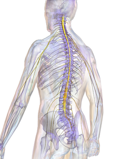
The central nervous system (CNS) is the part of the nervous system consisting primarily of the brain and spinal cord. The CNS is so named because the brain integrates the received information and coordinates and influences the activity of all parts of the bodies of bilaterally symmetric animals—i.e., all multicellular animals except sponges and jellyfish. It consists of a large nerve running from the anterior to the posterior, with the anterior end is enlarged into the brain. Not all animals with a central nervous system have a brain, although the large majority do.

A body cavity is any space or compartment, or potential space in the animal body. Cavities accommodate organs and other structures; cavities as potential spaces contain fluid.
Tetraplegia, also known as quadriplegia, is paralysis caused by illness or injury that results in the partial or total loss of use of all four limbs and torso; paraplegia is similar but does not affect the arms. The loss is usually sensory and motor, which means that both sensation and control are lost. The paralysis may be flaccid or spastic.
Paraplegia is an impairment in motor and/or sensory function of the lower extremities. The word comes from Ionic Greek (παραπληγίη) "half-stricken". It is usually caused by spinal cord injury or a congenital condition that affects the neural (brain) elements of the spinal canal. The area of the spinal canal that is affected in paraplegia is either the thoracic, lumbar, or sacral regions. If four limbs are affected by paralysis, tetraplegia or quadriplegia is the correct term. If only one limb is affected, the correct term is monoplegia. Spastic paraplegia is a form of paraplegia defined by spasticity of the affected muscles, rather than flaccid paralysis.

Afferent nerve fibers are the axons carried by a sensory nerve that relay sensory information from sensory receptors to regions of the brain. Afferent projections arrive at a particular brain region. Efferent nerve fibers are carried by efferent nerves and exit a region to act on muscles and glands.

The spinothalamic tract is a sensory pathway to the thalamus. From the ventral posterolateral nucleus in the thalamus, sensory information is relayed upward to the somatosensory cortex of the postcentral gyrus.
Neuromeres are morphologically or molecularly defined transient segments of the early developing brain. Rhombomeres are such segments that make up the rhombencephalon or hindbrain. More controversially, some argue that there exist early developmental segments that give rise to structures of the midbrain (mesomeres) and forebrain (prosomeres).
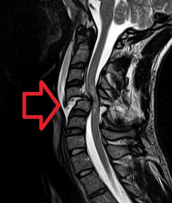
A spinal cord injury (SCI) is damage to the spinal cord that causes temporary or permanent changes in its function. Symptoms may include loss of muscle function, sensation, or autonomic function in the parts of the body served by the spinal cord below the level of the injury. Injury can occur at any level of the spinal cord and can be complete, with a total loss of sensation and muscle function at lower sacral segments, or incomplete, meaning some nervous signals are able to travel past the injured area of the cord up to the Sacral S4-5 spinal cord segments. Depending on the location and severity of damage, the symptoms vary, from numbness to paralysis, including bowel or bladder incontinence. Long term outcomes also range widely, from full recovery to permanent tetraplegia or paraplegia. Complications can include muscle atrophy, loss of voluntary motor control, spasticity, pressure sores, infections, and breathing problems.

The filum terminale is a delicate strand of fibrous tissue, about 20 cm in length, proceeding downward from the apex of the conus medullaris. It is one of the modifications of pia mater. It gives longitudinal support to the spinal cord and consists of two parts:

The central canal(, also known as ependymal canal

The stretch reflex, or more accurately "muscle stretch reflex", is a muscle contraction in response to stretching within the muscle. The reflex functions to maintain the muscle at a constant length. The term deep tendon reflex is often wrongfully used by many health workers and students to refer to this reflex. "Tendons have little to do with the response, other than being responsible for mechanically transmitting the sudden stretch from the reflex hammer to the muscle spindle. In addition, some muscles with stretch reflexes have no tendons ".

The apex of the posterior grey column, one of the three grey columns of the spinal cord, is capped by a V-shaped or crescentic mass of translucent, gelatinous neuroglia, termed the substantia gelatinosa of Rolando, which contains both neuroglia cells, and small nerve cells. The gelatinous appearance is due to a very low concentration of myelinated fibers. It extends the entire length of the spinal cord and into the medulla oblongata where it becomes the spinal nucleus of the trigeminal nerve.

The lateral corticospinal tract is the largest part of the corticospinal tract. It extends throughout the entire length of the spinal cord, and on transverse section appears as an oval area in front of the posterior column and medial to the posterior spinocerebellar tract.
Diastematomyelia is a congenital disorder in which a part of the spinal cord is split, usually at the level of the upper lumbar vertebra.

Anterior spinal veins are veins that receive blood from the anterior spinal cord.
Surfer's myelopathy is a rare nontraumatic injury causing paraplegia which is paralysis below the waist. It is a spinal cord injury caused by hyperextension of the back. When the back is hyperextended, a blood vessel leading to the spine can become kinked, depriving the spinal cord of oxygen The condition gets its name because the phenomenon is most often seen in those surfing for the first time, but it can be caused by any activity in which the back is hyperextended. In some cases the paralysis is permanent.

The spinal cord is a long, thin, tubular structure made up of nervous tissue, which extends from the medulla oblongata in the brainstem to the lumbar region of the vertebral column. It encloses the central canal of the spinal cord, which contains cerebrospinal fluid. The brain and spinal cord together make up the central nervous system (CNS). In humans, the spinal cord begins at the occipital bone, passing through the foramen magnum and entering the spinal canal at the beginning of the cervical vertebrae. The spinal cord extends down to between the first and second lumbar vertebrae, where it ends. The enclosing bony vertebral column protects the relatively shorter spinal cord. It is around 45 cm (18 in) in men and around 43 cm (17 in) long in women. The diameter of the spinal cord ranges from 13 mm in the cervical and lumbar regions to 6.4 mm in the thoracic area.

Chondroitin sulfate proteoglycans (CSPGs) are proteoglycans consisting of a protein core and a chondroitin sulfate side chain. They are known to be structural components of a variety of human tissues, including cartilage, and also play key roles in neural development and glial scar formation. They are known to be involved in certain cell processes, such as cell adhesion, cell growth, receptor binding, cell migration, and interaction with other extracellular matrix constituents. They are also known to interact with laminin, fibronectin, tenascin, and collagen. CSPGs are generally secreted from cells.

The vertebral column, also known as the backbone or spine, is part of the axial skeleton. The vertebral column is the defining characteristic of a vertebrate in which the notochord found in all chordates has been replaced by a segmented series of bone: vertebrae separated by intervertebral discs. The vertebral column houses the spinal canal, a cavity that encloses and protects the spinal cord.

In the vertebrate spinal column, each vertebra is an irregular bone with a complex structure composed of bone and some hyaline cartilage, the proportions of which vary according to the segment of the backbone and the species of vertebrate.
References
- ↑ "Neuroembryology: Spinal Cord: Development (1/2)". Neuroanatomy Lab Resource Appendices. Archived from the original on 14 July 2006. Retrieved 26 May 2021.
| | This developmental biology article is a stub. You can help Wikipedia by expanding it. |