Sympathetic ganglia

Last updated
Sympathetic ganglia
Gray839.png
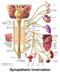
Autonomic nervous system innervation, showing the sympathetic (thoracolumbar) and parasympathetic (craniosacral) systems, in red and blue, respectively
Gray819.png
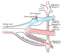
Diagram of the course and branches of a typical intercostal nerve. (Sympathetic ganglion visible at center top.)
Details
Identifiers
Latin ganglia sympathicum
MeSH D005728
TA98 A14.2.00.011
TA2 6169
FMA 5890
Anatomical terminology

The sympathetic ganglia, or paravertebral ganglia, are autonomic ganglia of the sympathetic nervous system. Ganglia are 20,000 to 30,000 afferent and efferent nerve cell bodies that run along on either side of the spinal cord. Afferent nerve cell bodies bring information from the body to the brain and spinal cord, while efferent nerve cell bodies bring information from the brain and spinal cord to the rest of the body. The cell bodies create long sympathetic chains that are on either side of the spinal cord. They also form para- or pre-vertebral ganglia of gross anatomy.

Contents

The efferent nerve cell bodies bring information from the brain to the body regarding perceptions of danger. This perception of danger can instigate the fight-or-flight response associated with the sympathetic nervous system. [1] The fight-or-flight response is adaptive when there is a real and present danger which can be avoided or diminished through increased sympathetic activity. Sympathetic activity could be increased heart rate, dilated pupils, or sweaty palms, for example. The fight-or-flight response is maladaptive when the danger is imagined, prolonged, or when it lasts after the threat is over. When the intensity or duration of the response is excessive, the individual may meet criteria for a variety of psychological disorders. [2] Neuroblastoma tumors can arise from the sympathetic ganglia tissue. [3]

Structure

Sympathetic chain ganglia

The bilaterally symmetric sympathetic chain ganglia, also called the paravertebral ganglia, are located just ventral and lateral to the spinal cord. [3] The chain extends from the upper neck down to the coccyx, forming the unpaired coccygeal ganglion. Each ganglion within this chain is either cervical, thoracic, lumbar, or sacral. Preganglionic nerves from the spinal cord synapse at one of the chain ganglia, and the postganglionic fiber extends to an effector, a visceral organ in the thoracic cavity, abdominal cavity, or pelvic cavity.

There are usually 22–23 pairs of these ganglia: 3 in the cervical region (cervical ganglia), 11 in the thoracic region (note the presence of the stellate cervicothoracic ganglia), 4 in the lumbar region and 4–5 in the sacral region. [3] Throughout human evolution, the first thoracic and inferior cervical ganglia merged - and this resulting ganglion is called the stellate ganglion (so called because of its radiating pattern similar in appearance to a star).

The general rule of interaction of the nerve fibers in the sympathetic nervous system begins at the spinal cord. Here they arise from the thoracolumbar (T1–L2) regions' lateral horn of grey and emerge via the ventral root. They enter their respective spinal nerve (e.g. T5), and thus enter the white ramus communicans. This myelinated division can then enter the sympathetic chain.

Here four options are available to the fibers: (1) they can run up the chain and synapse, (2) they can synapse at the level of entry, (3) they can pass straight through and synapse elsewhere – such as in the case of T5–12 (the splanchnic nerves), or (4) they can enter the chain and descend to synapse. It is this ability to move superiorly and inferiorly along the chain that results in the mass response to the sympathetic nervous system. A preganglionic fibre may synapse to 15–20 postganglionic fibres. The postganglionic neurons extend across most of the body. [4]

Upon exiting the sympathetic chain, the fibres enter a less-myelinated gray ramus communicans. There is still a myelin sheath present – just in far lower amounts compared to the white ramus communicans. This ramus then enters the spinal nerve and is sent to its synapsing target, or becomes a visceral branch to enter a plexus (e.g. the superficial or deep cardiac plexuses), or synapses directly onto a target.

Collateral ganglia

Neurons of the collateral ganglia, also called the prevertebral ganglia, receive input from the splanchnic nerves and innervate organs of the abdominal and pelvic region. These include the celiac ganglia, superior mesenteric ganglia, and inferior mesenteric ganglia.

Additional images

Related Research Articles

<span class="mw-page-title-main">Autonomic nervous system</span> Division of the nervous system supplying internal organs, smooth muscle and glands

The autonomic nervous system (ANS), formerly referred to as the vegetative nervous system, is a division of the nervous system that supplies internal organs, smooth muscle and glands. The autonomic nervous system is a control system that acts largely unconsciously and regulates bodily functions, such as the heart rate, its force of contraction, digestion, respiratory rate, pupillary response, urination, and sexual arousal. This system is the primary mechanism in control of the fight-or-flight response.

<span class="mw-page-title-main">Parasympathetic nervous system</span> Division of the autonomic nervous system

The parasympathetic nervous system (PSNS) is one of the three divisions of the autonomic nervous system, the others being the sympathetic nervous system and the enteric nervous system. The enteric nervous system is sometimes considered part of the autonomic nervous system, and sometimes considered an independent system.

<span class="mw-page-title-main">Sympathetic nervous system</span> Part of the autonomic nervous system which stimulates fight-or-flight responses

The sympathetic nervous system (SNS) is one of the three divisions of the autonomic nervous system, the others being the parasympathetic nervous system and the enteric nervous system. The enteric nervous system is sometimes considered part of the autonomic nervous system, and sometimes considered an independent system.

<span class="mw-page-title-main">Spinal nerve</span> Nerve that carries signals between the spinal cord and the body

A spinal nerve is a mixed nerve, which carries motor, sensory, and autonomic signals between the spinal cord and the body. In the human body there are 31 pairs of spinal nerves, one on each side of the vertebral column. These are grouped into the corresponding cervical, thoracic, lumbar, sacral and coccygeal regions of the spine. There are eight pairs of cervical nerves, twelve pairs of thoracic nerves, five pairs of lumbar nerves, five pairs of sacral nerves, and one pair of coccygeal nerves. The spinal nerves are part of the peripheral nervous system.

<span class="mw-page-title-main">Pterygopalatine ganglion</span> Parasympathetic ganglion in the pterygopalatine fossa

The pterygopalatine ganglion is a parasympathetic ganglion in the pterygopalatine fossa. It is one of four parasympathetic ganglia of the head and neck,.

<span class="mw-page-title-main">Ciliary ganglion</span> Bundle of nerves, parasympathetic ganglion

The ciliary ganglion is a bundle of nerves, parasympathetic ganglion located just behind the eye in the posterior orbit. It is 1–2 mm in diameter and in humans contains approximately 2,500 neurons. The ganglion contains postganglionic parasympathetic neurons. These neurons supply the pupillary sphincter muscle, which constricts the pupil, and the ciliary muscle which contracts to make the lens more convex. Both of these muscles are involuntary since they are controlled by the parasympathetic division of the autonomic nervous system.

<span class="mw-page-title-main">Superior cervical ganglion</span> Largest of the cervical ganglia

The superior cervical ganglion (SCG) is the upper-most and largest of the cervical sympathetic ganglia of the sympathetic trunk. It probably formed by the union of four sympathetic ganglia of the cervical spinal nerves C1-C4. It is the only ganglion of the sympathetic nervous system that innervates the head and neck. The SCG innervates numerous structures of the head and neck.

<span class="mw-page-title-main">Sympathetic trunk</span>

The sympathetic trunks are a paired bundle of nerve fibers that run from the base of the skull to the coccyx. They are a major component of the sympathetic nervous system.

<span class="mw-page-title-main">White ramus communicans</span>

The white ramus communicans from Latin ramus (branch) and communicans (communicating) is the preganglionic sympathetic outflow nerve tract from the spinal cord.

Each spinal nerve receives a branch called a gray ramus communicans from the adjacent paravertebral ganglion of the sympathetic trunk. The gray rami communicantes contain postganglionic nerve fibers of the sympathetic nervous system and are composed of largely unmyelinated neurons. This is in contrast to the white rami communicantes, in which heavily myelinated neurons give the rami their white appearance.

<span class="mw-page-title-main">Ramus communicans</span> Latin term used for a nerve which connects two other nerves

Ramus communicans is the Latin term used for a nerve which connects two other nerves, and can be translated as "communicating branch".

<span class="mw-page-title-main">Lateral grey column</span>

The lateral grey column is one of the three grey columns of the spinal cord ; the others being the anterior and posterior grey columns. The lateral grey column is primarily involved with activity in the sympathetic division of the autonomic motor system. It projects to the side as a triangular field in the thoracic and upper lumbar regions of the postero-lateral part of the anterior grey column.

<span class="mw-page-title-main">Aorticorenal ganglion</span>

The aorticorenal ganglion is composed of the superior mesenteric, renal, and inferior mesenteric ganglia. This is distinct from the celiac ganglia. However, they are part of the preaortic ganglia.

<span class="mw-page-title-main">Esophageal plexus</span>

The esophageal plexus is formed by nerve fibers from two sources, branches of the vagus nerve, and visceral branches of the sympathetic trunk. The esophageal plexus and the cardiac plexus contain the same types of fibers and are both considered thoracic autonomic plexus.

<span class="mw-page-title-main">Cervical ganglia</span>

The cervical ganglia are paravertebral ganglia of the sympathetic nervous system. Preganglionic nerves from the thoracic spinal cord enter into the cervical ganglions and synapse with its postganglionic fibers or nerves. The cervical ganglion has three paravertebral ganglia:

<span class="mw-page-title-main">General visceral afferent fiber</span> Part of the visceral nervous system

The general visceral afferent (GVA) fibers conduct sensory impulses from the internal organs, glands, and blood vessels to the central nervous system. They are considered to be part of the visceral nervous system, which is closely related to the autonomic nervous system, but 'visceral nervous system' and 'autonomic nervous system' are not direct synonyms and care should be taken when using these terms. Unlike the efferent fibers of the autonomic nervous system, the afferent fibers are not classified as either sympathetic or parasympathetic.

<span class="mw-page-title-main">Lumbar ganglia</span>

The lumbar ganglia are paravertebral ganglia located in the inferior portion of the sympathetic trunk. The lumbar portion of the sympathetic trunk typically has 4 lumbar ganglia. The lumbar splanchnic nerves arise from the ganglia here, and contribute sympathetic efferent fibers to the nearby plexuses. The first two lumbar ganglia have both white and gray rami communicates.

<span class="mw-page-title-main">Thoracic ganglia</span>

The thoracic ganglia are paravertebral ganglia. The thoracic portion of the sympathetic trunk typically has 12 thoracic ganglia. Emerging from the ganglia are thoracic splanchnic nerves that help provide sympathetic innervation to thoracic and abdominal structures. The thoracic part of sympathetic trunk lies posterior to the costovertebral pleura and is hence not a content of the posterior mediastinum

<span class="mw-page-title-main">Outline of the human nervous system</span> Overview of and topical guide to the human nervous system

The following diagram is provided as an overview of and topical guide to the human nervous system:

<span class="mw-page-title-main">Roots of the ciliary ganglion</span>

The ciliary ganglion is a parasympathetic ganglion located just behind the eye in the posterior orbit. Three types of axons enter the ciliary ganglion but only the preganglionic parasympathetic axons synapse there. The entering axons are arranged into three roots of the ciliary ganglion, which join enter the posterior surface of the ganglion.

References

  1. Life, the science of biology . Sadava, David E. (8th ed.). Sunderland, MA: Sinauer Associates. 2008. ISBN   9780716776710. OCLC   71632224.{{cite book}}: CS1 maint: others (link)
  2. American Psychiatric Association (2013-05-22). Diagnostic and Statistical Manual of Mental Disorders . American Psychiatric Association. doi:10.1176/appi.books.9780890425596. hdl:2027.42/138395. ISBN   978-0890425558.
  3. 1 2 3 "Structure of the Autonomic Nervous System | Boundless Anatomy and Physiology". courses.lumenlearning.com. Retrieved 2019-10-28.
  4. Drake, Richard L. (Richard Lee), 1950– (2005). Gray's anatomy for students. Vogl, Wayne., Mitchell, Adam W. M., Gray, Henry, 1825–1861. Philadelphia: Elsevier/Churchill Livingstone. ISBN   0443066124. OCLC   55139039.{{cite book}}: CS1 maint: multiple names: authors list (link)