Vascular access

Last updated

Vascular access refers to a rapid, direct method of introducing or removing devices or chemicals from the bloodstream. In hemodialysis, vascular access is used to remove the patient's blood so that it can be filtered through the dialyzer. Three primary methods are used to gain access to the blood: an intravenous catheter, an arteriovenous fistula (AV) or a synthetic graft. In the latter two, needles are used to puncture the graft or fistula each time dialysis is performed.

Contents

The type of vascular access created for patients on hemodialysis is influenced by factors such as the expected time course of a patient's kidney failure and the condition of his or her vasculature. Patients may have multiple accesses, usually because an AV fistula or graft is maturing and a catheter is still being used. The creation of all these three major types of vascular accesses requires surgery. [1]

Catheter

Catheter access, sometimes called a CVC (central venous catheter), consists of a plastic catheter with two lumens (or occasionally two separate catheters) which is inserted into a large vein (usually the vena cava, via the internal jugular vein or the femoral vein) to allow large flows of blood to be withdrawn from one lumen, to enter the dialysis circuit, and to be returned via the other lumen. However, blood flow is almost always less than that of a well functioning fistula or graft.

Catheters are usually found in two general varieties, tunnelled and non-tunnelled. [2]

Non-tunnelled catheter access is for short-term access (up to about 10 days, but often for one dialysis session only), and the catheter emerges from the skin at the site of entry into the vein.

Tunnelled catheter access involves a longer catheter, which is tunnelled under the skin from the point of insertion in the vein to an exit site some distance away. It is usually placed in the internal jugular vein in the neck and the exit site is usually on the chest wall. The tunnel acts as a barrier to invading microbes, and as such, tunnelled catheters are designed for short- to medium-term access (weeks to months only), because infection is still a frequent problem.

Aside from infection, venous stenosis is another serious problem with catheter access. The catheter is a foreign body in the vein and often provokes an inflammatory reaction in the vein wall. This results in scarring and narrowing of the vein, often to the point of occlusion. This can cause problems with severe venous congestion in the area drained by the vein and may also render the vein, and the veins drained by it, useless for creating a fistula or graft at a later date. Patients on long-term hemodialysis can literally 'run out' of access, so this can be a fatal problem.

Catheter access is usually used for rapid access for immediate dialysis, for tunnelled access in patients who are deemed likely to recover from acute kidney injury, and for patients with end-stage kidney failure who are either waiting for alternative access to mature or who are unable to have alternative access.

Catheter access is often popular with patients, because attachment to the dialysis machine doesn't require needles. However, the serious risks of catheter access noted above mean that such access should be contemplated only as a long-term solution in the most desperate access situation.

Permacath for dialysis Permacath.jpg
Permacath for dialysis

AV fistula

A radiocephalic fistula. Radiocephalic fistula.svg
A radiocephalic fistula.
Illustration depicting AV fistula during hemodialysis Blausen 0313 Dialysis.png
Illustration depicting AV fistula during hemodialysis

AV (arteriovenous) fistulas are recognized as the preferred access method. To create a fistula, a vascular surgeon joins an artery and a vein together through anastomosis. Since this bypasses the capillaries, blood flows rapidly through the fistula. One can feel this by placing one's finger over a mature fistula. This is called feeling for "thrill" and produces a distinct 'buzzing' feeling over the fistula. One can also listen through a stethoscope for the sound of the blood "whooshing" through the fistula, a sound called bruit .

Fistulas are usually created in the nondominant arm and may be situated on the hand (the 'snuffbox' fistula'), the forearm (usually a radiocephalic fistula, or so-called Brescia-Cimino fistula, in which the radial artery is anastomosed to the cephalic vein), or the elbow (usually a brachiocephalic fistula, where the brachial artery is anastomosed to the cephalic vein). Though less common, fistulas can also be created in the groin, though the creation process differs. Placement in the groin is usually done when options in the arm and hands are not available due to anatomy or the failure of fistulas previously created in the arms/hands. A fistula will take a number of weeks to mature, on average perhaps 4–6 weeks.

During treatment, two needles are inserted into the vein, one to draw blood and one to return it. The orientation of the needles takes the normal flow of the blood into account. The "arterial" needle draws blood from the "upstream" location while the "venous" needle returns blood "downstream". This sequence prevents partial recycling of the same blood through the dialysis machine, which would lead to less effective treatment.

The advantages of the AV fistula use are lower infection rates, because no foreign material is involved in their formation, higher blood flow rates (which translates to more effective dialysis), and a lower incidence of thrombosis. The complications are fewer than with other access methods. If a fistula has a very high blood flow and the vasculature that supplies the rest of the limb is poor, a steal syndrome can occur, where blood entering the limb is drawn into the fistula and returned to the general circulation without entering the limb's capillaries. This results in cold extremities of that limb, cramping pains, and, if severe, tissue damage. One long-term complication of an AV fistula can be the development of an aneurysm, a bulging in the wall of the vein where it is weakened by the repeated insertion of needles over time. To a large extent the risk of developing an aneurysm can be reduced by carefully rotating needle sites over the entire fistula ("rope-laddering" [3] ), or using the "buttonhole" (constant site) technique; button-holing is safe when a fistula is used, but runs the risk of infection or degradation of an arterio-venous graft. [3] Aneurysms may necessitate corrective surgery and may shorten the useful life of a fistula. Fistulas can also become blocked due to blood clotting or infected if sterile precautions are not followed during needle insertion at the start of dialysis. Because of the high volume of blood flowing through the fistula, excessive bleeding can also occur. This is most common soon after a dialysis treatment. Pressure must be applied to the needle holes to induce clotting. If that pressure is removed prematurely or a patient engages in physical activity too soon after dialysis, the needle holes can open up.

To prevent damage to the fistula and aneurysm or pseudoaneurysm formation, it is recommended that the needle be inserted at different points in a rotating fashion. Another approach is to cannulate the fistula with a blunted needle, in exactly the same place. This is called a 'buttonhole' approach. Often two or three buttonhole places are available on a given fistula. This also can prolong fistula life and help prevent damage to the fistula.

A recent study that was published in The American Journal of Pathology, provides information about the mechanisms underlying failure of the most common type of hemodialysis vascular access, the arteriovenous fistula. In spite of AV Fistula being one of the most preferred methods of Vascular access, the researchers observed that, up to 60% of newly created fistulas never become usable for dialysis because they fail to mature (meaning the vessels do not enlarge enough to support the dialysis blood circuit.). This study suggests that, the impairment in responsiveness to nitric oxide that occurs in some patients with end-stage renal disease may result in hyperplasia (excessive growth) of the innermost layer of the blood vessels or reduced ability of the vessels to dilate. Either abnormality can limit the maturation and viability of the arteriovenous fistula. This research raises the possibility that therapeutic restoration of nitric oxide responsiveness through manipulation of local mediators may prevent fistula maturation failure in patients and potentially contribute to their ability to remain on hemodialysis. [4]

AV graft

An arteriovenous graft. Blausen 0050 ArteriovenousGraft.png
An arteriovenous graft.

AV (arteriovenous) grafts are much like fistulas in most respects, except that an artificial vessel is used to join the artery and vein. The graft usually is made of a synthetic material, often PTFE, but sometimes chemically treated, sterilized veins from animals are used. Grafts are inserted when the patient's native vasculature does not permit a fistula. They mature faster than fistulas, and may be ready for use several weeks after formation (some newer grafts may be used even sooner). However, AV grafts are at high risk to develop narrowing, especially in the vein just downstream from where the graft has been sewn to the vein. Narrowing often leads to thrombosis (clotting). As foreign material, they are at greater risk for becoming infected. More options for sites to place a graft are available, because the graft can be made quite long. Thus a graft can be placed in the thigh or even the neck (the 'necklace graft').

Fistula First project

AV fistulas have a much better access patency and survival than do venous catheters or grafts. They also produce better patient survival and have far fewer complications compared to grafts or venous catheters. For this reason, the Centers for Medicare & Medicaid (CMS) has set up a Fistula First Initiative, [5] whose goal is to increase the use of AV fistulas in dialysis patients. This initiative has had many successes, but fistula is not always the superior strategy when it comes to the elderly. [6]

There is ongoing research to make bio-engineered blood vessels, which may be of immense importance in creating AV fistulas for patients on hemodialysis, who do not have good blood vessels for creation of one. It involves growing cells which produce collagen and other proteins on a biodegradable micromesh tube followed by removal of those cells to make the 'blood vessels' storable in refrigerators. [7]

See also

Related Research Articles

<span class="mw-page-title-main">Arteriovenous malformation</span> Vascular anomaly

An arteriovenous malformation (AVM) is an abnormal connection between arteries and veins, bypassing the capillary system. Usually congenital, this vascular anomaly is widely known because of its occurrence in the central nervous system, but can appear anywhere in the body. The symptoms of AVMs can range from none at all to intense pain or bleeding, and they can lead to other serious medical problems.

<span class="mw-page-title-main">Central venous catheter</span> A tubular device placed in a large vein used to administer medicines

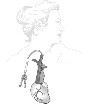
A central venous catheter (CVC), also known as a central line (c-line), central venous line, or central venous access catheter, is a catheter placed into a large vein. It is a form of venous access. Placement of larger catheters in more centrally located veins is often needed in critically ill patients, or in those requiring prolonged intravenous therapies, for more reliable vascular access. These catheters are commonly placed in veins in the neck, chest, groin, or through veins in the arms.

<span class="mw-page-title-main">Angiography</span> Medical imaging technique

Angiography or arteriography is a medical imaging technique used to visualize the inside, or lumen, of blood vessels and organs of the body, with particular interest in the arteries, veins, and the heart chambers. Modern angiography is performed by injecting a radio-opaque contrast agent into the blood vessel and imaging using X-ray based techniques such as fluoroscopy.

<span class="mw-page-title-main">Interventional radiology</span> Medical subspecialty

Interventional radiology (IR) is a medical specialty that performs various minimally-invasive procedures using medical imaging guidance, such as x-ray fluoroscopy, computed tomography, magnetic resonance imaging, or ultrasound. IR performs both diagnostic and therapeutic procedures through very small incisions or body orifices. Diagnostic IR procedures are those intended to help make a diagnosis or guide further medical treatment, and include image-guided biopsy of a tumor or injection of an imaging contrast agent into a hollow structure, such as a blood vessel or a duct. By contrast, therapeutic IR procedures provide direct treatment—they include catheter-based medicine delivery, medical device placement, and angioplasty of narrowed structures.

<span class="mw-page-title-main">Vascular surgery</span> Medical specialty, operative procedures for the treatment of vascular disorders

Vascular surgery is a surgical subspecialty in which vascular diseases involving the arteries, veins, or lymphatic vessels, are managed by medical therapy, minimally-invasive catheter procedures and surgical reconstruction. The specialty evolved from general and cardiovascular surgery where it refined the management of just the vessels, no longer treating the heart or other organs. Modern vascular surgery includes open surgery techniques, endovascular techniques and medical management of vascular diseases - unlike the parent specialities. The vascular surgeon is trained in the diagnosis and management of diseases affecting all parts of the vascular system excluding the coronaries and intracranial vasculature. Vascular surgeons also are called to assist other physicians to carry out surgery near vessels, or to salvage vascular injuries that include hemorrhage control, dissection, occlusion or simply for safe exposure of vascular structures.

<span class="mw-page-title-main">Hemodialysis</span> Medical procedure for purifying blood

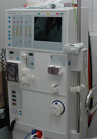
Hemodialysis, also spelled haemodialysis, or simply dialysis, is a process of filtering the blood of a person whose kidneys are not working normally. This type of dialysis achieves the extracorporeal removal of waste products such as creatinine and urea and free water from the blood when the kidneys are in a state of kidney failure. Hemodialysis is one of three renal replacement therapies. An alternative method for extracorporeal separation of blood components such as plasma or cells is apheresis.

<span class="mw-page-title-main">Pseudoaneurysm</span> Collection of blood between outer artery layers

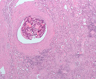
A pseudoaneurysm, also known as a false aneurysm, is a locally contained hematoma outside an artery or the heart due to damage to the vessel wall. The injury passes through all three layers of the arterial wall, causing a leak, which is contained by a new, weak "wall" formed by the products of the clotting cascade. A pseudoaneurysm does not contain any layer of the vessel wall.

<span class="mw-page-title-main">Dialysis catheter</span>

A dialysis catheter is a catheter used for exchanging blood to and from a hemodialysis machine and a patient.

<span class="mw-page-title-main">Embolization</span> Passage and lodging of an embolus within the bloodstream

Embolization refers to the passage and lodging of an embolus within the bloodstream. It may be of natural origin (pathological), in which sense it is also called embolism, for example a pulmonary embolism; or it may be artificially induced (therapeutic), as a hemostatic treatment for bleeding or as a treatment for some types of cancer by deliberately blocking blood vessels to starve the tumor cells.

<span class="mw-page-title-main">Arteriovenous fistula</span> Medical condition

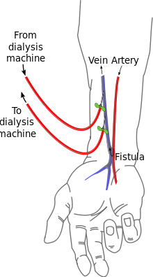
An arteriovenous fistula is an abnormal connection or passageway between an artery and a vein. It may be congenital, surgically created for hemodialysis treatments, or acquired due to pathologic process, such as trauma or erosion of an arterial aneurysm.

<span class="mw-page-title-main">Hemofiltration</span>

Hemofiltration, also haemofiltration, is a renal replacement therapy which is used in the intensive care setting. It is usually used to treat acute kidney injury (AKI), but may be of benefit in multiple organ dysfunction syndrome or sepsis. During hemofiltration, a patient's blood is passed through a set of tubing via a machine to a semipermeable membrane where waste products and water are removed by convection. Replacement fluid is added and the blood is returned to the patient.

Renal replacement therapy (RRT) is therapy that replaces the normal blood-filtering function of the kidneys. It is used when the kidneys are not working well, which is called kidney failure and includes acute kidney injury and chronic kidney disease. Renal replacement therapy includes dialysis, hemofiltration, and hemodiafiltration, which are various ways of filtration of blood with or without machines. Renal replacement therapy also includes kidney transplantation, which is the ultimate form of replacement in that the old kidney is replaced by a donor kidney.

<span class="mw-page-title-main">Cimino fistula</span>

A Cimino fistula, also Cimino-Brescia fistula, surgically created arteriovenous fistula and arteriovenous fistula, is a type of vascular access for hemodialysis. It is typically a surgically created connection between an artery and a vein in the arm, although there have been acquired arteriovenous fistulas which do not in fact demonstrate connection to an artery.

A vascular bypass is a surgical procedure performed to redirect blood flow from one area to another by reconnecting blood vessels. Often, this is done to bypass around a diseased artery, from an area of normal blood flow to another relatively normal area. It is commonly performed due to inadequate blood flow (ischemia) caused by atherosclerosis, as a part of organ transplantation, or for vascular access in hemodialysis. In general, someone's own vein (autograft) is the preferred graft material for a vascular bypass, but other types of grafts such as polytetrafluoroethylene (Teflon), polyethylene terephthalate (Dacron), or a different person's vein (allograft) are also commonly used. Arteries can also serve as vascular grafts. A surgeon sews the graft to the source and target vessels by hand using surgical suture, creating a surgical anastomosis.

<span class="mw-page-title-main">Dural arteriovenous fistula</span> Medical condition

A dural arteriovenous fistula (DAVF) or malformation is an abnormal direct connection (fistula) between a meningeal artery and a meningeal vein or dural venous sinus.

In nephrology, vascular access steal syndrome is a syndrome caused by ischemia resulting from a vascular access device that was installed to provide access for the inflow and outflow of blood during hemodialysis.

<span class="mw-page-title-main">Port (medical)</span> Small completely implantable medical appliance that is installed entirely beneath the skin

In medicine, a port is a small medical appliance that is installed beneath the skin. A catheter connects the port to a vein. Under the skin, the port has a septum through which drugs can be injected and blood samples can be drawn many times, usually with less discomfort for the patient than a more typical "needle stick".

The Nicoladoni–Branham sign is named after Carl Nicoladoni, who first noticed the phenomenon of the pulse slowing in a patient with right arm phlebarteriectasia when the brachialis artery proximal to it was compressed. In modern medicine, the sign is elicited when pressure is applied to an artery proximal to an arteriovenous fistula and said to be positive if the following occurs:

In medicine, vascular access is a means of accessing the bloodstream through the peripheral or central blood vessels in order to obtain blood or deliver medications including chemotherapy. A vascular access procedure involves insertion of a sterile plastic tube called a catheter into a blood vessel. Types of catheters can be either peripherally or centrally located. Peripheral catheters are approximately one inch (25 mm) long and are inserted into the small veins of the forearm. Central catheters are bigger and longer and are inserted into the large veins of the extremities, neck, or chest. Central venous catheters are the primary modality used for delivery of chemotherapeutic agents. The duration of central venous catheterization is dependent on the type of treatment given.

Ischemic monomelic neuropathy(IMN) is an uncommon vascular access complication in hemodialysis patients that manifests as multiple mononeuropathies without clinical ischemia. Ischemic monomelic neuropathy is most likely to affect patients who have had brachiocephalic vascular grafts, and it is characterized by symptoms of acute pain, numbness, and paresthesia in addition to motor weakness. The term "ischemic monomelic neuropathy" was first used in 1983 by Wilbourn, despite the fact that Bolton et al. had originally reported on it in 1979.

References

  1. Kallenbach J.Z.In: Review of hemodialysis for nurses and dialysis personnel. 7th ed. St. Louis, Missouri:Elsevier Mosby; 2005.
  2. Frankel, A. (2006-04-01). "Temporary Access and Central Venous Catheters". European Journal of Vascular and Endovascular Surgery. 31 (4): 417–422. doi: 10.1016/j.ejvs.2005.10.003 . PMID   16360326.
  3. 1 2 Buchanan, Michael (15 March 2024). "Kidney patients' health was put at risk, doctor tells BBC". BBC News.
  4. "New research shows promise for improving vascular access for hemodialysis patients". EurekAlert!. Retrieved 2017-09-06.
  5. Fistula First Initiative
  6. DeSilva, R. N.; Patibandla, B. K.; Vin, Y.; Narra, A.; Chawla, V.; Brown, R. S.; Goldfarb-Rumyantzev, A. S. (2013-08-01). "Fistula First Is Not Always the Best Strategy for the Elderly". Journal of the American Society of Nephrology. 24 (8): 1297–1304. doi:10.1681/ASN.2012060632. ISSN   1046-6673. PMC   3736704 . PMID   23813216.
  7. Seppa, Nathan (2 February 2011). "Bioengineering Better Blood Vessels". Science News . Retrieved 4 February 2011.