Lactococcin-like family | |||||||||
---|---|---|---|---|---|---|---|---|---|
Identifiers | |||||||||
Symbol | Lactococcin | ||||||||
Pfam | PF04369 | ||||||||
Pfam clan | CL0400 | ||||||||
InterPro | IPR007464 | ||||||||
TCDB | 1.C.22 | ||||||||
OPM superfamily | 141 | ||||||||
OPM protein | 6gnz | ||||||||
|
Bacteriocin (Lactococcin_972) | |||||||||
---|---|---|---|---|---|---|---|---|---|
Identifiers | |||||||||
Symbol | Lactococcin_972 | ||||||||
Pfam | PF09683 | ||||||||
InterPro | IPR006540 | ||||||||
TCDB | 1.C.37 | ||||||||
OPM superfamily | 457 | ||||||||
OPM protein | 2lgn | ||||||||
|
Bacteriocins are proteinaceous or peptidic toxins produced by bacteria to inhibit the growth of similar or closely related bacterial strain(s). They are similar to yeast and paramecium killing factors, and are structurally, functionally, and ecologically diverse. Applications of bacteriocins are being tested to assess their application as narrow-spectrum antibiotics. [1]
Bacteriocins were first discovered by André Gratia in 1925. [2] [3] He was involved in the process of searching for ways to kill bacteria, which also resulted in the development of antibiotics and the discovery of bacteriophage, all within a span of a few years. He called his first discovery a colicine because it was made by E. coli.
Bacteriocins are categorized in several ways, including producing strain, common resistance mechanisms, and mechanism of killing. There are several large categories of bacteriocin which are only phenomenologically related. These include the bacteriocins from gram-positive bacteria, the colicins, [4] the microcins, and the bacteriocins from Archaea. The bacteriocins from E. coli are called colicins (formerly called 'colicines', meaning 'coli killers'). They are the longest studied bacteriocins. They are a diverse group of bacteriocins and do not include all the bacteriocins produced by E. coli. In fact, one of the oldest known so-called colicins was called colicin V and is now known as microcin V . It is much smaller and produced and secreted in a different manner than the classic colicins.
This naming system is problematic for a number of reasons. First, naming bacteriocins by what they putatively kill would be more accurate if their killing spectrum were contiguous with genus or species designations. The bacteriocins frequently possess spectra that exceed the bounds of their named taxa and almost never kill the majority of the taxa for which they are named. Further, the original naming is generally derived not from the sensitive strain the bacteriocin kills, but instead the organism that produces the bacteriocin. This makes the use of this naming system a problematic basis for theory; thus the alternative classification systems.[ citation needed ]
Bacteriocins that contain the modified amino acid lanthionine as part of their structure are called lantibiotics. However, efforts to reorganize the nomenclature of the family of ribosomally synthesized and post-translationally modified peptide (RiPP) natural products have led to the differentiation of lantipeptides from bacteriocins based on biosynthetic genes. [5]
Alternative methods of classification include: method of killing (pore-forming, nuclease activity, peptidoglycan production inhibition, etc.), genetics (large plasmids, small plasmids, chromosomal), molecular weight and chemistry (large protein, peptide, with/without sugar moiety, containing atypical amino acids such as lanthionine), and method of production (ribosomal, post-ribosomal modifications, non-ribosomal).
Gram negative bacteriocins are typically classified by size. Microcins are less than 20 kDa in size, colicin-like bacteriocins are 20 to 90 kDa in size and tailocins or so called high molecular weight bacteriocins which are multi subunit bacteriocins that resemble the tails of bacteriophages. This size classification also coincides with genetic, structural and functional similarities.
See main article on microcins.
Colicins are bacteriocins found in the Gram-negative E. coli. Similar bacteriocins (CLBs, colicin-like bacteriocins) occur in other Gram-negative bacteria. CLBs typically target same species and have species-specific names: klebicins from Klebsiella and pesticins from Yersia pestis. [6] Pseudomonas -genus produces bacteriocins called pyocins. S-type pyocins belong to CLBs, but R- and F-type pyocins belong to tailocins. [7]
CLBs are distinct from Gram-positive bacteriocins. They are modular proteins between 20 and 90 kDa in size. They often consist of a receptor binding domain, a translocation domain and a cytotoxic domain. Combinations of these domains between different CLBs occur frequently in nature and can be created in the laboratory. Due to these combinations further subclassification can be based on either import mechanism (group A and B) or on cytotoxic mechanism (nucleases, pore forming, M-type, L-type). [4]
Most well studied are the tailocins of Pseudomonas aeruginosa . They can be further subdivided into R-type and F-type pyocins. [8] Some research was made to identify the pyocins and show how they are involved in the “cell-to-cell” competition of the closely related Pseudomonas bacteria.
The two types of tailocins differ by their structure; they are both composed of a sheath and a hollow tube forming a long helicoidal hexameric structure attached to a baseplate. There are multiple tail fibers that allow the viral particle to bind to the target cell. However, the R-pyocins are a large, rigid contractile tail-like structure whereas the F-pyocins are a small flexible, non-contractile tail-like structure.
The tailocins are coded by prophage sequences in the bacteria genome, and the production will happen when a kin bacteria is spotted in the environment of the competitive bacteria. The particles are synthesized in the center of the cells and after maturation they will migrate to the cell pole via tubulin structure. The tailocins will then be ejected in the medium with the cell lysis. They can be projected up to several tens of micrometers thanks to a very high turgor pressure of the cell. The tailocins released will then recognize and bind to the kin bacteria to kill them. [9]
Bacteriocins from Gram positive bacteria are typically classified into Class I, Class IIa/b/c, and Class III. [10]
The class I bacteriocins are small peptide inhibitors and include nisin and other lantibiotics.
The class II bacteriocins are small (<10 kDa) heat-stable proteins. This class is subdivided into five subclasses. The class IIa bacteriocins (pediocin-like bacteriocins) are the largest subgroup and contain an N-terminal consensus sequence -Tyr-Gly-Asn-Gly-Val-Xaa-Cys across this group. [11] [12] The C-terminal is responsible for species-specific activity, causing cell-leakage by permeabilizing the target cell wall.
The most recently proposed subclass is the Class IIe, which encompasses those bacteriocins composed of three or four non-pediocin like peptides. The best example is aureocin A70, a four-peptide bacteriocin, highly active against Listeria monocytogenes , with potential biotechnological applications. [16]
Class III bacteriocins are large, heat-labile (>10 kDa) protein bacteriocins. This class is subdivided in two subclasses: subclass IIIa (bacteriolysins) and subclass IIIb. Subclass IIIa comprises those peptides that kill bacterial cells by cell wall degradation, thus causing cell lysis. The best studied bacteriolysin is lysostaphin, a 27 kDa peptide that hydrolyzes the cell walls of several Staphylococcus species, principally S. aureus . [17] Subclass IIIb, in contrast, comprises those peptides that do not cause cell lysis, killing the target cells by disrupting plasma membrane potential.
Class IV bacteriocins are defined as complex bacteriocins containing lipid or carbohydrate moieties. Confirmation by experimental data was established with the characterisation of sublancin and glycocin F (GccF) by two independent groups. [18] [19]
Two databases of bacteriocins are available: BAGEL [20] and BACTIBASE. [21] [22]
As of 2016, nisin was the only bacteriocin generally recognized as safe by the FDA and was used as a food preservative in several countries. [23] Generally bacteriocins are not useful as food preservatives because they are expensive to make, are broken down in food products, they harm some proteins in food, and they target too narrow a range of microbes. [23]
Furthermore, bacteriocins active against E. coli , Salmonella and Pseudomonas aeruginosa have been produced in plants with the aim for them to be used as food additives. [24] [25] [26] The use of bacteriocins in food has been generally regarded as safe by the FDA. [24]
Moreover, has been recently demonstrated that bacteriocins active against plant pathogenic bacteria can be expressed in plants to provide robust resistance against plant disease. [27]
Bacteriocins are made by non-pathogenic Lactobacilli in the vagina and help maintain the stability of the vaginal microbiome. [28]
Bacteriocins have been proposed as a replacement for antibiotics to which pathogenic bacteria have become resistant. Potentially, the bacteriocins could be produced by bacteria intentionally introduced into the patient to combat infection. [1] There are several strategies by which new bacteriocins can be discovered. In the past, bacteriocins had to be identified by intensive culture-based screening for antimicrobial activity against suitable targets and subsequently purified using fastidious methods prior to testing. However, since the advent of the genomic era, the availability of the bacterial genome sequences has revolutionized the approach to identifying bacteriocins. Recently developed in silico -based methods can be applied to rapidly screen thousands of bacterial genomes in order to identify novel antimicrobial peptides. [29]
As of 2014 some bacteriocins had been studied in in vitro studies to see if they can stop viruses from replicating, namely staphylococcin 188 against Newcastle disease virus, influenza virus, and coliphage HSA virus; each of enterocin AAR-71 class IIa, enterocin AAR-74 class IIa, and erwiniocin NA4 against coliphage HSA virus; each of enterocin ST5Ha, enterocin NKR-5-3C, and subtilosin against HSV-1; each of enterocin ST4V and enterocin CRL35 class IIa against HSV-1 and HSV-2; labyrinthopeptin A1 against HIV-1 and HSV-1; and bacteriocin from Lactobacillus delbrueckii against influenza virus. [30]
As of 2009, some bacteriocins, cytolysin, pyocin S2, colicins A and E1, and the microcin MccE492 [31] had been tested on eukaryotic cell lines and in a mouse model of cancer. [32]
Beta-lactamases (β-lactamases) are enzymes produced by bacteria that provide multi-resistance to beta-lactam antibiotics such as penicillins, cephalosporins, cephamycins, monobactams and carbapenems (ertapenem), although carbapenems are relatively resistant to beta-lactamase. Beta-lactamase provides antibiotic resistance by breaking the antibiotics' structure. These antibiotics all have a common element in their molecular structure: a four-atom ring known as a beta-lactam (β-lactam) ring. Through hydrolysis, the enzyme lactamase breaks the β-lactam ring open, deactivating the molecule's antibacterial properties.
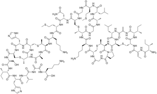
Nisin is a polycyclic antibacterial peptide produced by the bacterium Lactococcus lactis that is used as a food preservative. It has 34 amino acid residues, including the uncommon amino acids lanthionine (Lan), methyllanthionine (MeLan), didehydroalanine (Dha), and didehydroaminobutyric acid (Dhb). These unusual amino acids are introduced by posttranslational modification of the precursor peptide. In these reactions a ribosomally synthesized 57-mer is converted to the final peptide. The unsaturated amino acids originate from serine and threonine, and the enzyme-catalysed addition of cysteine residues to the didehydro amino acids result in the multiple (5) thioether bridges.
Lantibiotics are a class of polycyclic peptide antibiotics that contain the characteristic thioether amino acids lanthionine or methyllanthionine, as well as the unsaturated amino acids dehydroalanine, and 2-aminoisobutyric acid. They belong to ribosomally synthesized and post-translationally modified peptides.
The periplasm is a concentrated gel-like matrix in the space between the inner cytoplasmic membrane and the bacterial outer membrane called the periplasmic space in gram-negative bacteria. Using cryo-electron microscopy it has been found that a much smaller periplasmic space is also present in gram-positive bacteria, between cell wall and the plasma membrane. The periplasm may constitute up to 40% of the total cell volume of gram-negative bacteria, but is a much smaller percentage in gram-positive bacteria.
Polymyxins are antibiotics. Polymyxins B and E are used in the treatment of Gram-negative bacterial infections. They work mostly by breaking up the bacterial cell membrane. They are part of a broader class of molecules called nonribosomal peptides.
Antimicrobial peptides (AMPs), also called host defence peptides (HDPs) are part of the innate immune response found among all classes of life. Fundamental differences exist between prokaryotic and eukaryotic cells that may represent targets for antimicrobial peptides. These peptides are potent, broad spectrum antimicrobials which demonstrate potential as novel therapeutic agents. Antimicrobial peptides have been demonstrated to kill Gram negative and Gram positive bacteria, enveloped viruses, fungi and even transformed or cancerous cells. Unlike the majority of conventional antibiotics it appears that antimicrobial peptides frequently destabilize biological membranes, can form transmembrane channels, and may also have the ability to enhance immunity by functioning as immunomodulators.
Microcins are very small bacteriocins, composed of relatively few amino acids. For this reason, they are distinct from their larger protein cousins. The classic example is microcin V, of Escherichia coli. Subtilosin A is another bacteriocin from Bacillus subtilis. The peptide has a cyclized backbone and forms three cross-links between the sulphurs of Cys13, Cys7 and Cys4 and the alpha-positions of Phe22, Thr28 and Phe31.
Pseudomonas aeruginosa is a common encapsulated, Gram-negative, aerobic–facultatively anaerobic, rod-shaped bacterium that can cause disease in plants and animals, including humans. A species of considerable medical importance, P. aeruginosa is a multidrug resistant pathogen recognized for its ubiquity, its intrinsically advanced antibiotic resistance mechanisms, and its association with serious illnesses – hospital-acquired infections such as ventilator-associated pneumonia and various sepsis syndromes. P. aeruginosa is able to selectively inhibit various antibiotics from penetrating its outer membrane - and has high resistance to several antibiotics. According to the World Health Organization P. aeruginosa poses one of the greatest threats to humans in terms of antibiotic resistance.
Mutacin 1140 is a bacteriocin produced by Streptococcus mutans. It has activity against a broad spectrum of Gram-positive bacteria. It is a member of the class of compounds known as lantibiotics.
A colicin is a type of bacteriocin produced by and toxic to some strains of Escherichia coli. Colicins are released into the environment to reduce competition from other bacterial strains. Colicins bind to outer membrane receptors, using them to translocate to the cytoplasm or cytoplasmic membrane, where they exert their cytotoxic effect, including depolarisation of the cytoplasmic membrane, DNase activity, RNase activity, or inhibition of murein synthesis.
Class II bacteriocins are a class of small peptides that inhibit the growth of various bacteria.
Sakacins are bacteriocins produced by Lactobacillus sakei. They are often clustered with the other lactic acid bacteriocins. The best known sakacins are sakacin A, G, K, P, and Q. In particular, sakacin A and P have been well characterized.
ATP-dependent Clp protease proteolytic subunit (ClpP) is an enzyme that in humans is encoded by the CLPP gene. This protein is an essential component to form the protein complex of Clp protease.
Rhamnolipids are a class of glycolipid produced by Pseudomonas aeruginosa, amongst other organisms, frequently cited as bacterial surfactants. They have a glycosyl head group, in this case a rhamnose moiety, and a 3-(hydroxyalkanoyloxy)alkanoic acid (HAA) fatty acid tail, such as 3-hydroxydecanoic acid.
Bacterial morphological plasticity refers to changes in the shape and size that bacterial cells undergo when they encounter stressful environments. Although bacteria have evolved complex molecular strategies to maintain their shape, many are able to alter their shape as a survival strategy in response to protist predators, antibiotics, the immune response, and other threats.
Ribosomally synthesized and post-translationally modified peptides (RiPPs), also known as ribosomal natural products, are a diverse class of natural products of ribosomal origin. Consisting of more than 20 sub-classes, RiPPs are produced by a variety of organisms, including prokaryotes, eukaryotes, and archaea, and they possess a wide range of biological functions.
Resistance-nodulation-division (RND) family transporters are a category of bacterial efflux pumps, especially identified in Gram-negative bacteria and located in the cytoplasmic membrane, that actively transport substrates. The RND superfamily includes seven families: the heavy metal efflux (HME), the hydrophobe/amphiphile efflux-1, the nodulation factor exporter family (NFE), the SecDF protein-secretion accessory protein family, the hydrophobe/amphiphile efflux-2 family, the eukaryotic sterol homeostasis family, and the hydrophobe/amphiphile efflux-3 family. These RND systems are involved in maintaining homeostasis of the cell, removal of toxic compounds, and export of virulence determinants. They have a broad substrate spectrum and can lead to the diminished activity of unrelated drug classes if over-expressed. The first reports of drug resistant bacterial infections were reported in the 1940s after the first mass production of antibiotics. Most of the RND superfamily transport systems are made of large polypeptide chains. RND proteins exist primarily in gram-negative bacteria but can also be found in gram-positive bacteria, archaea, and eukaryotes.
Rhs toxins belong to the polymorphic toxin category of bacterial exotoxins. Rhs proteins are widespread and can be produced by both Gram-negative and Gram-positive bacteria. Rhs toxins are very large proteins of usually more than 1,500 aminoacids with variable C-terminal toxic domains. Their toxic activity can either target eukaryotes or other bacteria.
Bacterial secretion systems are protein complexes present on the cell membranes of bacteria for secretion of substances. Specifically, they are the cellular devices used by pathogenic bacteria to secrete their virulence factors to invade the host cells. They can be classified into different types based on their specific structure, composition and activity. Generally, proteins can be secreted through two different processes. One process is a one-step mechanism in which proteins from the cytoplasm of bacteria are transported and delivered directly through the cell membrane into the host cell. Another involves a two-step activity in which the proteins are first transported out of the inner cell membrane, then deposited in the periplasm, and finally through the outer cell membrane into the host cell.
Pyocins are bacteriocins produced by bacteria belonging to the Pseudomonas genus. François Jacob described the first pyocin in 1954. Pyocins can be divided into three distinct classes: S-type, R-type, and F-type pyocins. S-type pyocins are colicin-like bacteriocins as R-type and F-type pyocins belong to tailocins.