Biomarkers of aging

Last updated
Chromosomes during methylation Epigenetic mechanisms.png
Chromosomes during methylation

Biomarkers of aging are biomarkers that could predict functional capacity at some later age better than chronological age. [1] Stated another way, biomarkers of aging would give the true "biological age", which may be different from the chronological age.

Contents

Validated biomarkers of aging would allow for testing interventions to extend lifespan, because changes in the biomarkers would be observable throughout the lifespan of the organism. [1] Although maximum lifespan would be a means of validating biomarkers of aging, it would not be a practical means for long-lived species such as humans because longitudinal studies would take far too much time. [2] Ideally, biomarkers of aging should assay the biological process of aging and not a predisposition to disease, should cause a minimal amount of trauma to assay in the organism, and should be reproducibly measurable during a short interval compared to the lifespan of the organism. [1] An assemblage of biomarker data for an organism could be termed its "ageotype". [3]

Although graying of hair increases with age, [4] hair graying cannot be called a biomarker of ageing. Similarly, skin wrinkles and other common changes seen with aging are not better indicators of future functionality than chronological age. Biogerontologists have continued efforts to find and validate biomarkers of aging, but success thus far has been limited. Levels of CD4 and CD8 memory T cells and naive T cells have been used to give good predictions of the expected lifespan of middle-aged mice. [5]

Advances in big data analysis allowed for the new types of "aging clocks" to be developed. The epigenetic clock is a promising biomarker of aging and can accurately predict human chronological age. [6] Basic blood biochemistry and cell counts can also be used to accurately predict the chronological age. [7] Further studies of the hematological clock on the large datasets from South Korean, Canadian, and Eastern European populations demonstrated that biomarkers of aging may be population-specific and predictive of mortality. [8] It is also possible to predict the human chronological age using the transcriptomic clock [9] .

Epigenetic marks

Loss of histones

A new epigenetic mark found in studies of aging cells is the loss of histones. Most of the evidence shows that loss of histones is linked to cell division. In aging and dividing yeast MNase-seq (Micrococcal Nuclease sequencing) showed a loss of nucleosomes of ~50%. Proper histone dosage is important in yeast as shown from the extended lifespans seen in strains that are overexpressing histones. [10] A consequence of histone loss in yeast is the amplification of transcription. In younger cells, genes that are most induced with age have specific chromatin structures, such as fuzzy nuclear positioning, lack of a nucleosome depleted region (NDR) at the promoter, weak chromatin phasing, a higher frequency of TATA elements, and higher occupancy of repressive chromatin factors. In older cells, however, the same genes nucleosome loss at the promoter is more prevalent which leads to higher transcription of these genes. [10]

This phenomenon is not only seen in yeast, but has also been seen in aging worms, during aging of human diploid primary fibroblasts, and in senescent human cells. In human primary fibroblasts, reduced synthesis of new histones was seen to be a consequence of shortened telomeres that activate the DNA damage response. Loss of core histones may be a general epigenetic mark of aging across many organisms. [11]

Histone variants

In addition to the core histones, H2A, H2B, H3, and H4, there are other versions of the histone proteins that can be significantly different in their sequence and are important for regulating chromatin dynamics. Histone H3.3 is a variant of histone H3 that is incorporated into the genome independent of replication. It is the major form of histone H3 seen in the chromatin of senescent human cells, and it appears that excess H3.3 can drive senescence. [11]

There are multiple variants of histone 2, the one most notably implicated in aging is macroH2A. The function of macroH2A has generally been assumed to be transcriptional silencing; most recently, it has been suggested that macroH2A is important in repressing transcription at Senescence-Associated Heterochromatin Foci (SAHF). [11] Chromatin that contains macroH2A is impervious to ATP-dependent remodeling proteins and to the binding of transcription factors. [12]

Histone modifications

Increased acetylation of histones contributes to chromatin taking a more euchromatic state as an organism ages, similar to the increased transcription seen due to the loss of histones. [13] There is also a reduction in the levels of H3K56ac during aging and an increase in the levels of H4K16ac. [10] Increased H4K16ac in old yeast cells is associated with the decline in levels of the HDAC Sir2, which can increase the life span when overexpressed. [10]

Methylation of histones has been tied to life span regulation in many organisms, specifically H3K4me3, an activating mark, and H4K27me3, a repressing mark. In C. elegans, the loss of any of the three Trithorax proteins that catalyze the trimethylation of H3K4 such as, WDR-5 and the methyltransferases SET-2 and ASH-2, lowers the levels of H3K4me3 and increases lifespan. Loss of the enzyme that demethylates H3K4me3, RB-2, increases H3K4me3 levels in C. elegans and decreases their life spans. [13] In the rhesus macaque brain prefrontal cortex, H3K4me2 increases at promoters and enhancers during postnatal development and aging. [14] These increases reflect progressively more active and transcriptionally accessible (or open) chromatin structures that are often associated with stress responses such as the DNA damage response. These changes may form an epigenetic memory of stresses and damages experienced by the organism as it develops and ages. [14]

UTX-1, a H3K27me3 demethylase, plays a critical role in the aging of C.elegans: increased utx-1 expression correlates with a decrease in H3K27me3 and a decrease in lifespan. Utx-1 knockdowns showed an increase in lifespan [13] Changes in H3K27me3 levels also have affects on aging cells in Drosophila and humans.

DNA methylation

Methylation of DNA is a common modification in mammalian cells. The cytosine base is methylated and becomes 5-methylcytosine, most often when in the CpG context. Hypermethylation of CpG islands is associated with transcriptional repression and hypomethylation of these sites is associated with transcriptional activation. Many studies have shown that there is a loss of DNA methylation during ageing in many species such as, rats, mice, cows, hamsters, and humans. It has also been shown that DNMT1 and DNMT3a decrease with aging and DNMT3b increases. [15]

Hypomethylation of DNA can lower genomic stability, induce the reactivation of transposable elements, and cause the loss of imprinting, all of which can contribute to cancer progression and pathogenesis. [15]

Immune biomarkers

Recent data suggests that an increased frequency of senescent CD8+ T cells in the peripheral blood is associated with the development of hyperglycemia from a pre-diabetic state suggestive of senescence playing a role in metabolic aging. Senescent Cd8+ T cells could be utilized as a biomarker to signal the transition from pre-diabetes to overt hyperglycemia. [16]

Recently, Hashimoto and coworkers profiled thousands of circulating immune cells from supercentenarians at single-cell resolution. They identified a unique increase in cytotoxic CD4 T cells in these supercentenarians. Generally, CD4 T-cells have helper, but not cytotoxic, functions under physiological conditions however these supercentenarians, subjected to single cell profiling of their T-cell receptors, revealed accumulations of cytotoxic CD4 T-cells through clonal expansion. The conversion of helper CD4 T-cells to a cytotoxic variety might be an adaptation to the late stage of aging aiding in the fighting infections and potentially enhancing tumor surveillance. [17]

Applications of aging biomarkers

The main mechanisms identified as potential biomarkers of aging are DNA methylation, loss of histones, and histone modification. The uses for biomarkers of aging are ubiquitous and identifying a physical parameter of biological aging would allow humans to determine our true age, mortality, and morbidity. [10] The change in the physical biomarker should be proportional to the change in the age of the species. Thus after establishing a biomarker of aging, humans would be able to dive into research on extending life spans and finding timelines for the arise of potential genetic diseases.

One of the applications of this finding would allow for identification of the biological age of a person. DNA methylation uses the structure of dna at different stages of life to determine an age. DNA methylation is the methylation of the cysteine in the CG or Cpg region. The hypermethylation of this region is associated with decreased transcriptional activity and the opposite for hypomethylation. In other words, the more "tightly" held the DNA region then the more stable and "younger" the species. Looking at DNA methylation's properties in tissues, it was found to be almost zero for embryonic tissues, it can be used to determine acceleration of age and the results can be reproduced in chimpanzee tissue. [18]

More recently, biomarkers of aging has been used in multiple clinical trials to measure slowing or reversing of age-related decline or biological aging. [19] The Biomarkers of Aging Consortium (https://www.agingconsortium.org) is currently examining the application of these biomarkers to identify longevity interventions and ways to validate them. [20] Moreover, open-source resources, such as the R package methylCIPHER [21] and the Python package pyaging [22] are available to the public as hubs for several biomarkers of aging.

See also

Related Research Articles

<span class="mw-page-title-main">Histone</span> Protein family around which DNA winds to form nucleosomes

In biology, histones are highly basic proteins abundant in lysine and arginine residues that are found in eukaryotic cell nuclei and in most Archaeal phyla. They act as spools around which DNA winds to create structural units called nucleosomes. Nucleosomes in turn are wrapped into 30-nanometer fibers that form tightly packed chromatin. Histones prevent DNA from becoming tangled and protect it from DNA damage. In addition, histones play important roles in gene regulation and DNA replication. Without histones, unwound DNA in chromosomes would be very long. For example, each human cell has about 1.8 meters of DNA if completely stretched out; however, when wound about histones, this length is reduced to about 90 micrometers (0.09 mm) of 30 nm diameter chromatin fibers.

<span class="mw-page-title-main">Cellular differentiation</span> Developmental biology

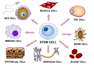
Cellular differentiation is the process in which a stem cell changes from one type to a differentiated one. Usually, the cell changes to a more specialized type. Differentiation happens multiple times during the development of a multicellular organism as it changes from a simple zygote to a complex system of tissues and cell types. Differentiation continues in adulthood as adult stem cells divide and create fully differentiated daughter cells during tissue repair and during normal cell turnover. Some differentiation occurs in response to antigen exposure. Differentiation dramatically changes a cell's size, shape, membrane potential, metabolic activity, and responsiveness to signals. These changes are largely due to highly controlled modifications in gene expression and are the study of epigenetics. With a few exceptions, cellular differentiation almost never involves a change in the DNA sequence itself. However, metabolic composition does get altered quite dramatically where stem cells are characterized by abundant metabolites with highly unsaturated structures whose levels decrease upon differentiation. Thus, different cells can have very different physical characteristics despite having the same genome.

<span class="mw-page-title-main">Histone H4</span> One of the five main histone proteins involved in the structure of chromatin

Histone H4 is one of the five main histone proteins involved in the structure of chromatin in eukaryotic cells. Featuring a main globular domain and a long N-terminal tail, H4 is involved with the structure of the nucleosome of the 'beads on a string' organization. Histone proteins are highly post-translationally modified. Covalently bonded modifications include acetylation and methylation of the N-terminal tails. These modifications may alter expression of genes located on DNA associated with its parent histone octamer. Histone H4 is an important protein in the structure and function of chromatin, where its sequence variants and variable modification states are thought to play a role in the dynamic and long term regulation of genes.

Histone methylation is a process by which methyl groups are transferred to amino acids of histone proteins that make up nucleosomes, which the DNA double helix wraps around to form chromosomes. Methylation of histones can either increase or decrease transcription of genes, depending on which amino acids in the histones are methylated, and how many methyl groups are attached. Methylation events that weaken chemical attractions between histone tails and DNA increase transcription because they enable the DNA to uncoil from nucleosomes so that transcription factor proteins and RNA polymerase can access the DNA. This process is critical for the regulation of gene expression that allows different cells to express different genes.

<span class="mw-page-title-main">Histone-modifying enzymes</span> Type of enzymes

Histone-modifying enzymes are enzymes involved in the modification of histone substrates after protein translation and affect cellular processes including gene expression. To safely store the eukaryotic genome, DNA is wrapped around four core histone proteins, which then join to form nucleosomes. These nucleosomes further fold together into highly condensed chromatin, which renders the organism's genetic material far less accessible to the factors required for gene transcription, DNA replication, recombination and repair. Subsequently, eukaryotic organisms have developed intricate mechanisms to overcome this repressive barrier imposed by the chromatin through histone modification, a type of post-translational modification which typically involves covalently attaching certain groups to histone residues. Once added to the histone, these groups elicit either a loose and open histone conformation, euchromatin, or a tight and closed histone conformation, heterochromatin. Euchromatin marks active transcription and gene expression, as the light packing of histones in this way allows entry for proteins involved in the transcription process. As such, the tightly packed heterochromatin marks the absence of current gene expression.

Chromatin remodeling is the dynamic modification of chromatin architecture to allow access of condensed genomic DNA to the regulatory transcription machinery proteins, and thereby control gene expression. Such remodeling is principally carried out by 1) covalent histone modifications by specific enzymes, e.g., histone acetyltransferases (HATs), deacetylases, methyltransferases, and kinases, and 2) ATP-dependent chromatin remodeling complexes which either move, eject or restructure nucleosomes. Besides actively regulating gene expression, dynamic remodeling of chromatin imparts an epigenetic regulatory role in several key biological processes, egg cells DNA replication and repair; apoptosis; chromosome segregation as well as development and pluripotency. Aberrations in chromatin remodeling proteins are found to be associated with human diseases, including cancer. Targeting chromatin remodeling pathways is currently evolving as a major therapeutic strategy in the treatment of several cancers.

An epigenetic clock is a biochemical test that can be used to measure age. The test is based on DNA methylation levels, measuring the accumulation of methyl groups to one's DNA molecules.

H3K4me3 is an epigenetic modification to the DNA packaging protein Histone H3 that indicates tri-methylation at the 4th lysine residue of the histone H3 protein and is often involved in the regulation of gene expression. The name denotes the addition of three methyl groups (trimethylation) to the lysine 4 on the histone H3 protein.

H3K27me3 is an epigenetic modification to the DNA packaging protein Histone H3. It is a mark that indicates the tri-methylation of lysine 27 on histone H3 protein.

H3K9me3 is an epigenetic modification to the DNA packaging protein Histone H3. It is a mark that indicates the tri-methylation at the 9th lysine residue of the histone H3 protein and is often associated with heterochromatin.

H3K9me2 is an epigenetic modification to the DNA packaging protein Histone H3. It is a mark that indicates the di-methylation at the 9th lysine residue of the histone H3 protein. H3K9me2 is strongly associated with transcriptional repression. H3K9me2 levels are higher at silent compared to active genes in a 10kb region surrounding the transcriptional start site. H3K9me2 represses gene expression both passively, by prohibiting acetylation as therefore binding of RNA polymerase or its regulatory factors, and actively, by recruiting transcriptional repressors. H3K9me2 has also been found in megabase blocks, termed Large Organised Chromatin K9 domains (LOCKS), which are primarily located within gene-sparse regions but also encompass genic and intergenic intervals. Its synthesis is catalyzed by G9a, G9a-like protein, and PRDM2. H3K9me2 can be removed by a wide range of histone lysine demethylases (KDMs) including KDM1, KDM3, KDM4 and KDM7 family members. H3K9me2 is important for various biological processes including cell lineage commitment, the reprogramming of somatic cells to induced pluripotent stem cells, regulation of the inflammatory response, and addiction to drug use.

H3K4me1 is an epigenetic modification to the DNA packaging protein Histone H3. It is a mark that indicates the mono-methylation at the 4th lysine residue of the histone H3 protein and often associated with gene enhancers.

H3K36me3 is an epigenetic modification to the DNA packaging protein Histone H3. It is a mark that indicates the tri-methylation at the 36th lysine residue of the histone H3 protein and often associated with gene bodies.

H4K20me is an epigenetic modification to the DNA packaging protein Histone H4. It is a mark that indicates the mono-methylation at the 20th lysine residue of the histone H4 protein. This mark can be di- and tri-methylated. It is critical for genome integrity including DNA damage repair, DNA replication and chromatin compaction.

H4K16ac is an epigenetic modification to the DNA packaging protein Histone H4. It is a mark that indicates the acetylation at the 16th lysine residue of the histone H4 protein.

H3K36me2 is an epigenetic modification to the DNA packaging protein Histone H3. It is a mark that indicates the di-methylation at the 36th lysine residue of the histone H3 protein.

H3K36me is an epigenetic modification to the DNA packaging protein Histone H3, specifically, the mono-methylation at the 36th lysine residue of the histone H3 protein.

H3R42me is an epigenetic modification to the DNA packaging protein histone H3. It is a mark that indicates the mono-methylation at the 42nd arginine residue of the histone H3 protein. In epigenetics, arginine methylation of histones H3 and H4 is associated with a more accessible chromatin structure and thus higher levels of transcription. The existence of arginine demethylases that could reverse arginine methylation is controversial.

H3R2me2 is an epigenetic modification to the DNA packaging protein histone H3. It is a mark that indicates the di-methylation at the 2nd arginine residue of the histone H3 protein. In epigenetics, arginine methylation of histones H3 and H4 is associated with a more accessible chromatin structure and thus higher levels of transcription. The existence of arginine demethylases that could reverse arginine methylation is controversial.

H3T11P is an epigenetic modification to the DNA packaging protein histone H3. It is a mark that indicates the phosphorylation the 11th threonine residue of the histone H3 protein.

References

  1. 1 2 3 Baker GT, Sprott RL (1988). "Biomarkers of aging". Experimental Gerontology. 23 (4–5): 223–39. doi:10.1016/0531-5565(88)90025-3. PMID   3058488. S2CID   31039588.
  2. Harrison, Ph.D., David E. (November 11, 2011). "V. Life span as a biomarker". Jackson Laboratory. Archived from the original on April 26, 2012. Retrieved 2011-12-03.
  3. Ahadi, Sara; Zhou, Wenyu; Schüssler-Fiorenza Rose, Sophia Miryam; Sailani, M. Reza; Contrepois, Kévin; Avina, Monika; Ashland, Melanie; Brunet, Anne; Snyder, Michael (2020). "Personal aging markers and ageotypes revealed by deep longitudinal profiling". Nature Medicine. 26 (1): 83–90. doi:10.1038/s41591-019-0719-5. PMC   7301912 . PMID   31932806.
  4. Van Neste D, Tobin DJ (2004). "Hair cycle and hair pigmentation: dynamic interactions and changes associated with aging". Micron. 35 (3): 193–200. doi:10.1016/j.micron.2003.11.006. PMID   15036274.
  5. Miller RA (April 2001). "Biomarkers of aging: prediction of longevity by using age-sensitive T-cell subset determinations in a middle-aged, genetically heterogeneous mouse population". The Journals of Gerontology. Series A, Biological Sciences and Medical Sciences. 56 (4): B180-6. doi: 10.1093/gerona/56.4.b180 . PMC   7537444 . PMID   11283189.
  6. Horvath S (2013). "DNA methylation age of human tissues and cell types". Genome Biology. 14 (10): R115. doi: 10.1186/gb-2013-14-10-r115 . PMC   4015143 . PMID   24138928.
  7. Putin E, Mamoshina P, Aliper A, Korzinkin M, Moskalev A, Kolosov A, Ostrovskiy A, Cantor C, Vijg J, Zhavoronkov A (May 2016). "Deep biomarkers of human aging: Application of deep neural networks to biomarker development". Aging. 8 (5): 1021–33. doi:10.18632/aging.100968. PMC   4931851 . PMID   27191382.
  8. Mamoshina P, Kochetov K, Putin E, Cortese F, Aliper A, Lee WS, Ahn SM, Uhn L, Skjodt N, Kovalchuk O, Scheibye-Knudsen M, Zhavoronkov A (October 2018). "Population Specific Biomarkers of Human Aging: A Big Data Study Using South Korean, Canadian, and Eastern European Patient Populations". The Journals of Gerontology. Series A, Biological Sciences and Medical Sciences. 73 (11): 1482–1490. doi:10.1093/gerona/gly005. PMC   6175034 . PMID   29340580.
  9. Peters MJ, Joehanes R, Pilling LC, Schurmann C, Conneely KN, Powell J, et al. (October 2015). "The transcriptional landscape of age in human peripheral blood". Nature Communications. 6: 8570. Bibcode:2015NatCo...6.8570.. doi:10.1038/ncomms9570. PMC   4639797 . PMID   26490707.
  10. 1 2 3 4 5 Sen P, Shah PP, Nativio R, Berger SL (August 2016). "Epigenetic Mechanisms of Longevity and Aging". Cell. 166 (4): 822–839. doi:10.1016/j.cell.2016.07.050. PMC   5821249 . PMID   27518561.
  11. 1 2 3 Pal S, Tyler JK (July 2016). "Epigenetics and aging". Science Advances. 2 (7): e1600584. Bibcode:2016SciA....2E0584P. doi:10.1126/sciadv.1600584. PMC   4966880 . PMID   27482540.
  12. Zhang R, Poustovoitov MV, Ye X, Santos HA, Chen W, Daganzo SM, et al. (January 2005). "Formation of MacroH2A-containing senescence-associated heterochromatin foci and senescence driven by ASF1a and HIRA". Developmental Cell. 8 (1): 19–30. doi: 10.1016/j.devcel.2004.10.019 . PMID   15621527.
  13. 1 2 3 McCauley BS, Dang W (December 2014). "Histone methylation and aging: lessons learned from model systems". Biochimica et Biophysica Acta (BBA) - Gene Regulatory Mechanisms. 1839 (12): 1454–62. doi:10.1016/j.bbagrm.2014.05.008. PMC   4240748 . PMID   24859460.
  14. 1 2 Han Y, Han D, Yan Z, Boyd-Kirkup JD, Green CD, Khaitovich P, Han JD (December 2012). "Stress-associated H3K4 methylation accumulates during postnatal development and aging of rhesus macaque brain". Aging Cell. 11 (6): 1055–64. doi: 10.1111/acel.12007 . PMID   22978322. S2CID   17523080.
  15. 1 2 Lillycrop KA, Hoile SP, Grenfell L, Burdge GC (August 2014). "DNA methylation, ageing and the influence of early life nutrition". The Proceedings of the Nutrition Society. 73 (3): 413–21. doi: 10.1017/S0029665114000081 . PMID   25027290.
  16. Lee, Yong-ho; Kim, So Ra; Han, Dai Hoon; Yu, Hee Tae; Han, Yoon Dae; Kim, Jin Hee; Kim, Soo Hyun; Lee, Chan Joo; Min, Byoung-Hoon; Kim, Dong-Hyun; Kim, Kyung Hwan (2018-11-02). "Senescent T Cells Predict the Development of Hyperglycemia in Humans". Diabetes. 68 (1): 156–162. doi: 10.2337/db17-1218 . ISSN   0012-1797. PMID   30389747.
  17. Hashimoto, Kosuke; Kouno, Tsukasa; Ikawa, Tomokatsu; Hayatsu, Norihito; Miyajima, Yurina; Yabukami, Haruka; Terooatea, Tommy; Sasaki, Takashi; Suzuki, Takahiro (2019-05-20). "Single-cell transcriptomics reveals expansion of cytotoxic CD4 T-cells in supercentenarians". Proceedings of the National Academy of Sciences of the United States of America. 116 (48): 24242–24251. Bibcode:2019PNAS..11624242H. bioRxiv   10.1101/643528 . doi: 10.1073/pnas.1907883116 . PMC   6883788 . PMID   31719197.
  18. Horvath, Steve (2013). "DNA methylation age of human tissues and cell types". Genome Biology. 14 (10): R115. doi: 10.1186/gb-2013-14-10-r115 . ISSN   1465-6906. PMC   4015143 . PMID   24138928.
  19. Moqri, Mahdi; Herzog, Chiara; Poganik, Jesse R.; Justice, Jamie; Belsky, Daniel W.; Higgins-Chen, Albert; Moskalev, Alexey; Fuellen, Georg; Cohen, Alan A.; Bautmans, Ivan; Widschwendter, Martin; Ding, Jingzhong; Fleming, Alexander; Mannick, Joan; Han, Jing-Dong Jackie; Zhavoronkov, Alex; Barzilai, Nir; Kaeberlein, Matt; Cummings, Steven; Kennedy, Brian K.; Ferrucci, Luigi; Horvath, Steve; Verdin, Eric; Maier, Andrea B.; Snyder, Michael P.; Sebastiano, Vittorio; Gladyshev, Vadim N.; Gladyshev, V. N. (2023). "Biomarkers of aging for the identification and evaluation of longevity interventions". Cell. 186 (18): 3758–3775. doi:10.1016/j.cell.2023.08.003. PMID   37657418.
  20. Moqri, Mahdi; Herzog, Chiara; Poganik, Jesse R.; Ying, Kejun; Justice, Jamie N.; Belsky, Daniel W.; Higgins-Chen, Albert T.; Chen, Brian H.; Cohen, Alan A.; Fuellen, Georg; Hägg, Sara; Marioni, Riccardo E.; Widschwendter, Martin; Fortney, Kristen; Fedichev, Peter O. (February 2024). "Validation of biomarkers of aging". Nature Medicine. 30 (2): 360–372. doi:10.1038/s41591-023-02784-9. ISSN   1546-170X. PMID   38355974.
  21. Thrush, Kyra L.; Higgins-Chen, Albert T.; Liu, Zuyun; Levine, Morgan E. (2022). "bioRxiv". doi:10.1101/2022.07.13.499978.{{cite journal}}: Cite journal requires |journal= (help)
  22. Camillo, Lucas Paulo de Lima (2024). "pyaging: a Python-based compendium of GPU-optimized aging clocks". Bioinformatics. 40 (btae200): btae200. doi: 10.1093/bioinformatics/btae200 . PMC   11058068 . PMID   38603598.