Blastocyst

Last updated
Blastocyst
Diagram of Blastocyst stage.png
Blastocyst just before implantation
Human blastocyst.jpg
A human blastocyst, with inner cell mass at upper right
Details
Carnegie stage 3
Days5–9
Gives rise to Gastrula
Identifiers
Latin blastocystis
MeSH D001755
TE E2.0.1.2.0.0.12
FMA 83041
Anatomical terminology

The blastocyst is a structure formed in the early embryonic development of mammals. It possesses an inner cell mass (ICM) also known as the embryoblast which subsequently forms the embryo, and an outer layer of trophoblast cells called the trophectoderm. [1] [2] This layer surrounds the inner cell mass and a fluid-filled cavity known as the blastocoel. [3] In the late blastocyst, the trophectoderm is known as the trophoblast. [2] The trophoblast gives rise to the chorion and amnion, the two fetal membranes that surround the embryo. The placenta derives from the embryonic chorion (the portion of the chorion that develops villi) and the underlying uterine tissue of the mother. [4] [5]

Contents

The name "blastocyst" arises from the Greek βλαστόςblastós ("a sprout") and κύστιςkýstis ("bladder, capsule").

In non-mammalian animals, this is a structure consisting of an undifferentiated ball of cells and is called a blastula.

In humans, blastocyst formation begins about five days after fertilization when a fluid-filled cavity opens up in the morula, the early embryonic stage of a ball of 16 cells. The blastocyst has a diameter of about 0.1–0.2 mm and comprises 200–300 cells (32 mitotic divisions ) following rapid cleavage (cell division). About seven days after fertilization, [6] the blastocyst undergoes implantation, embedding into the endometrium of the uterine wall where it will undergo further developmental processes, including gastrulation. Embedding of the blastocyst into the endometrium requires that it hatches from the zona pellucida, the egg coat that prevents adherence to the fallopian tube as the pre-embryo makes its way to the uterus.

The use of blastocysts in in vitro fertilization (IVF) involves culturing a fertilized egg for five days before transferring it into the uterus. It can be a more viable method of fertility treatment than traditional IVF. The inner cell mass of blastocysts is the source of embryonic stem cells, which are broadly applicable in stem cell therapies including cell repair, replacement and regeneration. Assisted zona hatching may also be used in IVF, and other fertility treatments.

Development cycle

The blastocyst stage occurs between 5 and 9 days after conception. During embryonic development, after fertilization (approximately 5–6 days in the human), the cells of the morula begin to undergo cell differentiation, and the morula changes into the blastocyst. In the uterus the zona pellucida surrounding the blastocyst breaks down, allowing it to implant into the uterine wall. Implantation marks the end of the germinal stage of embryogenesis, and the beginning of gestation.[ medical citation needed ]

Blastocyst formation

Early development of the human embryo from ovulation through implantation 2904 Preembryonic Development-02.jpg
Early development of the human embryo from ovulation through implantation

The zygote develops by mitosis, and when it has developed into a compacted ball of 8-16 cells becomes known as the morula. Until this stage in development, all cells (blastomeres) are autonomous and not specified to any particular subsequent functional development. The morula then develops by cavitation to become the blastocyst, or in many other animals the blastula. Cellular differentiation then develops the morula's cells into two types: trophoblast cells that surround the blastocoel and an inner mass of cells (the embryoblast). The inner cell mass consists of embryonic stem cells. [7] The conceptus is then known as the blastocyst. [8]

Before cell differentiation takes place there are two transcription factors, Oct-4 and nanog that are uniformly expressed on all of the cells, but both of these transcription factors are turned off in the trophoblast once it has formed. [9] The side of the blastocyst where the inner cell mass forms is called the embryonic pole, and the opposite side is the abembryonic pole. The outer layer of trophoblast cells, resulting from compaction, pumps sodium ions into the blastocyst, which causes water to enter through osmosis and form the internal fluid-filled blastocyst cavity (blastocoel). The blastocoel, trophoblast cells, and inner cell mass are hallmarks of the blastocyst. [10]

Implantation

Implantation is critical to the survival and development of the early human embryo. It establishes a connection between the mother and the early embryo which will continue through the remainder of the pregnancy. Implantation is made possible through structural changes in both the blastocyst and endometrial wall. [11] The zona pellucida surrounding the blastocyst breaches, referred to as hatching. This removes the constraint on the physical size of the embryonic mass and exposes the outer cells of the blastocyst to the interior of the uterus. Furthermore, hormonal changes in the mother, specifically a peak in luteinizing hormone (LH), prepare the endometrium to receive and envelop the blastocyst. The immune system is also modulated to allow for the invasion of the foreign embryonic cells. Once bound to the extracellular matrix of the endometrium, trophoblast cells secrete enzymes and other factors to embed the blastocyst into the uterine wall. The enzymes released degrade the endometrial lining, while autocrine growth factors such as human chorionic gonadotropin (hCG) and insulin-like growth factor (IGF) allow the blastocyst to further invade the endometrium. [12]

Implantation in the uterine wall allows for the next step in embryogenesis, gastrulation, which includes the formation of the placenta from trophoblastic cells and differentiation of the inner cell mass into the amniotic sac and epiblast.

Structure

There are two types of blastomere cells: [13]

The blastocoel fluid cavity contains amino acids, growth factors, and other molecules necessary for cellular differentiation. [17]

Cell specification

Multiple processes control cell lineage specification in the blastocyst to produce the trophoblast, epiblast, and primitive endoderm. These processes include gene expression, cell signaling, cell-cell contact and positional relationships, and epigenetics.

Once the inner cell mass has been established within the blastocyst, it prepares for further specification into the epiblast and primitive endoderm. This process of specification known as cell fate determination is carried out in part by fibroblast growth factor (FGF) signaling which generates a MAP kinase pathway to alter cellular genomes. [18] Further segregation of blastomeres into the trophectoderm and inner cell mass are regulated by the homeodomain protein, Cdx2. This transcription factor represses the expression of Oct4 and Nanog transcription factors in the trophoblast. [19] These genomic alterations allow for the progressive specification of both epiblast and primitive endoderm lineages at the end of the blastocyst phase of development preceding gastrulation. Much of the research conducted on these early embryonic stages is on mouse embryos and specific factors may differ between mammals.

During implantation, the trophoblast gives rise to extraembryonic membranes and cell types that will eventually form most of the fetal placenta, the specialized organ through which the embryo obtains maternal nourishment necessary for subsequent exponential growth. [20] The specification of the trophoblast is controlled by the combination of morphological cues arising from cell polarity with differential activity of signaling pathways such as Hippo and Notch, and the restriction to outer cells of lineage specifiers such as CDX2. [21]

In the mouse, primordial germ cells are specified from epiblast cells, a process that is accompanied by extensive genome-wide epigenetic reprogramming. [22] Reprogramming involves global DNA demethylation and chromatin reorganization resulting in cellular totipotency. [22] The process of genome-wide demethylation involves the DNA base excision repair pathway. [23]

Trophoblasts express integrin on their cell surfaces which allow for adhesion to the extracellular matrix of the uterine wall. This interaction allows for implantation and triggers further specification into the three different cell types, preparing the blastocyst for gastrulation. [24]

Clinical implications

Pregnancy tests

The level of human chorionic gonadotropin (hCG) secreted by the blastocyst during implantation is the factor measured in a pregnancy test. hCG can be measured in both blood and urine to determine whether a woman is pregnant. More hCG is secreted in a multiple pregnancy. Blood tests of hCG can also be used to check for abnormal pregnancies.

In vitro fertilization

In vitro fertilization (IVF) is an alternative to traditional in vivo fertilization for fertilizing an egg with sperm and implanting that embryo into a female's womb. For many years the embryo was inserted into the uterus two to three days after fertilization. However at this stage of development it is very difficult to predict which embryos will develop best, and several embryos were typically implanted. Several implanted embryos increased the likelihood of a developing fetus but also led to the development of multiple fetuses. This was a major problem and drawback for using embryos in IVF.

The use of blastocysts for human IVF has proved successful. A blastocyst is implanted five to six days after the eggs have been fertilized. [25] After five or six days it is much easier to determine which embryos will result in healthy live births. Knowing which embryos will succeed allows just one blastocyst to be implanted, cutting down dramatically on the health risk and expense of multiple births. Now that the nutrient requirements for embryonic and blastocyst development have been determined, it is much easier to give embryos the correct nutrients to sustain them into the blastocyst phase.

Embryo transfer following in vitro fertilization is a procedure in which a catheter is inserted into the vagina, guided through the cervix via ultrasound, and into the uterine cavity where the blastocysts are inserted into the womb.

Blastocysts also offer an advantage because they can be used to genetically test the cells to check for genetic problems. There are enough cells in a blastocyst that a few trophectoderm cells can be removed without disturbing the developing blastocyst. These cells can be tested for chromosome aneuploidy using preimplantation genetic screening (PGS), or specific conditions such as cystic fibrosis, often known as preimplantation genetic diagnosis (PGD). [26]

Embryo transfer process

In an embryo transfer procedure following an initial ultrasound, a speculum is used to open the walls of the vagina, and using a catheter an embryo is passed through the tube for placement into the womb.

See also

Related Research Articles

<span class="mw-page-title-main">Pregnancy (mammals)</span> Period of reproduction

In mammals, pregnancy is the period of reproduction during which a female carries one or more live offspring from implantation in the uterus through gestation. It begins when a fertilized zygote implants in the female's uterus, and ends once it leaves the uterus.

<span class="mw-page-title-main">Embryo</span> Multicellular diploid eukaryote in its earliest stage of development

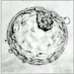
An embryo is an initial stage of development of a multicellular organism. In organisms that reproduce sexually, embryonic development is the part of the life cycle that begins just after fertilization of the female egg cell by the male sperm cell. The resulting fusion of these two cells produces a single-celled zygote that undergoes many cell divisions that produce cells known as blastomeres. The blastomeres are arranged as a solid ball that when reaching a certain size, called a morula, takes in fluid to create a cavity called a blastocoel. The structure is then termed a blastula, or a blastocyst in mammals.

The amniotic sac, also called the bag of waters or the membranes, is the sac in which the embryo and later fetus develops in amniotes. It is a thin but tough transparent pair of membranes that hold a developing embryo until shortly before birth. The inner of these membranes, the amnion, encloses the amniotic cavity, containing the amniotic fluid and the embryo. The outer membrane, the chorion, contains the amnion and is part of the placenta. On the outer side, the amniotic sac is connected to the yolk sac, the allantois, and via the umbilical cord, the placenta.

<span class="mw-page-title-main">Blastulation</span> Sphere of cells formed during early embryonic development in animals

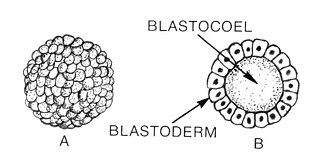
Blastulation is the stage in early animal embryonic development that produces the blastula. In mammalian development the blastula develops into the blastocyst with a differentiated inner cell mass and an outer trophectoderm. The blastula is a hollow sphere of cells known as blastomeres surrounding an inner fluid-filled cavity called the blastocoel. Embryonic development begins with a sperm fertilizing an egg cell to become a zygote, which undergoes many cleavages to develop into a ball of cells called a morula. Only when the blastocoel is formed does the early embryo become a blastula. The blastula precedes the formation of the gastrula in which the germ layers of the embryo form.

<span class="mw-page-title-main">Blastocoel</span> Fluid-filled or yolk-filled cavity that forms in the blastula

The blastocoel, also spelled blastocoele and blastocele, and also called cleavage cavity, or segmentation cavity is a fluid-filled or yolk-filled cavity that forms in the blastula during very early embryonic development. At this stage in mammals the blastula develops into the blastocyst containing an inner cell mass, and outer trophectoderm.

<span class="mw-page-title-main">Trophoblast</span> Early embryonic structure that gives rise to the placenta

The trophoblast is the outer layer of cells of the blastocyst. Trophoblasts are present four days after fertilization in humans. They provide nutrients to the embryo and develop into a large part of the placenta. They form during the first stage of pregnancy and are the first cells to differentiate from the fertilized egg to become extraembryonic structures that do not directly contribute to the embryo. After blastulation, the trophoblast is contiguous with the ectoderm of the embryo and is referred to as the trophectoderm. After the first differentiation, the cells in the human embryo lose their totipotency because they can no longer form a trophoblast. They become pluripotent stem cells.

A germ layer is a primary layer of cells that forms during embryonic development. The three germ layers in vertebrates are particularly pronounced; however, all eumetazoans produce two or three primary germ layers. Some animals, like cnidarians, produce two germ layers making them diploblastic. Other animals such as bilaterians produce a third layer between these two layers, making them triploblastic. Germ layers eventually give rise to all of an animal's tissues and organs through the process of organogenesis.

<span class="mw-page-title-main">Animal embryonic development</span> Process by which the embryo forms and develops

In developmental biology, animal embryonic development, also known as animal embryogenesis, is the developmental stage of an animal embryo. Embryonic development starts with the fertilization of an egg cell (ovum) by a sperm cell, (spermatozoon). Once fertilized, the ovum becomes a single diploid cell known as a zygote. The zygote undergoes mitotic divisions with no significant growth and cellular differentiation, leading to development of a multicellular embryo after passing through an organizational checkpoint during mid-embryogenesis. In mammals, the term refers chiefly to the early stages of prenatal development, whereas the terms fetus and fetal development describe later stages.

<span class="mw-page-title-main">Cytotrophoblast</span>

"Cytotrophoblast" is the name given to both the inner layer of the trophoblast or the cells that live there. It is interior to the syncytiotrophoblast and external to the wall of the blastocyst in a developing embryo.

In embryology, Carnegie stages are a standardized system of 23 stages used to provide a unified developmental chronology of the vertebrate embryo.

<span class="mw-page-title-main">Inner cell mass</span> Early embryonic mass that gives rise to the fetus

The inner cell mass (ICM) or embryoblast is a structure in the early development of an embryo. It is the mass of cells inside the blastocyst that will eventually give rise to the definitive structures of the fetus. The inner cell mass forms in the earliest stages of embryonic development, before implantation into the endometrium of the uterus. The ICM is entirely surrounded by the single layer of trophoblast cells of the trophectoderm.

<span class="mw-page-title-main">Epiblast</span> Embryonic inner cell mass tissue that forms the embryo itself, through the three germ layers

In amniote embryonic development, the epiblast is one of two distinct cell layers arising from the inner cell mass in the mammalian blastocyst, or from the blastula in reptiles and birds, the other layer is the hypoblast. It drives the embryo proper through its differentiation into the three primary germ layers, ectoderm, mesoderm and endoderm, during gastrulation. The amniotic ectoderm and extraembryonic mesoderm also originate from the epiblast.

<span class="mw-page-title-main">Implantation (embryology)</span> First stage of pregnancy

Implantation, also known as nidation, is the stage in the embryonic development of mammals in which the blastocyst hatches, attaches, adheres, and invades into the wall of the female's uterus. Implantation is the first stage of gestation, and, when successful, the female is considered to be pregnant. An implanted embryo is detected by the presence of increased levels of human chorionic gonadotropin (hCG) in a pregnancy test. The implanted embryo will receive oxygen and nutrients in order to grow.

<span class="mw-page-title-main">Bilaminar embryonic disc</span>

The bilaminar embryonic disc, bilaminar blastoderm or embryonic disc is the distinct two-layered structure of cells formed in an embryo. In the development of the human embryo this takes place by day eight. It is formed when the inner cell mass, also known as the embryoblast, forms a bilaminar disc of two layers, an upper layer called the epiblast and a lower layer called the hypoblast, which will eventually form into fetus. These two layers of cells are stretched between two fluid-filled cavities at either end: the primitive yolk sac and the amniotic sac.

<span class="mw-page-title-main">Human embryonic development</span> Development and formation of the human embryo

Human embryonic development or human embryogenesis is the development and formation of the human embryo. It is characterised by the processes of cell division and cellular differentiation of the embryo that occurs during the early stages of development. In biological terms, the development of the human body entails growth from a one-celled zygote to an adult human being. Fertilization occurs when the sperm cell successfully enters and fuses with an egg cell (ovum). The genetic material of the sperm and egg then combine to form the single cell zygote and the germinal stage of development commences. Embryonic development in the human, covers the first eight weeks of development; at the beginning of the ninth week the embryo is termed a fetus. The eight weeks has 23 stages.

<span class="mw-page-title-main">Hypoblast</span> Embryonic inner cell mass tissue that forms the yolk sac and, later, chorion

In amniote embryology, the hypoblast is one of two distinct layers arising from the inner cell mass in the mammalian blastocyst, or from the blastodisc in reptiles and birds. The hypoblast gives rise to the yolk sac, which in turn gives rise to the chorion.

Morphokinetics (‘morpho’’ form/shape and ‘kinetics’ movement) refers to time specific morphological changes during embryo development providing dynamic information on a fertilized egg. The detailed information eases morphological selection of embryos with high implantation potential to be used in In-Vitro Fertilisation treatment.

<span class="mw-page-title-main">Preimplantation factor</span> Peptide involved in placental development

Preimplantation factor(PIF) is a peptide secreted by trophoblast cells prior to placenta formation in early embryonic development. Human embryos begin to express PIF at the 4-cell stage, with expression increasing by the morula stage and continuing to do so throughout the first trimester. Expression of preimplantation factor in the blastocyst was discovered as an early correlate of the viability of the eventual pregnancy. Preimplantation factor was identified in 1994 by a lymphocyte platelet-binding assay, where it was thought to be an early biomarker of pregnancy. It has a simple primary structure with a short sequence of fifteen amino acids without any known quaternary structure. A synthetic analogue of preimplantation factor (commonly abbreviated in studies as sPIF or PIF*) that has an identical amino acid sequence and mimics the normal biological activity of PIF has been developed and is commonly used in research studies, particularly those that aim to study potential adult therapeutics.

Reichert's membrane is an extraembryonic membrane that forms during early mammalian embryonic development. It forms as a thickened basement membrane to cover the embryo immediately following implantation to give protection to the embryo from the uterine pressures exerted. Reichert's membrane is also important for the maternofetal exchange of nutrients. The membrane collapses once the placenta has fully developed.

This glossary of developmental biology is a list of definitions of terms and concepts commonly used in the study of developmental biology and related disciplines in biology, including embryology and reproductive biology, primarily as they pertain to vertebrate animals and particularly to humans and other mammals. The developmental biology of invertebrates, plants, fungi, and other organisms is treated in other articles; e.g terms relating to the reproduction and development of insects are listed in Glossary of entomology, and those relating to plants are listed in Glossary of botany.

References

PD-icon.svgThis article incorporates text in the public domain from the 20th edition of Gray's Anatomy (1918)

  1. "27.2C: Blastocyst Formation". Medicine LibreTexts. 24 July 2018. Retrieved 11 October 2022.
  2. 1 2 Standring, Susan (2016). Gray's anatomy : the anatomical basis of clinical practice (Forty-first ed.). [Philadelphia]: Elsevier Limited. p. 167. ISBN   9780702052309.
  3. Gilbert, Scott F. (2000). "Early Mammalian Development". Developmental Biology. 6th edition. Retrieved 13 May 2022.
  4. "trophoblast | embryology". Encyclopedia Britannica. Retrieved 2021-11-01.
  5. Solomon, Eldra (2018). Biology 11th Edition. Cengage Learning. ISBN   978-1337392938.
  6. VanPutte C (2020). Seeley's Anatomy & Physiology. New York: McGraw-Hill. p. 1092. ISBN   978-1-260-56596-6. OCLC   1099344977.
  7. Molnar, Charles; Gair, Jane (14 May 2015). "24.6. Fertilization and Early Embryonic Development" . Retrieved 19 October 2022.
  8. Nissen SB, Perera M, Gonzalez JM, Morgani SM, Jensen MH, Sneppen K, et al. (July 2017). "Four simple rules that are sufficient to generate the mammalian blastocyst". PLOS Biology. 15 (7): e2000737. doi: 10.1371/journal.pbio.2000737 . PMC   5507476 . PMID   28700688.
  9. Schoenwolf, Gary C. (2015). Larsen's human embryology (Fifth ed.). Philadelphia, PA. pp. 35–37. ISBN   9781455706846.{{cite book}}: CS1 maint: location missing publisher (link)
  10. Gilbert SF (2000). "Early Mammalian Development". Developmental Biology (6th ed.). Sunderland (MA): Sinauer Associates.
  11. Zhang S, Lin H, Kong S, Wang S, Wang H, Wang H, Armant DR (October 2013). "Physiological and molecular determinants of embryo implantation". Molecular Aspects of Medicine. 34 (5): 939–80. doi:10.1016/j.mam.2012.12.011. PMC   4278353 . PMID   23290997.
  12. Srisuparp S, Strakova Z, Fazleabas AT (2001). "The role of chorionic gonadotropin (CG) in blastocyst implantation". Archives of Medical Research. 32 (6): 627–34. doi:10.1016/S0188-4409(01)00330-7. PMID   11750740.
  13. Gilbert SF (15 July 2013). Developmental Biology. Sinauer Associates, Incorporated. ISBN   978-1-60535-173-5.[ page needed ]
  14. Schoenwolf GC, Larsen WJ (2009). Larsen's Human Embryology (4th ed.). Philadelphia: Churchill Livingstone/Elsevier.[ page needed ]
  15. James JL, Stone PR, Chamley LW (July 2005). "Cytotrophoblast differentiation in the first trimester of pregnancy: evidence for separate progenitors of extravillous trophoblasts and syncytiotrophoblast". Reproduction. 130 (1): 95–103. doi: 10.1530/rep.1.00723 . PMID   15985635.
  16. Vićovac L, Aplin JD (1996). "Epithelial-mesenchymal transition during trophoblast differentiation". Acta Anatomica. 156 (3): 202–16. doi:10.1159/000147847. PMID   9124037.
  17. Gasperowicz M, Natale DR (April 2011). "Establishing three blastocyst lineages--then what?". Biology of Reproduction. 84 (4): 621–30. doi: 10.1095/biolreprod.110.085209 . PMID   21123814.
  18. Yamanaka Y, Lanner F, Rossant J (March 2010). "FGF signal-dependent segregation of primitive endoderm and epiblast in the mouse blastocyst". Development. 137 (5): 715–24. doi:10.1242/dev.043471. PMID   20147376. S2CID   28481311.
  19. Strumpf D, Mao CA, Yamanaka Y, Ralston A, Chawengsaksophak K, Beck F, Rossant J (May 2005). "Cdx2 is required for correct cell fate specification and differentiation of trophectoderm in the mouse blastocyst". Development. 132 (9): 2093–102. doi: 10.1242/dev.01801 . hdl: 2381/1521 . PMID   15788452.
  20. Menchero S, Sainz de Aja J, Manzanares M (2018). "Our First Choice: Cellular and Genetic Underpinnings of Trophectoderm Identity and Differentiation in the Mammalian Embryo". Current Topics in Developmental Biology. Elsevier. 128: 59–80. doi:10.1016/bs.ctdb.2017.10.009. hdl: 20.500.12105/10490 . ISBN   978-0-12-804252-6. PMID   29477171.
  21. Menchero S, Rollan I, Lopez-Izquierdo A, Andreu MJ, Sainz de Aja J, Kang M, et al. (April 2019). "Transitions in cell potency during early mouse development are driven by Notch". eLife. 8: e42930. doi: 10.7554/eLife.42930 . PMC   6486152 . PMID   30958266.
  22. 1 2 Hackett JA, Sengupta R, Zylicz JJ, Murakami K, Lee C, Down TA, Surani MA (January 2013). "Germline DNA demethylation dynamics and imprint erasure through 5-hydroxymethylcytosine". Science. 339 (6118): 448–52. Bibcode:2013Sci...339..448H. doi:10.1126/science.1229277. PMC   3847602 . PMID   23223451.
  23. Hajkova P, Jeffries SJ, Lee C, Miller N, Jackson SP, Surani MA (July 2010). "Genome-wide reprogramming in the mouse germ line entails the base excision repair pathway". Science. 329 (5987): 78–82. Bibcode:2010Sci...329...78H. doi:10.1126/science.1187945. PMC   3863715 . PMID   20595612.
  24. Damsky CH, Librach C, Lim KH, Fitzgerald ML, McMaster MT, Janatpour M, et al. (December 1994). "Integrin switching regulates normal trophoblast invasion". Development. 120 (12): 3657–66. doi:10.1242/dev.120.12.3657. PMID   7529679. Archived from the original on 2020-05-27. Retrieved 2013-12-10.
  25. Fong CY, Bongso A, Ng SC, Anandakumar C, Trounson A, Ratnam S (March 1997). "Ongoing normal pregnancy after transfer of zona-free blastocysts: implications for embryo transfer in the human". Human Reproduction. 12 (3): 557–60. doi: 10.1093/humrep/12.3.557 . PMID   9130759.
  26. Wang J, Sauer MV (December 2006). "In vitro fertilization (IVF): a review of 3 decades of clinical innovation and technological advancement". Therapeutics and Clinical Risk Management. 2 (4): 355–64. doi: 10.2147/tcrm.2006.2.4.355 . PMC   1936357 . PMID   18360648.