Related Research Articles

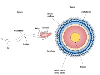
A spermatozoon is a motile sperm cell, or moving form of the haploid cell that is the male gamete. A spermatozoon joins an ovum to form a zygote.
Fertilisation or fertilization, also known as generative fertilisation, syngamy and impregnation, is the fusion of gametes to give rise to a zygote and initiate its development into a new individual organism or offspring. While processes such as insemination or pollination, which happen before the fusion of gametes, are also sometimes informally referred to as fertilisation, these are technically separate processes. The cycle of fertilisation and development of new individuals is called sexual reproduction. During double fertilisation in angiosperms, the haploid male gamete combines with two haploid polar nuclei to form a triploid primary endosperm nucleus by the process of vegetative fertilisation.
Inositol trisphosphate or inositol 1,4,5-trisphosphate abbreviated InsP3 or Ins3P or IP3 is an inositol phosphate signaling molecule. It is made by hydrolysis of phosphatidylinositol 4,5-bisphosphate (PIP2), a phospholipid that is located in the plasma membrane, by phospholipase C (PLC). Together with diacylglycerol (DAG), IP3 is a second messenger molecule used in signal transduction in biological cells. While DAG stays inside the membrane, IP3 is soluble and diffuses through the cell, where it binds to its receptor, which is a calcium channel located in the endoplasmic reticulum. When IP3 binds its receptor, calcium is released into the cytosol, thereby activating various calcium regulated intracellular signals.

For fertilization to happen between a sperm and egg cell, a sperm must first fuse with the plasma membrane and then penetrate the female egg cell to fertilize it. While the fusion of the sperm cell with the egg cell's plasma membrane is relatively straightforward, penetrating the egg's protective layers, such as the zona pellucida, presents a significant challenge. Therefore, sperm cells go through a process known as the acrosome reaction, which is the reaction that occurs in the acrosome of the sperm as it approaches the egg.

The zona pellucida is the specialized area surrounding mammalian oocytes (eggs). It is also known as an egg coat. The zona pellucida is essential for oocyte growth and fertilization.
Second messengers are intracellular signaling molecules released by the cell in response to exposure to extracellular signaling molecules—the first messengers. Second messengers trigger physiological changes at cellular level such as proliferation, differentiation, migration, survival, apoptosis and depolarization.
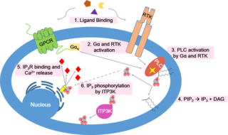
Calcium signaling is the use of calcium ions (Ca2+) to communicate and drive intracellular processes often as a step in signal transduction. Ca2+ is important for cellular signalling, for once it enters the cytosol of the cytoplasm it exerts allosteric regulatory effects on many enzymes and proteins. Ca2+ can act in signal transduction resulting from activation of ion channels or as a second messenger caused by indirect signal transduction pathways such as G protein-coupled receptors.
Human fertilization is the union of an egg and sperm, occurring primarily in the ampulla of the fallopian tube. The result of this union leads to the production of a fertilized egg called a zygote, initiating embryonic development. Scientists discovered the dynamics of human fertilization in the 19th century.
In biology, polyspermy describes the fertilization of an egg by more than one sperm. Diploid organisms normally contain two copies of each chromosome, one from each parent. The cell resulting from polyspermy, on the other hand, contains three or more copies of each chromosome—one from the egg and one each from multiple sperm. Usually, the result is an unviable zygote. This may occur because sperm are too efficient at reaching and fertilizing eggs due to the selective pressures of sperm competition. Such a situation is often deleterious to the female: in other words, the male–male competition among sperm spills over to create sexual conflict.

Phosphoinositide phospholipase C is a family of eukaryotic intracellular enzymes that play an important role in signal transduction processes. These enzymes belong to a larger superfamily of Phospholipase C. Other families of phospholipase C enzymes have been identified in bacteria and trypanosomes. Phospholipases C are phosphodiesterases.
The cortical reaction is a process initiated during fertilization that prevents polyspermy, the fusion of multiple sperm with one egg. In contrast to the fast block of polyspermy which immediately but temporarily blocks additional sperm from fertilizing the egg, the cortical reaction gradually establishes a permanent barrier to sperm entry and functions as the main part of the slow block of polyspermy in many animals.
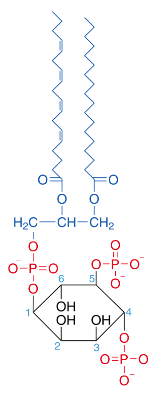
Phosphatidylinositol 4,5-bisphosphate or PtdIns(4,5)P2, also known simply as PIP2 or PI(4,5)P2, is a minor phospholipid component of cell membranes. PtdIns(4,5)P2 is enriched at the plasma membrane where it is a substrate for a number of important signaling proteins. PIP2 also forms lipid clusters that sort proteins.
Hyalin is a protein released from the cortical granules of a fertilized animal egg. The released hyalin modifies the extracellular matrix of the fertilized egg to block other sperm from binding to the egg, and is known as the slow-block to polyspermy. All animals have this slow-block mechanism.
The vitelline membrane or vitelline envelope is a structure surrounding the outer surface of the plasma membrane of an ovum or, in some animals, the extracellular yolk and the oolemma. It is composed mostly of protein fibers, with protein receptors needed for sperm binding which, in turn, are bound to sperm plasma membrane receptors. The species-specificity between these receptors contributes to prevention of breeding between different species. It is called zona pellucida in mammals. Between the vitelline membrane and zona pellucida is a fluid-filled perivitelline space.
Gq protein alpha subunit is a family of heterotrimeric G protein alpha subunits. This family is also commonly called the Gq/11 (Gq/G11) family or Gq/11/14/15 family to include closely related family members. G alpha subunits may be referred to as Gq alpha, Gαq, or Gqα. Gq proteins couple to G protein-coupled receptors to activate beta-type phospholipase C (PLC-β) enzymes. PLC-β in turn hydrolyzes phosphatidylinositol 4,5-bisphosphate (PIP2) to diacyl glycerol (DAG) and inositol trisphosphate (IP3). IP3 acts as a second messenger to release stored calcium into the cytoplasm, while DAG acts as a second messenger that activates protein kinase C (PKC).

Phospholipase C (PLC) is a class of membrane-associated enzymes that cleave phospholipids just before the phosphate group (see figure). It is most commonly taken to be synonymous with the human forms of this enzyme, which play an important role in eukaryotic cell physiology, in particular signal transduction pathways. Phospholipase C's role in signal transduction is its cleavage of phosphatidylinositol 4,5-bisphosphate (PIP2) into diacyl glycerol (DAG) and inositol 1,4,5-trisphosphate (IP3), which serve as second messengers. Activators of each PLC vary, but typically include heterotrimeric G protein subunits, protein tyrosine kinases, small G proteins, Ca2+, and phospholipids.

Zona pellucida sperm-binding protein 2 is a protein that in humans is encoded by the ZP2 gene.

Cortical granules are regulatory secretory organelles found within oocytes and are most associated with polyspermy prevention after the event of fertilization. Cortical granules are found among all mammals, many vertebrates, and some invertebrates. Within the oocyte, cortical granules are located along the cortex, the region furthest from the cell's center. Following fertilization, a signaling pathway induces the cortical granules to fuse with the oocyte's cell membrane and release their contents into the oocyte's extracellular matrix. This exocytosis of cortical granules is known as the cortical reaction. In mammals, the oocyte's extracellular matrix includes a surrounding layer of perivitelline space, zona pellucida, and finally cumulus cells. Experimental evidence has demonstrated that the released contents of the cortical granules modify the oocyte's extracellular matrix, particularly the zona pellucida. This alteration of the zona pellucida components is known as the zona reaction. The cortical reaction does not occur in all mammals, suggesting the likelihood of other functional purposes for cortical granules. In addition to modifying the oocyte's extracellular matrix and establishing a block to polyspermy, the exocytosis of cortical granules may also contribute towards protection and support of the developing embryo during preimplantation. Once the cortical granules complete their functions, the oocyte does not replenish them.
Egg jelly is a gelatinous layer that surrounds the oocytes of many organisms and releases species-specific chemoattractants that activate and guide sperm to the oocyte. The release of chemoattractants is species dependent. For example, sperm in Lytechinus variegatus, the green sea urchin, are not chemotactically attracted to the jelly or the egg. The egg jelly is located immediately surrounding the vitelline envelope and consists primarily of a network of short peptides and sulfated fucan glycoproteins. These short peptides diffuse into the surrounding area and stimulate respiration and movement of the sperm to the egg. An example of such a peptide is resact, which has been studied as the primary means of attracting and orientating sperm to the eggs in sea urchins. The sulfated fucan glycoproteins play an important role in binding to sperm receptors and triggering the acrosomal reaction.
Calcium plays a crucial role in regulating the events of cellular division. Calcium acts both to modulate intracellular signaling as a secondary messenger and to facilitate structural changes as cells progress through division. Exquisite control of intracellular calcium dynamics are required, as calcium appears to play a role at multiple cell cycle checkpoints.
References
- ↑ Saleh A, Kashir J, Thanassoulas A, Safieh-Garabedian B, Lai FA, Nomikos M (2020). "Essential Role of Sperm-Specific PLC-Zeta in Egg Activation and Male Factor Infertility: An Update". Front. Cell Dev. Biol. 8 (28). doi: 10.3389/fcell.2020.00028 . PMC 7000359 .
- ↑ Bianchi E, Doe B, Goulding D, Wright GJ (2014). "Juno is the egg Izumo receptor and is essential for mammalian fertilization". Nature. 508 (7497): 483–7. Bibcode:2014Natur.508..483B. doi:10.1038/nature13203. PMC 3998876 . PMID 24739963.
- ↑ Saunders C, Larman M, Parrington J, Cox L, Royse J, Blayney L, Swann K, Lai F (2002). "PLC zeta: a sperm-specific trigger of Ca(2+) oscillations in eggs and embryo development". Development. 129 (15): 3533–44. PMID 12117804.
- ↑ Yu, Yuansong; Nomikos, Michail; Theodoridou, Maria; Nounesis, George; Lai, F. Anthony; Swann, Karl (2012-01-15). "PLCζ causes Ca2+ oscillations in mouse eggs by targeting intracellular and not plasma membrane PI(4,5)P2". Molecular Biology of the Cell. 23 (2): 371–380. doi:10.1091/mbc.E11-08-0687. ISSN 1059-1524. PMC 3258180 . PMID 22114355.
- 1 2 3 4 5 6 Johnson, M. (2007). Essential Reproduction (6th ed.). Oxford: Blackwell. ISBN 9781405118668.
- ↑ Kashir, J.; Heindryckx, B.; Jones, C.; De Sutter, P.; Parrington, J.; Coward, K. (2010). "Oocyte activation, phospholipase C zeta and human infertility". Human Reproduction Update. 16 (6): 690–703. doi: 10.1093/humupd/dmq018 . PMID 20573804.
- ↑ Iba, T; Yano, Y; Umeno, M; Hinokio, K; Kuwahara, A; Irahara, M; Yamano, S; Yasui, T (2011). "Roscovitine in combination with calcium ionophore induces oocyte activation through reduction of M-phase promoting factor activity in mice". Zygote. 20 (4): 321–325. doi:10.1017/S0967199411000591. PMID 22008472.