Primitive streak

Last updated
Primitive streak
Gray13a.jpg
Surface view of embryo of a rabbit.
arg. Embryonic disk.
pr. Primitive streak. gc. germinal crescent.
Details
Carnegie stage 6b
Days15
Identifiers
Latin linea primitiva
MeSH D054240
Anatomical terminology

The primitive streak is a structure that forms in the early embryo in amniotes. [1] In amphibians, the equivalent structure is the blastopore. [2] During early embryonic development, the embryonic disc becomes oval shaped, and then pear-shaped with the broad end towards the anterior, and the narrower region projected to the posterior. The primitive streak forms a longitudinal midline structure in the narrower posterior (caudal) region of the developing embryo on its dorsal side. [3] At first formation, the primitive streak extends for half the length of the embryo. In the human embryo, this appears by stage 6, about 17 days. [4]

Contents

The primitive streak establishes bilateral symmetry, determines the site of gastrulation, and initiates germ layer formation. To form the primitive streak, mesenchymal stem cells are arranged along the prospective midline, establishing the second embryonic axis, and the site where cells will ingress and migrate during the process of gastrulation and germ layer formation. [5]

The primitive streak extends through this midline and creates the left–right and cranial–caudal body axes. [6] [7] Gastrulation involves the ingression of mesoderm progenitors and their migration to their ultimate position, [6] [8] where they will differentiate into the mesoderm germ layer [5] that, together with endoderm and ectoderm germ layers, will give rise to all the tissues of the adult organism.

Structure

Cellular differentiation arising from the development of the primitive streak during gastrulation following implantation 2908 Germ Layers-02.jpg
Cellular differentiation arising from the development of the primitive streak during gastrulation following implantation

The epiblast, a single epithelial layer of the bilaminar embryonic disc, is the source of all embryonic material in amniotes, and some of its cells will give rise to the primitive streak. [5] [8] In amphibians, the equivalent structure is the blastopore. [2] The primitive streak forms a longitudinal midline structure in the narrower caudal (posterior) region of the developing embryo on its dorsal side. [3] At first formation, the primitive streak extends for half the length of the embryo. In the human embryo, this appears by Carnegie stage 6, about 17 days. [4]

Towards the cranial (anterior) end of the disc, the primitive streak expands into an area known as the primitive node which is the organizer for gastrulation. [9] In birds, including the chick, this organizing node is called Hensen's node. In amphibians, where it was first identified, it is known as the Spemann-Mangold organizer.

In the middle of the node is a circular depression termed the primitive pit. The primitive pit extends towards the caudal end in a narrow depression in the primitive streak called the primitive groove [9] (Latin : sulcus primitivus). The groove is created by infolding of epiblastic cells. [10] [11]

Following its appearance and formation of the node, pit, and groove, the streak starts to regress caudally. Around day 20 in the human embryo, the remaining parts of the streak enlarge to produce a midline caudal cell mass termed the tail bud or caudal eminence. [9] Also at that time, the notochord develops cranially from the primitive node. By day 22, the primitive streak has regressed to between 10 and 20% of the embryo's length, and by day 26, has seemingly disappeared. [9]

Primitive streak in relation to later developed notochord shown on black crested gibbon embryo Gray14.png
Primitive streak in relation to later developed notochord shown on black crested gibbon embryo

Formation

The chick embryo as a model organism has provided much information about the formation of the primitive streak. In the chick blastula, its formation involves the coordinated movement and re-arrangement of cells in the epiblast. [12] Two counter-rotating flows of cells meet at the posterior end, where the streak forms. [12] There is little movement in the center of these flows, while the greatest movement is observed at the periphery of the vortices. [7] The vortex movements likened to polonaise movement is key for the formation of the primitive streak. [13] Cells overlaying Koller's sickle in the posterior end of the chick embryo move towards the midline, meet and change direction towards the center of the epiblast. Cells from the lateral posterior marginal zone replace those cells that left Koller's Sickle by meeting at the center of this region, changing direction and extending anteriorly. [8] [14] As these cells move and concentrate at the posterior end of the embryo, the streak undergoes a single- to multi-layered epithelial sheet transition that makes it a macroscopically visible structure. [8] Several mechanisms, including active proliferation, oriented cell division, cell-cell intercalation and chemotactic cell movement, have been proposed to explain the nature of the cellular movements required to form the primitive streak. [15] [8]

The marginal zone of a chick embryo contains cells that will contribute to the streak. [8] This region has a defined anterior-to-posterior gradient in its ability to induce the primitive streak, with the posterior end having the highest potential. [16] All cells in the epiblast can respond to signals from the marginal zone, [5] but once a given region is induced by these signals and undergoes streak formation, the remaining cells in the epiblast are no longer responsive to these inductive signals and prevent the formation of another streak. [16]

Underlying the epiblast is the hypoblast, where the extraembryonic tissue originates. [8] In the chick, the absence of the hypoblast results in multiple streaks, [17] suggesting that its presence is important for regulating the formation of a single primitive streak. In mice and other mammals, this structure is known as the anterior visceral endoderm (AVE). [17] The AVE migrates from the visceral endoderm (hypoblast). [18] The hypoblast also plays an important role in the regulation of streak formation. Removal of the hypoblast in the chick results in correctly patterned ectopic streaks, suggesting that the hypoblast serves to inhibit formation of the primitive streak. [19]

Signaling pathways

An intricate network of signaling pathways regulate the formation of the primitive streak. Formation of the Primitive Streak.pdf
An intricate network of signaling pathways regulate the formation of the primitive streak.

The formation of the primitive streak in the chick, is highly regulated by a complex network of signaling pathways. Activation of various secreted factors (Vg1, Nodal, Wnt8C, FGF8 and Chordin) and transcription factors (Brachyury and Goosecoid) adjacent to the site of streak formation is required for this process. [20] [21] [22] [23] [19]

Vg1 and Wnt signaling

Similarly, Vg1 (a transforming growth factor beta family member (TGF-β)) misexpression and grafts of the posterior marginal zone [16] in chicks can also induce ectopic streaks, but only within the marginal zone of the embryo, [22] indicating a specific characteristic of this region in its ability to induce streak formation. Several lines of evidence point to Wnt expression as the determinant of this ability. Deletion of Wnt3 in mouse embryos results in the absence of a streak formation, similarly to the phenotype of B-catenin mutant embryos. [24] In addition, mutating the intracellular negative regulator of Wnt signaling, Axin, [25] and misexpression of the chick cWnt8C [26] produces multiple streaks in mouse embryos. Localization of Wnt and components of its pathway, Lef1 and B-catenin, further supports streak-inducing role in the marginal zone. [22] Furthermore, it is expressed as a gradient decreasing from posterior to anterior, [22] [23] corresponding to the streak-inducing ability of the marginal zone. Misexpression of Vg1 or Wnt1 alone failed to induce an ectopic streak in the chick, but together their misexpression resulted in ectopic streak formation, confirming that the streak-inducing ability of the posterior marginal zone could be attributed to Wnt signaling [22] and that Vg1 and Wnt must cooperate to induce this process. Misexpression of Vg1 along with Wnt antagonists, Crescent or Dkk-1, prevents the formation of ectopic streaks, [22] demonstrating the importance of Wnt activity in the formation of Vg1-induced ectopic streaks and hence its implication in normal primitive streak formation.

Nodal signaling

Nodal, a known mesodermal inducer of the transforming growth factor beta family (TGFB family), [19] has been implicated in streak formation. Mouse embryos mutant for Nodal fail to gastrulate and lack most mesoderm, [27] but more than playing a role in mesoderm induction, Nodal regulates the induction and/or maintenance of the primitive streak. [27] In the presence of hypoblast, Nodal is unable to induce ectopic streaks in the chick embryo, while its removal, induces expression of Nodal, Chordin and Brachyury, [19] suggesting that the hypoblast must have a certain inhibitory effect on Nodal signaling. Indeed, the multifunctional antagonist of Nodal, Wnt and BMP signaling, Cerberus (produced in the hypoblast) and Cerberus-Short (which inhibits only Nodal), through its effect on Nodal signaling, inhibits streak formation. [19] Eventually, the hypoblast gets displaced anteriorly by the moving endoblast, allowing streak formation at the posterior end. At the anterior end, the presence of the hypoblast and the antagonists it secretes, such as Cerberus, inhibit the expression of Nodal and hence restrict streak formation to the posterior end only. [19] Similarly to the hypoblast in chick, the AVE in the mouse secretes two antagonists of Nodal signaling, Cerberus-like, Cerl, and Lefty1. [19] [28] In mouse, Cer-/-; Lefty1-/- double mutants develop multiple streaks [17] as indicated by ectopic expression of Brachyury and can be partially rescued by the removal of one copy of the Nodal gene. [17] In the mouse, the AVE restricts streak formation through the redundant functions of Cer1 and Lefty1, which negatively regulate Nodal signaling. [17] The role of the mouse's AVE in ensuring the formation of a single primitive streak is evolutionarily conserved in the hypoblast of the chick. [17] [19]

FGF signaling

Another important pathway in modulating formation of the primitive streak is FGF, which is thought to work together with Nodal to regulate this process. [29] Inhibition of FGF signaling through expression of a dominant negative receptor, using a FGF receptor inhibitor (SU5402) or depletion of FGF ligands, inhibit mesoderm formation [7] and this in turn, inhibits streak formation. [8] Furthermore, ectopic streak formation induced by Vg1 required FGF signaling. [29]

BMP signaling

Finally, BMP signaling is also important for regulating the process of streak formation in the chick embryo. The site of streak formation is characterized by low BMP signals, while the rest of the epiblast displays high levels of BMP activation. [30] In addition, misexpression of either BMP4 or BMP7 prevents streak formation, while the BMP inhibitor Chordin induces ectopic streak formation in the chick, [31] suggesting that streak formation is likely to require BMP inhibition.

Ethical implications

The primitive streak is an important concept in bioethics, where some experts have argued that experimentation with human embryos is permissible, but only before the primitive streak develops, generally around the fourteenth day of existence. The development of the primitive streak is taken, by such bioethicists, to signify the creation of a unique, human being. [32] In some countries, it is illegal to develop a human embryo for more than 14 days outside a woman's body. [33]

See also

Additional images

Related Research Articles

<span class="mw-page-title-main">Mesoderm</span> Middle germ layer of embryonic development

The mesoderm is the middle layer of the three germ layers that develops during gastrulation in the very early development of the embryo of most animals. The outer layer is the ectoderm, and the inner layer is the endoderm.

<span class="mw-page-title-main">Gastrulation</span> Stage in embryonic development in which germ layers form

Gastrulation is the stage in the early embryonic development of most animals, during which the blastula, or in mammals the blastocyst, is reorganized into a two-layered or three-layered embryo known as the gastrula. Before gastrulation, the embryo is a continuous epithelial sheet of cells; by the end of gastrulation, the embryo has begun differentiation to establish distinct cell lineages, set up the basic axes of the body, and internalized one or more cell types including the prospective gut.

<span class="mw-page-title-main">Somitogenesis</span>

Somitogenesis is the process by which somites form. Somites are bilaterally paired blocks of paraxial mesoderm that form along the anterior-posterior axis of the developing embryo in segmented animals. In vertebrates, somites give rise to skeletal muscle, cartilage, tendons, endothelium, and dermis.

The Wnt signaling pathways are a group of signal transduction pathways which begin with proteins that pass signals into a cell through cell surface receptors. The name Wnt is a portmanteau created from the names Wingless and Int-1. Wnt signaling pathways use either nearby cell-cell communication (paracrine) or same-cell communication (autocrine). They are highly evolutionarily conserved in animals, which means they are similar across animal species from fruit flies to humans.

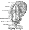
The primitive node is the organizer for gastrulation in most amniote embryos. In birds it is known as Hensen's node, and in amphibians it is known as the Spemann-Mangold organizer. It is induced by the Nieuwkoop center in amphibians, or by the posterior marginal zone in amniotes including birds.

<span class="mw-page-title-main">Intermediate mesoderm</span> Layer of cells in mammalian embryos

Intermediate mesoderm or intermediate mesenchyme is a narrow section of the mesoderm located between the paraxial mesoderm and the lateral plate of the developing embryo. The intermediate mesoderm develops into vital parts of the urogenital system.

<span class="mw-page-title-main">Epiblast</span> Embryonic inner cell mass tissue that forms the embryo itself, through the three germ layers

In amniote embryonic development, the epiblast is one of two distinct cell layers arising from the inner cell mass in the mammalian blastocyst, or from the blastula in reptiles and birds, the other layer is the hypoblast. It drives the embryo proper through its differentiation into the three primary germ layers, ectoderm, mesoderm and endoderm, during gastrulation. The amniotic ectoderm and extraembryonic mesoderm also originate from the epiblast.

<span class="mw-page-title-main">Apical ectodermal ridge</span>

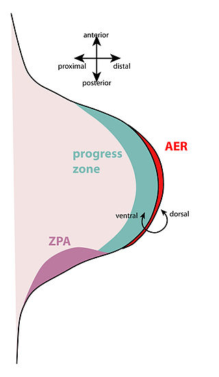
The apical ectodermal ridge (AER) is a structure that forms from the ectodermal cells at the distal end of each limb bud and acts as a major signaling center to ensure proper development of a limb. After the limb bud induces AER formation, the AER and limb mesenchyme—including the zone of polarizing activity (ZPA)—continue to communicate with each other to direct further limb development.

In developmental biology, the cells that give rise to the gametes are often set aside during embryonic cleavage. During development, these cells will differentiate into primordial germ cells, migrate to the location of the gonad, and form the germline of the animal.

The limb bud is a structure formed early in vertebrate limb development. As a result of interactions between the ectoderm and underlying mesoderm, formation occurs roughly around the fourth week of development. In the development of the human embryo the upper limb bud appears in the third week and the lower limb bud appears four days later.

In the field of developmental biology, regional differentiation is the process by which different areas are identified in the development of the early embryo. The process by which the cells become specified differs between organisms.

<span class="mw-page-title-main">Cripto</span> Protein-coding gene in the species Homo sapiens

Cripto is an EGF-CFC or epidermal growth factor-CFC, which is encoded by the Cryptic family 1 gene. Cryptic family protein 1B is a protein that in humans is encoded by the CFC1B gene. Cryptic family protein 1B acts as a receptor for the TGF beta signaling pathway. It has been associated with the translation of an extracellular protein for this pathway. The extracellular protein which Cripto encodes plays a crucial role in the development of left and right division of symmetry.

Growth differentiation factors (GDFs) are a subfamily of proteins belonging to the transforming growth factor beta superfamily that have functions predominantly in development.

<span class="mw-page-title-main">GDF3</span> Protein-coding gene in humans

Growth differentiation factor-3 (GDF3), also known as Vg-related gene 2 (Vgr-2) is protein that in humans is encoded by the GDF3 gene. GDF3 belongs to the transforming growth factor beta (TGF-β) superfamily. It has high similarity to other TGF-β superfamily members including Vg1 and GDF1.

<span class="mw-page-title-main">Fish development</span>

The development of fishes is unique in some specific aspects compared to the development of other animals.

<span class="mw-page-title-main">Cerberus (protein)</span> Protein found in humans

Cerberus is a protein that in humans is encoded by the CER1 gene. Cerberus is a signaling molecule which contributes to the formation of the head, heart and left-right asymmetry of internal organs. This gene varies slightly from species to species but its overall functions seem to be similar.

<span class="mw-page-title-main">Hypoblast</span> Embryonic inner cell mass tissue that forms the yolk sac and, later, chorion

In amniote embryology, the hypoblast is one of two distinct layers arising from the inner cell mass in the mammalian blastocyst, or from the blastodisc in reptiles and birds. The hypoblast gives rise to the yolk sac, which in turn gives rise to the chorion.

<span class="mw-page-title-main">Nodal homolog</span> Mammalian protein found in Homo sapiens

Nodal homolog is a secretory protein that in humans is encoded by the NODAL gene which is located on chromosome 10q22.1. It belongs to the transforming growth factor beta superfamily. Like many other members of this superfamily it is involved in cell differentiation in early embryogenesis, playing a key role in signal transfer from the primitive node, in the anterior primitive streak, to lateral plate mesoderm (LPM).

<span class="mw-page-title-main">Koller's sickle</span>

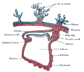
In avian gastrulation, Koller's sickle is a local thickening of cells at the posterior edge of the upper layer of the area pellucida called the epiblast. Koller's sickle is crucial for avian development, due to its critical role in inducing the differentiation of various avian body parts. Koller's sickle induces primitive streak and Hensen's node, which are major components of avian gastrulation. Avian gastrulation is a process by which developing cells in an avian embryo move relative to one another in order to form the three germ layers.

The Nodal signaling pathway is a signal transduction pathway important in regional and cellular differentiation during embryonic development.

References

  1. "Definition of AMNIOTE". www.merriam-webster.com. Retrieved 11 May 2022.
  2. 1 2 Wolpert, Lewis (2015). Principles of development (Fifth ed.). Oxford, United Kingdom. p. 113. ISBN   9780199678143.{{cite book}}: CS1 maint: location missing publisher (link)
  3. 1 2 Schoenwolf, Gary C. (2015). Larsen's human embryology (Fifth ed.). Philadelphia, PA. p. 57. ISBN   9781455706846.{{cite book}}: CS1 maint: location missing publisher (link)
  4. 1 2 Standring, Susan (2016). Gray's anatomy : the anatomical basis of clinical practice (Forty-first ed.). [Philadelphia]. p. 181. ISBN   9780702052309.{{cite book}}: CS1 maint: location missing publisher (link)
  5. 1 2 3 4 Mikawa T, Poh AM, Kelly KA, Ishii Y, Reese DE (2004). "Induction and patterning of the primitive streak, an organizing center of gastrulation in the amniote". Dev Dyn. 229 (3): 422–32. doi: 10.1002/dvdy.10458 . PMID   14991697. S2CID   758473.
  6. 1 2 Downs KM (2009). "The enigmatic primitive streak: prevailing notions and challenges concerning the body axis of mammals". BioEssays. 31 (8): 892–902. doi:10.1002/bies.200900038. PMC   2949267 . PMID   19609969.
  7. 1 2 3 Chuai M, Zeng W, Yang X, Boychenko V, Glazier JA, Weijer CJ (2006). "Cell movement during chick primitive streak formation". Dev. Biol. 296 (1): 137–49. doi:10.1016/j.ydbio.2006.04.451. PMC   2556955 . PMID   16725136.
  8. 1 2 3 4 5 6 7 8 Chuai M, Weijer CJ (2008). "The mechanisms underlying primitive streak formation in the chick embryo". Curr Top Dev Biol. Current Topics in Developmental Biology. 81: 135–56. doi:10.1016/S0070-2153(07)81004-0. ISBN   978-0-12-374253-7. PMID   18023726.
  9. 1 2 3 4 Schoenwolf, Gary C. (2015). Larsen's human embryology (Fifth ed.). Philadelphia, PA. pp. 78–80. ISBN   9781455706846.{{cite book}}: CS1 maint: location missing publisher (link)
  10. Moore, Keith L.; Persaud, T. V. N.; Torchia, Mark G. (2018). "4 - Third Week of Human Development". The Developing Human (11th ed.). Saunders. pp. 47–63. ISBN   9780323611541.
  11. Standring, Susan (2020). "10 - Cell populations at gastrulation". Gray's Anatomy (42nd ed.). Elsevier. pp. 188–195. ISBN   9780702077050.
  12. 1 2 Cui C, Yang X, Chuai M, Glazier JA, Weijer CJ (2005). "Analysis of tissue flow patterns during primitive streak formation in the chick embryo". Dev. Biol. 284 (1): 37–47. doi: 10.1016/j.ydbio.2005.04.021 . PMID   15950214.
  13. Saadaoui, M; Rocancourt, D; Roussel, J; Corson, F; Gros, J (24 January 2020). "A tensile ring drives tissue flows to shape the gastrulating amniote embryo". Science. 367 (6476): 453–458. Bibcode:2020Sci...367..453S. doi: 10.1126/science.aaw1965 . PMID   31974255. S2CID   92787648.
  14. Hatada Y, Stern CD (1994). "A fate map of the epiblast of the early chick embryo". Development. 120 (10): 2879–89. doi:10.1242/dev.120.10.2879. PMID   7607078.
  15. Mikawa, Takashi; Poh, Alisa M.; Kelly, Kristine A.; Ishii, Yasuo; Reese, David E. (2004). "Induction and patterning of the primitive streak an organizing centre of gastrulation in the amniote". Developmental Dynamics. 229 (3): 422–432. doi: 10.1002/dvdy.10458 . PMID   14991697. S2CID   758473.
  16. 1 2 3 Khaner O, Eyal-Giladi H (1989). "The chick's marginal zone and primitive streak formation. I. Coordinative effect of induction and inhibition". Dev. Biol. 134 (1): 206–14. doi:10.1016/0012-1606(89)90090-0. PMID   2731648.
  17. 1 2 3 4 5 6 Perea-Gomez A, Vella FD, Shawlot W, Oulad-Abdelghani M, Chazaud C, Meno C, Pfister V, Chen L, Robertson E, Hamada H, Behringer RR, Ang SL (2002). "Nodal antagonists in the anterior visceral endoderm prevent the formation of multiple primitive streaks". Dev Cell. 3 (5): 745–56. doi: 10.1016/S1534-5807(02)00321-0 . PMID   12431380.
  18. Gilbert, Scott F. (2006). Developmental biology (8th ed.). Sunderland, Mass.: Sinauer Associates Publishers. p. 358. ISBN   9780878932504.
  19. 1 2 3 4 5 6 7 8 Bertocchini F, Stern CD (2002). "The hypoblast of the chick embryo positions the primitive streak by antagonizing nodal signaling". Dev Cell. 3 (5): 735–44. doi: 10.1016/S1534-5807(02)00318-0 . PMID   12431379.
  20. Shah SB, Skromne I, Hume CR, Kessler DS, Lee KJ, Stern CD, Dodd J (1997). "Misexpression of chick Vg1 in the marginal zone induces primitive streak formation". Development. 124 (24): 5127–38. doi:10.1242/dev.124.24.5127. PMID   9362470.
  21. Bachvarova RF, Skromne I, Stern CD (1998). "Induction of primitive streak and Hensen's node by the posterior marginal zone in the early chick embryo". Development. 125 (17): 3521–34. doi:10.1242/dev.125.17.3521. PMID   9693154.
  22. 1 2 3 4 5 6 Skromne I, Stern CD (2001). "Interactions between Wnt and Vg1 signalling pathways initiate primitive streak formation in the chick embryo". Development. 128 (15): 2915–27. doi:10.1242/dev.128.15.2915. PMID   11532915.
  23. 1 2 Skromne I, Stern CD (2002). "A hierarchy of gene expression accompanying induction of the primitive streak by Vg1 in the chick embryo". Mech. Dev. 114 (1–2): 115–8. doi: 10.1016/S0925-4773(02)00034-5 . PMID   12175495.
  24. Liu P, Wakamiya M, Shea MJ, Albrecht U, Behringer RR, Bradley A (1999). "Requirement for Wnt3 in vertebrate axis formation". Nat. Genet. 22 (4): 361–5. doi:10.1038/11932. PMID   10431240. S2CID   22195563.
  25. Zeng L, Fagotto F, Zhang T, Hsu W, Vasicek TJ, Perry WL 3rd, Lee JJ, Tilghman SM, Gumbiner BM, Costantini F (1997). "The mouse Fused locus encodes Axin, an inhibitor of the Wnt signaling pathway that regulates embryonic axis formation". Cell. 90 (1): 181–92. doi: 10.1016/S0092-8674(00)80324-4 . PMID   9230313.
  26. Pöpperl H, Schmidt C, Wilson V, Hume CR, Dodd J, Krumlauf R, Beddington RS (1997). "Misexpression of Cwnt8C in the mouse induces an ectopic embryonic axis and causes a truncation of the anterior neuroectoderm". Development. 124 (15): 2997–3005. doi:10.1242/dev.124.15.2997. PMID   9247341.
  27. 1 2 Conlon FL, Lyons KM, Takaesu N, Barth KS, Kispert A, Herrmann B, Robertson EJ (1994). "A primary requirement for nodal in the formation and maintenance of the primitive streak in the mouse". Development. 120 (7): 1919–28. doi:10.1242/dev.120.7.1919. PMID   7924997.
  28. C Perea-Gomez A, Rhinn M, Ang SL (2001). "Role of the anterior visceral endoderm in restricting posterior signals in the mouse embryo". Int J Dev Biol. 45 (1): 311–20. PMID   11291861.
  29. 1 2 Bertocchini F, Skromne I, Wolpert L, Stern CD (2004). "Determination of embryonic polarity in a regulative system: evidence for endogenous inhibitors acting sequentially during primitive streak formation in the chick embryo". Development. 131 (14): 3381–90. doi: 10.1242/dev.01178 . PMID   15226255. S2CID   942758.
  30. Faure S, de Santa Barbara P, Roberts DJ, Whitman M (2002). "Endogenous patterns of BMP signaling during early chick development". Dev. Biol. 244 (1): 44–65. doi: 10.1006/dbio.2002.0579 . PMID   11900458.
  31. Streit A, Lee KJ, Woo I, Roberts C, Jessell TM, Stern CD (1998). "Chordin regulates primitive streak development and the stability of induced neural cells, but is not sufficient for neural induction in the chick embryo". Development. 125 (3): 507–19. doi:10.1242/dev.125.3.507. PMID   9425145.
  32. "The President's Council on Bioethics, Human Cloning and Human Dignity: An Ethical Inquiry. Chapter 6". July 2002.
  33. Government of Australia Department of Health and Ageing, 22 Dec 2008