Human fertilization

Last updated
Human fertilization
The sperm and ovum during fertilization.svg
Sperm about to enter the ovum with acrosomal head ready to penetrate the zona pellucida and fertilize the egg
Blausen 0404 Fertilization.png
Illustration depicting ovulation and fertilization
Details
Days0
Precursor Gametes
Gives rise to Zygote
Anatomical terminology

Human fertilization is the union of an egg and sperm, occurring primarily in the ampulla of the fallopian tube. [1] The result of this union leads to the production of a fertilized egg called a zygote, initiating embryonic development. Scientists discovered the dynamics of human fertilization in the 19th century. [2]

Contents

The process of fertilization involves a sperm fusing with an ovum. The most common sequence begins with ejaculation during copulation, follows with ovulation, and finishes with fertilization. Various exceptions to this sequence are possible, including artificial insemination, in vitro fertilization, external ejaculation without copulation, or copulation shortly after ovulation. [3] [4] Upon encountering the secondary oocyte, the acrosome of the sperm produces enzymes which allow it to burrow through the outer shell called the zona pellucida of the egg. The sperm plasma then fuses with the egg's plasma membrane and their nuclei fuse, triggering the sperm head to disconnect from its flagellum as the egg travels down the fallopian tube to reach the uterus.

In vitro fertilization (IVF) is a process by which egg cells are fertilized by sperm outside the womb, in vitro .

History

In Antiquity, Aristotle depicted the formation of new individuals occurring through fusion of male and female fluids, with form and function emerging gradually, in a mode called by him as epigenetic. [5] .

Sperm and oocyte meet

Ampulla

Fertilization occurs in the ampulla of the fallopian tube, the section that curves around the ovary. Capacitated sperm are attracted to progesterone, which is secreted from the cumulus cells surrounding the oocyte. [6] Progesterone binds to the CatSper receptor on the sperm membrane and increases intracellular calcium levels, causing hyperactive motility. The sperm will continue to swim towards higher concentrations of progesterone, effectively guiding it to the oocyte. [7] Around 200 out of 200 million spermatozoa reach the ampulla.

Sperm preparation

The sperm entering the ovum using acrosomal enzymes to dissolve the gelatinous envelope (zona pellucida)of the oocyte. Egg cell fertilization - Zygote.png
The sperm entering the ovum using acrosomal enzymes to dissolve the gelatinous envelope (zona pellucida)of the oocyte.

At the beginning of the process, the sperm undergoes a series of changes, as freshly ejaculated sperm is unable or poorly able to fertilize. [8] The sperm must undergo capacitation in the female's reproductive tract, which increases its motility and hyperpolarizes its membrane, preparing it for the acrosome reaction, the enzymatic penetration of the egg's tough membrane, the zona pellucida, which surrounds the oocyte. [9]

Corona radiata

The sperm binds through the corona radiata, a layer of follicle cells on the outside of the secondary oocyte. The corona radiata sends out chemicals that attract the sperm in the fallopian tube to the oocyte. It lies above the zona pellucida, a membrane of glycoproteins that surrounds the oocyte. [10]

Cone of attraction and perivitelline membrane

Where the spermatozoan is about to pierce, the yolk (ooplasm) is drawn out into a conical elevation, termed the cone of attraction or reception cone. Once the spermatozoon has entered, the peripheral portion of the yolk changes into a membrane, the perivitelline membrane, which prevents the passage of additional spermatozoa. [11]

Zona pellucida and acrosome reaction

After binding to the corona radiata the sperm reaches the zona pellucida, which is an extracellular matrix of glycoproteins. A ZP3 glycoprotein on the zona pellucida binds to a receptor on the cell surface of the sperm head. This binding triggers the acrosome to burst, releasing acrosomal enzymes that help the sperm penetrate through the thick zona pellucida layer surrounding the oocyte, ultimately gaining access to the egg's cell membrane. [12]

Some sperm cells consume their acrosome prematurely on the surface of the egg cell, facilitating the penetration by other sperm cells. As a population, mature haploid sperm cells have on average 50% genome similarity, so the premature acrosomal reactions aid fertilization by a member of the same cohort. [13] It may be regarded as a mechanism of kin selection.

Recent studies have shown that the egg is not passive during this process. In other words, they too appear to undergo changes that help facilitate such interaction. [14] [15]

Fusion

Fertilization and implantation in humans. 2904 Preembryonic Development-02.jpg
Fertilization and implantation in humans.

Cortical reaction

After the sperm enters the cytoplasm of the oocyte, the tail and the outer coating of the sperm disintegrate. The fusion of sperm and oocyte membranes causes cortical reaction to occur. [16] Cortical granules inside the secondary oocyte fuse with the plasma membrane of the cell, causing enzymes inside these granules to be expelled by exocytosis to the zona pellucida. This in turn causes the glycoproteins in the zona pellucida to cross-link with each other — i.e. the enzymes cause the ZP2 to hydrolyse into ZP2f — making the whole matrix hard and impermeable to sperm. This prevents fertilization of an egg by more than one sperm. [17]

Fusion of genetic material

Preparation

In preparation for the fusion of their genetic material both the oocyte and the sperm undergo transformations as a reaction to the fusion of cell membranes.

The oocyte completes its second meiotic division. This results in a mature haploid ovum and the release of a polar body. [18] The nucleus of the oocyte is called a pronucleus in this process, to distinguish it from the nuclei that are the result of fertilization.

Drawing of an ovum Ovum Diagram.svg
Drawing of an ovum

The sperm's tail and mitochondria degenerate with the formation of the male pronucleus. This is why all mitochondria in humans are of maternal origin. Still, a considerable amount of RNA from the sperm is delivered to the resulting embryo and likely influences embryo development and the phenotype of the offspring. [19]

Fusion

The sperm nucleus then fuses with the ovum, enabling fusion of their genetic material.

Blocks of polyspermy

When the sperm enters the perivitelline space, a sperm-specific protein Izumo on the head binds to Juno receptors on the oocyte membrane. [20] Once it is bound, two blocks to polyspermy then occur. After approximately 40 minutes, the other Juno receptors on the oocyte are lost from the membrane, causing it to no longer be fusogenic. Additionally, the cortical reaction will happen which is caused by ovastacin binding and cleaving ZP2 receptors on the zona pellucida. [21] These two blocks of polyspermy are what prevent the zygote from having too much DNA.

Replication

The pronuclei migrate toward the center of the oocyte, rapidly replicating their DNA as they do so to prepare the zygote for its first mitotic division. [22]

Mitosis

Usually 23 chromosomes from spermatozoon and 23 chromosomes from egg cell fuse (approximately half of spermatozoons carry X chromosome and the other half Y chromosome [23] ). Their membranes dissolve, leaving no barriers between the male and female chromosomes. During this dissolution, a mitotic spindle forms between them. The spindle captures the chromosomes before they disperse in the egg cytoplasm. Upon subsequently undergoing mitosis (which includes pulling of chromatids towards centrioles in anaphase) the cell gathers genetic material from the male and female together. Thus, the first mitosis of the union of sperm and oocyte is the actual fusion of their chromosomes. [22]

Each of the two daughter cells resulting from that mitosis has one replica of each chromatid that was replicated in the previous stage. Thus, they are genetically identical.[ citation needed ]

Fertilization age

Fertilization is the event most commonly used to mark the beginning point of life, in descriptions of prenatal development of the embryo or fetus. [24] The resultant age is known as fertilization age, fertilizational age, conceptional age, embryonic age, fetal age or (intrauterine) developmental (IUD) [25] age.

Gestational age, in contrast, takes the beginning of the last menstrual period (LMP) as the start point. By convention, gestational age is calculated by adding 14 days to fertilization age and vice versa. [26] Fertilization though usually occurs within a day of ovulation, which, in turn, occurs on average 14.6 days after the beginning of the preceding menstruation (LMP). [27] There is also considerable variability in this interval, with a 95% prediction interval of the ovulation of 9 to 20 days after menstruation even for an average woman who has a mean LMP-to-ovulation time of 14.6. [28] In a reference group representing all women, the 95% prediction interval of the LMP-to-ovulation is 8.2 to 20.5 days. [27]

The average time to birth has been estimated to be 268 days (38 weeks and two days) from ovulation, with a standard deviation of 10 days or coefficient of variation of 3.7%. [29]

Fertilization age is sometimes used postnatally (after birth) as well to estimate various risk factors. For example, it is a better predictor than postnatal age for risk of intraventricular hemorrhage in premature babies treated with extracorporeal membrane oxygenation. [30]

Diseases affecting human fertility

Various disorders can arise from defects in the fertilization process. Whether that results in the process of contact between the sperm and egg, or the state of health of the biological parent carrying the zygote cell. The following are a few of the diseases that can occur and be present during the process.

See also

Related Research Articles

<span class="mw-page-title-main">Spermatozoon</span> Motile sperm cell

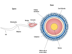
A spermatozoon is a motile sperm cell, or moving form of the haploid cell that is the male gamete. A spermatozoon joins an ovum to form a zygote.

<span class="mw-page-title-main">Zygote</span> Diploid eukaryotic cell formed by fertilization between two gametes

A zygote is a eukaryotic cell formed by a fertilization event between two gametes. The zygote's genome is a combination of the DNA in each gamete, and contains all of the genetic information of a new individual organism. The sexual fusion of haploid cells is called karyogamy, the result of which is the formation of a diploid cell called the zygote or zygospore.

<span class="mw-page-title-main">Fertilisation</span> Union of gametes of opposite sexes during the process of sexual reproduction to form a zygote

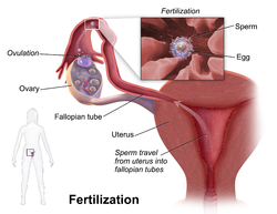
Fertilisation or fertilization, also known as generative fertilisation, syngamy and impregnation, is the fusion of gametes to give rise to a zygote and initiate its development into a new individual organism or offspring. While processes such as insemination or pollination, which happen before the fusion of gametes, are also sometimes informally referred to as fertilisation, these are technically separate processes. The cycle of fertilisation and development of new individuals is called sexual reproduction. During double fertilisation in angiosperms, the haploid male gamete combines with two haploid polar nuclei to form a triploid primary endosperm nucleus by the process of vegetative fertilisation.

<span class="mw-page-title-main">Acrosome</span> Organelle of sperm cells

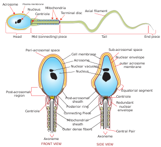
The acrosome is an organelle that develops over the anterior (front) half of the head in the spermatozoa of humans, and many other animals. It is a cap-like structure derived from the Golgi apparatus. In placental mammals, the acrosome contains degradative enzymes. These enzymes break down the outer membrane of the ovum, called the zona pellucida, allowing the haploid nucleus in the sperm cell to join with the haploid nucleus in the ovum. This shedding of the acrosome, or acrosome reaction, can be stimulated in vitro by substances a sperm cell may encounter naturally such as progesterone or follicular fluid, as well as the more commonly used calcium ionophore A23187. This can be done to serve as a positive control when assessing the acrosome reaction of a sperm sample by flow cytometry or fluorescence microscopy. This is usually done after staining with a fluoresceinated lectin such as FITC-PNA, FITC-PSA, FITC-ConA, or fluoresceinated antibody such as FITC-CD46.

<span class="mw-page-title-main">Acrosome reaction</span> Sperm-meets-egg process

During fertilization, a sperm must first fuse with the plasma membrane and then penetrate the female egg cell to fertilize it. Fusing to the egg cell usually causes little problem, whereas penetrating through the egg's hard shell or extracellular matrix can be more difficult. Therefore, sperm cells go through a process known as the acrosome reaction, which is the reaction that occurs in the acrosome of the sperm as it approaches the egg.

<span class="mw-page-title-main">Female reproductive system</span> Reproductive system of human females

The female reproductive system is made up of the internal and external sex organs that function in the reproduction of new offspring. The human female reproductive system is immature at birth and develops to maturity at puberty to be able to produce gametes, and to carry a fetus to full term. The internal sex organs are the vagina, uterus, fallopian tubes, and ovaries. The female reproductive tract includes the vagina, uterus, and fallopian tubes and is prone to infections. The vagina allows for sexual intercourse and childbirth, and is connected to the uterus at the cervix. The uterus or womb accommodates the embryo, which develops into the fetus. The uterus also produces secretions, which help the transit of sperm to the fallopian tubes, where sperm fertilize ova produced by the ovaries. The external sex organs are also known as the genitals and these are the organs of the vulva including the labia, clitoris, and vaginal opening.

<span class="mw-page-title-main">Zona pellucida</span> Glycoprotein layer surrounding the plasma membrane of mammalian oocytes

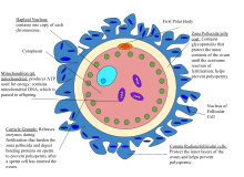
The zona pellucida is the specialized area surrounding mammalian oocytes (eggs). It is also known as an egg coat. The zona pellucida is essential for oocyte growth and fertilization.

Hyperactivation is a type of sperm motility. Hyperactivated sperm motility is characterised by a high amplitude, asymmetrical beating pattern of the sperm tail (flagellum). This type of motility may aid in sperm penetration of the zona pellucida, which encloses the ovum.

In biology, polyspermy describes the fertilization of an egg by more than one sperm. Diploid organisms normally contain two copies of each chromosome, one from each parent. The cell resulting from polyspermy, on the other hand, contains three or more copies of each chromosome—one from the egg and one each from multiple sperm. Usually, the result is an unviable zygote. This may occur because sperm are too efficient at reaching and fertilizing eggs due to the selective pressures of sperm competition. Such a situation is often deleterious to the female: in other words, the male–male competition among sperm spills over to create sexual conflict.

<span class="mw-page-title-main">Cortical reaction</span> Biological process that prevents polyspermy

The cortical reaction is a process initiated during fertilization that prevents polyspermy, the fusion of multiple sperm with one egg. In contrast to the fast block of polyspermy which immediately but temporarily blocks additional sperm from fertilizing the egg, the cortical reaction gradually establishes a permanent barrier to sperm entry and functions as the main part of the slow block of polyspermy in many animals.

<span class="mw-page-title-main">Polar body</span> Byproduct of oogenesis

A polar body is a small haploid cell that is formed at the same time as an egg cell during oogenesis, but generally does not have the ability to be fertilized. It is named from its polar position in the egg.

<span class="mw-page-title-main">Human reproduction</span> Procreative biological processes of humanity

Human reproduction is sexual reproduction that results in human fertilization to produce a human offspring. It typically involves sexual intercourse between a sexually mature human male and female. During sexual intercourse, the interaction between the male and female reproductive systems results in fertilization of the ovum by the sperm to form a zygote. While normal cells contain 46 chromosomes, gamete cells only contain 23 single chromosomes, and it is when these two cells merge into one zygote cell that genetic recombination occurs and the new zygote contains 23 chromosomes from each parent, giving it 46 chromosomes. The zygote then undergoes a defined development process that is known as human embryogenesis, and this starts the typical 9-month gestation period that is followed by childbirth. The fertilization of the ovum may be achieved by artificial insemination methods, which do not involve sexual intercourse. Assisted reproductive technology also exists.

<span class="mw-page-title-main">Corona radiata (embryology)</span>

The corona radiata is the innermost layer of the cells of the cumulus oophorus and is directly adjacent to the zona pellucida, the inner protective glycoprotein layer of the ovum. Cumulus oophorus are the cells surrounding corona radiata, and are the cells between corona radiata and follicular antrum. Its main purpose in many animals is to supply vital proteins to the cell. It is formed by follicle cells adhering to the oocyte before it leaves the ovarian follicle, and originates from the squamous granulosa cells present at the primordial stage of follicular development. The corona radiata is formed when the granulosa cells enlarge and become cuboidal, which occurs during the transition from the primordial to primary stage. These cuboidal granulosa cells, also known as the granulosa radiata, form more layers throughout the maturation process, and remain attached to the zona pellucida after the ovulation of the Graafian follicle. For fertilization to occur, sperm cells rely on hyaluronidase to disperse the corona radiata from the zona pellucida of the secondary (ovulated) oocyte, thus permitting entry into the perivitelline space and allowing contact between the sperm cell and the nucleus of the oocyte.

The vitelline membrane or vitelline envelope is a structure surrounding the outer surface of the plasma membrane of an ovum or, in some animals, the extracellular yolk and the oolemma. It is composed mostly of protein fibers, with protein receptors needed for sperm binding which, in turn, are bound to sperm plasma membrane receptors. The species-specificity between these receptors contributes to prevention of breeding between different species. It is called zona pellucida in mammals. Between the vitelline membrane and zona pellucida is a fluid-filled perivitelline space.

The hamster zona-free ovum test, or hamster egg-penetration test, or sometimes just hamster test, is an in-vitro test used to study physiological profile of spermatozoa. The primary application of the test is to diagnose male infertility caused by sperm unable to penetrate the ova. The test has limited value, due to expense and a high false negative rate.

<span class="mw-page-title-main">Human embryonic development</span> Development and formation of the human embryo

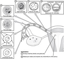
Human embryonic development or human embryogenesis is the development and formation of the human embryo. It is characterised by the processes of cell division and cellular differentiation of the embryo that occurs during the early stages of development. In biological terms, the development of the human body entails growth from a one-celled zygote to an adult human being. Fertilization occurs when the sperm cell successfully enters and fuses with an egg cell (ovum). The genetic material of the sperm and egg then combine to form the single cell zygote and the germinal stage of development commences. Embryonic development in the human, covers the first eight weeks of development; at the beginning of the ninth week the embryo is termed a fetus. The eight weeks has 23 stages.

<span class="mw-page-title-main">ZP2</span> Protein-coding gene in the species Homo sapiens

Zona pellucida sperm-binding protein 2 is a protein that in humans is encoded by the ZP2 gene.

<span class="mw-page-title-main">Fallopian tube</span> Tubes in the human female reproductive system

The fallopian tubes, also known as uterine tubes, oviducts or salpinges, are paired tubes in the human female body that stretch from the uterus to the ovaries. The fallopian tubes are part of the female reproductive system. In other vertebrates, they are only called oviducts.

Oocyteactivation is a series of processes that occur in the oocyte during fertilization.

<span class="mw-page-title-main">Cortical granule</span>

Cortical granules are regulatory secretory organelles found within oocytes and are most associated with polyspermy prevention after the event of fertilization. Cortical granules are found among all mammals, many vertebrates, and some invertebrates. Within the oocyte, cortical granules are located along the cortex, the region furthest from the cell's center. Following fertilization, a signaling pathway induces the cortical granules to fuse with the oocyte's cell membrane and release their contents into the oocyte's extracellular matrix. This exocytosis of cortical granules is known as the cortical reaction. In mammals, the oocyte's extracellular matrix includes a surrounding layer of perivitelline space, zona pellucida, and finally cumulus cells. Experimental evidence has demonstrated that the released contents of the cortical granules modify the oocyte's extracellular matrix, particularly the zona pellucida. This alteration of the zona pellucida components is known as the zona reaction. The cortical reaction does not occur in all mammals, suggesting the likelihood of other functional purposes for cortical granules. In addition to modifying the oocyte's extracellular matrix and establishing a block to polyspermy, the exocytosis of cortical granules may also contribute towards protection and support of the developing embryo during preimplantation. Once the cortical granules complete their functions, the oocyte does not replenish them.

References

  1. Spermatogenesis — Fertilization — Contraception: Molecular, Cellular and Endocrine Events in Male Reproduction. Ernst Schering Foundation Symposium Proceedings. Springer-Verlag. 1992. ISBN   978-3-662-02817-9.[ page needed ]
  2. Garrison FH (1921). An Introduction to the History of Medicine. Saunders. pp. 566–567.
  3. "Go Ask Alice!: Pregnant without intercourse?". Archived from the original on 2011-12-22. Retrieved 2016-01-24.
  4. Knight B (1998). Lawyer's guide to forensic medicine (2nd ed.). London: Cavendish Pub. Ltd. p. 188. ISBN   978-1-85941-159-9. Pregnancy is well known to occur from such external ejaculation ...
  5. Maienschein J (2017). "The first century of cell theory: From structural units to complex living systems.". In Stadler F (ed.). Integrated History and Philosophy of Science. Vienna Circle Institute Yearbook, Institute Vienna Circle, University of Vienna, Vienna Circle Society, Society for the Advancement of Scientific World Conceptions. Vol. 20. Cham.: Springer. ISBN   978-3-319-53258-5.
  6. Oren-Benaroya R, Orvieto R, Gakamsky A, Pinchasov M, Eisenbach M (October 2008). "The sperm chemoattractant secreted from human cumulus cells is progesterone". Human Reproduction. 23 (10): 2339–2345. doi: 10.1093/humrep/den265 . PMID   18621752.
  7. Publicover S, Barratt C (March 2011). "Reproductive biology: Progesterone's gateway into sperm". Nature. 471 (7338): 313–314. Bibcode:2011Natur.471..313P. doi:10.1038/471313a. PMID   21412330. S2CID   205062974.
  8. "Fertilization". Archived from the original on 24 June 2010. Retrieved 28 July 2010.
  9. Puga Molina LC, Luque GM, Balestrini PA, Marín-Briggiler CI, Romarowski A, Buffone MG (2018). "Molecular Basis of Human Sperm Capacitation". Frontiers in Cell and Developmental Biology. 6: 72. doi: 10.3389/fcell.2018.00072 . PMC   6078053 . PMID   30105226.
  10. Miles, Linda. "LibGuides: BIO 140 - Human Biology I - Textbook: Chapter 45 - Fertilization". guides.hostos.cuny.edu. Retrieved 2022-11-28.
  11. "Fertilization of the Ovum". Gray's Anatomy. Archived from the original on 2010-12-02. Retrieved 2010-10-16.
  12. Alberts, Bruce; Johnson, Alexander; Lewis, Julian; Raff, Martin; Roberts, Keith; Walter, Peter (2002). "Fertilization". Molecular Biology of the Cell. 4th Edition.
  13. Angier N (2007-06-12). "Sleek, Fast and Focused: The Cells That Make Dad Dad". The New York Times. Archived from the original on 2017-04-29.
  14. Wymelenberg S (1990). Science and babies: private decisions, public dilemmas. Washington, DC: National Academy Press. p. 17. ISBN   978-0-309-04136-2.
  15. Jones RE, Lopez KH (2006). Human Reproductive Biology (Third ed.). Elsevier. p. 238. ISBN   978-0-08-050836-8.
  16. Flaws, Jodi A.; Spencer, Thomas E. (2018), "Content and Volume Overview", Encyclopedia of Reproduction, Elsevier, pp. 1–2, doi:10.1016/b978-0-12-811899-3.64622-0, ISBN   9780128151457 , retrieved 2022-11-28
  17. "Fertilization: The Cortical Reaction". Boundless. Boundless. Archived from the original on 10 April 2013. Retrieved 14 March 2013.
  18. "Prefertilization Events". www.med.umich.edu. Retrieved 2022-11-28.
  19. Jodar M, Selvaraju S, Sendler E, Diamond MP, Krawetz SA (November 2013). "The presence, role and clinical use of spermatozoal RNAs". Human Reproduction Update. 19 (6): 604–624. doi:10.1093/humupd/dmt031. PMC   3796946 . PMID   23856356.
  20. Bianchi E, Wright GJ (1 July 2014). "Izumo meets Juno: preventing polyspermy in fertilization". Cell Cycle. 13 (13): 2019–2020. doi:10.4161/cc.29461. PMC   4111690 . PMID   24906131.
  21. Burkart AD, Xiong B, Baibakov B, Jiménez-Movilla M, Dean J (April 2012). "Ovastacin, a cortical granule protease, cleaves ZP2 in the zona pellucida to prevent polyspermy". The Journal of Cell Biology. 197 (1): 37–44. doi:10.1083/jcb.201112094. PMC   3317803 . PMID   22472438.
  22. 1 2 Marieb EN (2001). Human anatomy & physiology (5th ed.). San Francisco: Benjamin Cummings. pp. 1119–1122. ISBN   978-0-8053-4989-4.
  23. "Five Facts about XX or XY | Gender Selection Authority". Archived from the original on 2016-10-06. Retrieved 2016-07-31.
  24. "BRIEF OF BIOLOGISTS AS AMICI CURIAE IN SUPPORT OF NEITHER PARTY" (PDF). United States Supreme Court. Retrieved 20 April 2023.
  25. Wagner F, Erdösová B, Kylarová D (December 2004). "Degradation phase of apoptosis during the early stages of human metanephros development". Biomedical Papers of the Medical Faculty of the University Palacky, Olomouc, Czechoslovakia. 148 (2): 255–256. doi: 10.5507/bp.2004.054 . PMID   15744391.
  26. Robinson HP, Fleming JE (September 1975). "A critical evaluation of sonar "crown-rump length" measurements". British Journal of Obstetrics and Gynaecology. 82 (9): 702–710. doi:10.1111/j.1471-0528.1975.tb00710.x. PMID   1182090. S2CID   31663686.
  27. 1 2 Geirsson RT (May 1991). "Ultrasound instead of last menstrual period as the basis of gestational age assignment". Ultrasound in Obstetrics & Gynecology. 1 (3): 212–219. doi:10.1046/j.1469-0705.1991.01030212.x. PMID   12797075. S2CID   29063110.
  28. Derived from a standard deviation in this interval of 2.6, as given in: Fehring RJ, Schneider M, Raviele K (May 2006). "Variability in the phases of the menstrual cycle". Journal of Obstetric, Gynecologic, and Neonatal Nursing. 35 (3): 376–384. doi:10.1111/j.1552-6909.2006.00051.x. PMID   16700687.
  29. Jukic AM, Baird DD, Weinberg CR, McConnaughey DR, Wilcox AJ (October 2013). "Length of human pregnancy and contributors to its natural variation". Human Reproduction. 28 (10): 2848–2855. doi:10.1093/humrep/det297. PMC   3777570 . PMID   23922246.
  30. Jobe AH (2004). "Post-conceptional age and IVH in ECMO patients". The Journal of Pediatrics. 145 (2): A2. doi:10.1016/j.jpeds.2004.07.010.
  31. Gould M (2012). Biology of Fertilization V3 : the Fertilization Response Of the Egg. Oxford: Elsevier Science. ISBN   978-0-323-14843-6.
  32. 1 2 3 4 5 "Diseases That Cause Infertility". Center of Reproductive Medicine. Retrieved 6 March 2021.