MERRF syndrome | |
---|---|
Other names | Fukuhara syndrome |
"ragged red fibers" in MERRF syndrome | |
Specialty | Neurology |
MERRF syndrome (or myoclonic epilepsy with ragged red fibers) is a mitochondrial disease. It is extremely rare, and has varying degrees of expressivity owing to heteroplasmy. [1] MERRF syndrome affects different parts of the body, particularly the muscles and nervous system. [2] The signs and symptoms of this disorder appear at an early age, generally childhood or adolescence. The causes of MERRF syndrome are difficult to determine, but because it is a mitochondrial disorder, it can be caused by the mutation of nuclear DNA or mitochondrial DNA. [3] The classification of this disease varies from patient to patient, since many individuals do not fall into one specific disease category. The primary features displayed on a person with MERRF include myoclonus, seizures, cerebellar ataxia, myopathy, [3] and ragged red fibers (RRF) on muscle biopsy, leading to the disease's name. Secondary features include dementia, optic atrophy, bilateral deafness, peripheral neuropathy, spasticity, or multiple lipomata. Mitochondrial disorders, including MERRFS, may present at any age. [4]
An individual displaying MERRFs syndrome will manifest not only a single symptom, but patients regularly display more than one affected body part at a time. It has been observed that patients with MERRF syndrome will primarily display myoclonus as a first symptom. There may also be seizures, cerebellar ataxia and myopathy. [3] Secondary features can include dementia, optic atrophy, bilateral deafness, peripheral neuropathy, spasticity, multiple lipomata, and/or cardiomyopathy with Wolff Parkinson-White syndrome. Most patients will not exhibit all of these symptoms, but more than one of these symptoms will be present in a patient who has been diagnosed with MERRF disease. Mitochondrial disorders, including MERRF, may present at any age. [4] Due to the multiple symptoms presented by the individual, the severity of the syndrome is very difficult to evaluate. [5]
The cause of MERRF disorder is due to mutations in the mitochondrial genome. This means that it is a pathological variant in mtDNA (mitochondrial DNA) and is transmitted by maternal inheritance. Four point mutations in the genome can be identified that are associated with MERRF: m.A8344G, m.T8356C, m.G8361A, and m.G8363A. The point mutation m.A8344G is most commonly associated with MERRF, [6] in a study published by Paul Jose Lorenzoni from the Department of neurology at University of Panama [7] stated that 80% of the patients with MERRF disease exhibited this point mutation. This point mutation disrupts the mitochondrial gene for tRNA-Lys. This disrupts the synthesis of proteins. The remaining mutations only account for 10% of cases, and the remaining 10% of the patients with MERRF did not have an identifiable mutation in the mitochondrial DNA.[ citation needed ]
Many genes are involved. [8] These genes include:
It involves the following characteristics:
There is currently no cure for MERRF.[ citation needed ]
The mechanism by which MERRFs syndrome occur is not yet well understood. The human mitochondrial tRNA mutations are associated with a variety of diseases including mitochondrial myopathies. [12] However, it is understood that defects in the mitochondrial DNA (mtDNA) have been associated with these diseases, and studies have been able to assign biochemical defects. [13] One of these defects has to do with the decreased energy available for cell processes. As muscles are stained with Gömöri trichrome, characteristic ragged red fibers are visible under the microscope. This appearance is due to the accumulation of abnormal mitochondria below the plasma membrane of the muscle fiber. [6] These may extend throughout the muscle fiber as the disease severity increases. The mitochondrial aggregates cause the contour of the muscle fiber to become irregular, leading to the "ragged" appearance. [3]
The diagnosis varies from individual to individual. Each is evaluated and diagnosed according to age, clinical phenotype, and pressed inheritance pattern. [14] If the individual has been experiencing myoclonus, the doctor will run a series of genetic studies to determine if it is a mitochondrial disorder.[ citation needed ]
The molecular genetic studies are run to identify the reason of for the mutations underlying the mitochondrial dysfunction. This approach will avoid the need for a muscle biopsy or an exhaustive metabolic evaluation. After sequencing the mitochondrial genomes, four points mutations in the genome can be identified which are associated with MERRF: A8344G, T8356C, G8361A, and G8363A. The point mutation [9] A8344G is mostly associated with MERRF, [6] in a study published by Paul Jose Lorenzoni from the Department of neurology at University of Panama [7] stated that 80% of the patients with MERRF disease exhibited this point mutation. The remaining mutations only account for 10% of cases, and the remaining 10% of the patients with MERRF did not have an identifiable mutation in the mitochondrial DNA. [12]
If a patient does not exhibit mitochondrial DNA mutations, there are other ways that they can be diagnosed with MERRF. They can go through computed tomography (CT) or magnetic resonance imaging (MRI).The classification for the severity of MERRF syndrome is difficult to distinguish since most individuals will exhibit multi-symptoms. [12] This is often necessary for children with complex neurologic or multi-system involvement, as described below. [4]
A detailed family history should be obtained from at least three generations, particularly if there have been any neonatal and childhood deaths. A family history may also indicate if any family members exhibit features of the multi-system disease, specifically if there has been maternal inheritance. This would show transmission of the disease only to females, or if there is a family member who experienced a multi-system involvement such as: [14] brain condition that a family member has been record to have such as seizures, dystonia, ataxia, or stroke-like episodes. There may also be optic atrophy, skeletal muscle with a history of myalgia, weakness, or ptosis. Family history may also include neuropathy and dysautonomia, or heart conditions such as cardiomyopathy. The patient's history might also exhibit kidney problems, such as proximal nephron dysfunction. There may also be endocrine conditions, such as diabetes or hypoparathyroidism. The patient might have also had a gastrointestinal condition which could have been due to liver disease, as well as episodes of nausea or vomiting. Multiple lipomas in the skin, sideroblastic anemia and pancytopenia in the metabolic system, or short stature might all be examples of patients with possible symptoms of MERRF disease.[ citation needed ]
Like many mitochondrial diseases, there is no cure for MERRF, no matter the means for diagnosis of the disease. The treatment is primarily symptomatic. High doses of coenzyme Q10, B complex vitamins, and L-Carnitine are used for the altered metabolic processing that results in the disease. [9] There is very little success with these treatments as therapies in hopes of improving mitochondrial function. [15] The treatment only alleviates symptoms, and these do not prevent the disease from progressing. Patients with concomitant disease, such as diabetes, deafness, or cardiac disease, are treated in combination to manage symptoms.[ citation needed ]
The Journal of Child Neurology published a paper in 2011 that discusses possible new methods to test for MERRF and other mitochondrial diseases through a simple swabbing technique. This is a less invasive technique which allows for an analysis of buccal mitochondrial DNA, and showed significant amounts of the common 5 kb and 7.4 kb mitochondrial DNA deletions, which are also detectable in blood. [16] This study suggests that a buccal swab approach can be used to informatively examine mitochondrial dysfunction in children with seizures and may be applicable to screening mitochondrial disease with other clinical presentations.[ citation needed ]
The Proceedings of the National Academy of Science of the United States of America published an article in 2007 investigating the human mitochondrial tRNA (hmt-tRNA) mutations which are associated with mitochondrial myopathies. Since the current understanding of the precise molecular mechanisms of these mutations is limited, there is no efficient method to treat their associated mitochondrial diseases. All pathogenic mutants displayed pleiotropic phenotypes, with the exception of the G34A anticodon mutation, which solely affected aminoacylation. [12]
MERRF syndrome was the final diagnosis of seventh episode of third season on the show House, M.D..
Myoclonus is a brief, involuntary, irregular twitching of a muscle, a joint, or a group of muscles, different from clonus, which is rhythmic or regular. Myoclonus describes a medical sign and, generally, is not a diagnosis of a disease. It belongs to the hyperkinetic movement disorders, among tremor and chorea for example. These myoclonic twitches, jerks, or seizures are usually caused by sudden muscle contractions or brief lapses of contraction. The most common circumstance under which they occur is while falling asleep. Myoclonic jerks occur in healthy people and are experienced occasionally by everyone. However, when they appear with more persistence and become more widespread they can be a sign of various neurological disorders. Hiccups are a kind of myoclonic jerk specifically affecting the diaphragm. When a spasm is caused by another person it is known as a provoked spasm. Shuddering attacks in babies fall in this category.
Mitochondrial myopathies are types of myopathies associated with mitochondrial disease. Adenosine triphosphate (ATP), the chemical used to provide energy for the cell, cannot be produced sufficiently by oxidative phosphorylation when the mitochondrion is either damaged or missing necessary enzymes or transport proteins. With ATP production deficient in mitochondria, there is an over-reliance on anaerobic glycolysis which leads to lactic acidosis either at rest or exercise-induced.
Kearns–Sayre syndrome (KSS), oculocraniosomatic disorder or oculocranionsomatic neuromuscular disorder with ragged red fibers is a mitochondrial myopathy with a typical onset before 20 years of age. KSS is a more severe syndromic variant of chronic progressive external ophthalmoplegia, a syndrome that is characterized by isolated involvement of the muscles controlling movement of the eyelid and eye. This results in ptosis and ophthalmoplegia respectively. KSS involves a combination of the already described CPEO as well as pigmentary retinopathy in both eyes and cardiac conduction abnormalities. Other symptoms may include cerebellar ataxia, proximal muscle weakness, deafness, diabetes mellitus, growth hormone deficiency, hypoparathyroidism, and other endocrinopathies. In both of these diseases, muscle involvement may begin unilaterally but always develops into a bilateral deficit, and the course is progressive. This discussion is limited specifically to the more severe and systemically involved variant.
MELAS is one of the family of mitochondrial diseases, which also include MIDD, MERRF syndrome, and Leber's hereditary optic neuropathy. It was first characterized under this name in 1984. A feature of these diseases is that they are caused by defects in the mitochondrial genome which is inherited purely from the female parent. The most common MELAS mutation is mitochondrial mutation, mtDNA, referred to as m.3243A>G.
Myoclonic epilepsy refers to a family of epilepsies that present with myoclonus. When myoclonic jerks are occasionally associated with abnormal brain wave activity, it can be categorized as myoclonic seizure. If the abnormal brain wave activity is persistent and results from ongoing seizures, then a diagnosis of myoclonic epilepsy may be considered.
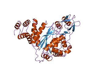
Mitochondrial neurogastrointestinal encephalopathy syndrome (MNGIE) is a rare autosomal recessive mitochondrial disease. It has been previously referred to as polyneuropathy, ophthalmoplegia, leukoencephalopathy, and intestinal pseudoobstruction. The disease presents in childhood, but often goes unnoticed for decades. Unlike typical mitochondrial diseases caused by mitochondrial DNA (mtDNA) mutations, MNGIE is caused by mutations in the TYMP gene, which encodes the enzyme thymidine phosphorylase. Mutations in this gene result in impaired mitochondrial function, leading to intestinal symptoms as well as neuro-ophthalmologic abnormalities. A secondary form of MNGIE, called MNGIE without leukoencephalopathy, can be caused by mutations in the POLG gene.
Progressive Myoclonic Epilepsies (PME) are a rare group of inherited neurodegenerative diseases characterized by myoclonus, resistance to treatment, and neurological deterioration. The cause of PME depends largely on the type of PME. Most PMEs are caused by autosomal dominant or recessive and mitochondrial mutations. The location of the mutation also affects the inheritance and treatment of PME. Diagnosing PME is difficult due to their genetic heterogeneity and the lack of a genetic mutation identified in some patients. The prognosis depends largely on the worsening symptoms and failure to respond to treatment. There is no current cure for PME and treatment focuses on managing myoclonus and seizures through antiepileptic medication (AED).
Chronic progressive external ophthalmoplegia (CPEO) is a type of eye disorder characterized by slowly progressive inability to move the eyes and eyebrows. It is often the only feature of mitochondrial disease, in which case the term CPEO may be given as the diagnosis. In other people suffering from mitochondrial disease, CPEO occurs as part of a syndrome involving more than one part of the body, such as Kearns–Sayre syndrome. Occasionally CPEO may be caused by conditions other than mitochondrial diseases.
Mitochondrially encoded tRNA leucine 1 (UUA/G) also known as MT-TL1 is a transfer RNA which in humans is encoded by the mitochondrial MT-TL1 gene.
Mitochondrially encoded tRNA histidine, also known as MT-TH, is a transfer RNA which, in humans, is encoded by the mitochondrial MT-TH gene.
Mitochondrially encoded tRNA aspartic acid also known as MT-TD is a transfer RNA which in humans is encoded by the mitochondrial MT-TD gene.
Mitochondrially encoded tRNA glutamic acid also known as MT-TE is a transfer RNA which in humans is encoded by the mitochondrial MT-TE gene. MT-TE is a small 69 nucleotide RNA that transfers the amino acid glutamic acid to a growing polypeptide chain at the ribosome site of protein synthesis during translation.
Mitochondrially encoded tRNA phenylalanine also known as MT-TF is a transfer RNA which in humans is encoded by the mitochondrial MT-TF gene.
Mitochondrially encoded tRNA glycine also known as MT-TG is a transfer RNA which in humans is encoded by the mitochondrial MT-TG gene.
Mitochondrially encoded tRNA isoleucine also known as MT-TI is a transfer RNA which in humans is encoded by the mitochondrial MT-TI gene.
Mitochondrially encoded tRNA lysine also known as MT-TK is a transfer RNA which in humans is encoded by the mitochondrial MT-TK gene.
Mitochondrially encoded tRNA leucine 2 (CUN) also known as MT-TL2 is a transfer RNA which in humans is encoded by the mitochondrial MT-TL2 gene.
Mitochondrially encoded tRNA proline also known as MT-TP is a transfer RNA that in humans is encoded by the mitochondrial MT-TP gene.
Mitochondrially encoded tRNA threonine also known as MT-TT is a transfer RNA which in humans is encoded by the mitochondrial MT-TT gene.
Mitochondrially encoded tRNA tyrosine, also known as MT-TY, is a transfer RNA which in humans is encoded by the mitochondrial MT-TY gene.