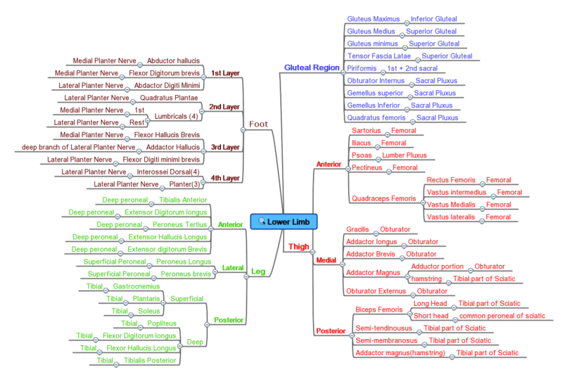
 Anterior view
 Posterior view
 A more detailed overview
Innervation overview


This is a table of skeletal muscles of the human anatomy, with muscle counts and other information.


The muscles are described using anatomical terminology. The columns are as follows:
| Name | Explanation |
|---|---|
| Muscle | US English [lower-alpha 1] name of the muscle per Terminologia Anatomica (TA), [13] minus the term "muscle", with the words reordered occasionally for better sorting. Parts and bellies are listed out as separate rows, as they are sometimes considered separate muscles. - Anything denoting the muscles relationship to another muscle such as e.g. superior, inferior etc. should always be used as a suffix and not a prefix, to create better sortability of the list. |
| Location | The location of the muscle in a standard human body. The location first specifies a group such as head, neck, torso, upper limbs, or lower limbs, then may have more specific information. However this additional information must be describing location not function. |
| Origin | The bone or other structure the muscle is attached to that remains immobile during the action. The term "bone" is omitted from bone names. |
| Insertion | The attachment point of the muscle, on a bone or otherwise, that moves during the action. |
| Artery | The artery which supplies the muscle with blood. The term "artery" is included to avoid confusing columns. |
| Nerve | The nerve(s) which tell the muscle to act (innervates the muscle). The term "nerve" is included for clarity. |
| Action | The movement performed by the muscle from the standard anatomical position. In other positions, other actions may be performed. |
| Antagonist | The muscle which can 'cancel' or to some degree reverse the action of the muscle. Muscle synergies are noted in parentheses when relevant. |
| O (Occurrences) | Number of times that the named muscle row occurs in a standard human body. Here it may also be denoted when a given muscles only occurs in a male or a female body. By (F) for female and (M) for male, if nothing is denoted. The muscle can be assumed to occur in both genders. This gender denomination should always be behind the number, not in front of it. |
| TA | The number of rows in the table for the relevant Terminologia Anatomica muscle. For example, TA splits the nasalis muscle into transverse and alar parts, so their TA column entries are 2. |
For Origin, Insertion and Action please name a specific Rib, Thoracic vertebrae or Cervical vertebrae, by using C1-7, T1-12 or R1-12.
There does not appear to be a definitive source counting all skeletal muscles. Different sources group muscles differently, regarding what is defined as different parts of a single muscle or as several muscles. There are also vestigial muscles that are present in some people but absent in others, such as the palmaris longus muscle. [14] [15] There are between 600 and 840 muscles within the typical human body, depending on how they are counted. [16] [17] [18] In the present table, using statistical counts of the instances of each muscle, and ignoring gender-specific muscles, there are 761 skeletal muscles. Sometimes male and females have the same muscle but with different purposes [19]
Numbers based on the list above:
| Total number | |
|---|---|
| Types of skeletal muscles - in a standard human 2023 | Male / Female [20] | ??(M) / ??(F) |
| Total number of skeletal muscles - in a standard human 2023 | Male / Female | ??(M) / ??(F) |
| Types of skeletal muscles - Non gender specific - listed | ?? |
| Total number of skeletal muscles - Non gender specific - listed | 761 |
| Types of skeletal muscles - Gender specific - listed | 4? |
| Total number of skeletal muscles - Gender specific - listed | 8? |
| Types of skeletal muscles listed | 290 |
| Total number of skeletal muscles represented in table (inclusive) | 791 |
| Total Number of entries in Terminologia Anatomica represented in table | 337 |
| Muscles listed which do not appear in a standard human 2023(gender specific muscles that appear in a standard male or female are not counted here) | 18 |
Muscles are often paired as agonistic and antagonistic muscles. [21] This can be a bit misleading as, in general, it is groups of muscles working together to either make or cancel a movement. [22] The present table lists some well-known relationships but is not at all complete.

The bladder is a hollow organ in humans and other vertebrates that stores urine from the kidneys before disposal by urination. In placental mammals, urine enters the bladder via the ureters and exits via the urethra. In humans, the bladder is a distensible organ that sits on the pelvic floor. The typical adult human bladder will hold between 300 and 500 ml before the urge to empty occurs, but can hold considerably more.
The muscular system is an organ system consisting of skeletal, smooth, and cardiac muscle. It permits movement of the body, maintains posture, and circulates blood throughout the body. The muscular systems in vertebrates are controlled through the nervous system although some muscles can be completely autonomous. Together with the skeletal system in the human, it forms the musculoskeletal system, which is responsible for the movement of the body.

The lumbricals are intrinsic muscles of the hand that flex the metacarpophalangeal joints, and extend the interphalangeal joints.

Skeletal muscles are organs of the vertebrate muscular system and typically are attached by tendons to bones of a skeleton. The muscle cells of skeletal muscles are much longer than in the other types of muscle tissue, and are often known as muscle fibers. The muscle tissue of a skeletal muscle is striated – having a striped appearance due to the arrangement of the sarcomeres.
Exercise physiology is the physiology of physical exercise. It is one of the allied health professions, and involves the study of the acute responses and chronic adaptations to exercise. Exercise physiologists are the highest qualified exercise professionals and utilise education, lifestyle intervention and specific forms of exercise to rehabilitate and manage acute and chronic injuries and conditions.

The somatic nervous system (SNS) is made up of nerves that link the brain and spinal cord to voluntary or skeletal muscles that are under conscious control as well as to skin sensory receptors. Specialized nerve fiber ends called sensory receptors are responsible for detecting information within and outside of the body.
Weakness is a symptom of many different medical conditions. The causes are many and can be divided into conditions that have true or perceived muscle weakness. True muscle weakness is a primary symptom of a variety of skeletal muscle diseases, including muscular dystrophy and inflammatory myopathy. It occurs in neuromuscular junction disorders, such as myasthenia gravis.

The deltoid muscle is the muscle forming the rounded contour of the human shoulder. It is also known as the 'common shoulder muscle', particularly in other animals such as the domestic cat. Anatomically, the deltoid muscle appears to be made up of three distinct sets of muscle fibers, namely the
Dystrophin is a rod-shaped cytoplasmic protein, and a vital part of a protein complex that connects the cytoskeleton of a muscle fiber to the surrounding extracellular matrix through the cell membrane. This complex is variously known as the costamere or the dystrophin-associated protein complex (DAPC). Many muscle proteins, such as α-dystrobrevin, syncoilin, synemin, sarcoglycan, dystroglycan, and sarcospan, colocalize with dystrophin at the costamere. It has a molecular weight of 427 kDa
Muscle contraction is the activation of tension-generating sites within muscle cells. In physiology, muscle contraction does not necessarily mean muscle shortening because muscle tension can be produced without changes in muscle length, such as when holding something heavy in the same position. The termination of muscle contraction is followed by muscle relaxation, which is a return of the muscle fibers to their low tension-generating state.

The posterior cricoarytenoid muscle is a intrinsic muscle of the larynx. It arises from the cricoid cartilage; it inserts onto the arytenoid cartilage of the same side. It is innervated by the recurrent laryngeal nerve. Each acts to open the vocal folds by pulling the vocal fold of the same side laterally. It participates in the production of sounds.

In humans and some other mammals, the soleus is a powerful muscle in the back part of the lower leg. It runs from just below the knee to the heel and is involved in standing and walking. It is closely connected to the gastrocnemius muscle, and some anatomists consider this combination to be a single muscle, the triceps surae. Its name is derived from the Latin word "solea", meaning "sandal".

The rectus capitis posterior minor is a muscle in the upper back part of the neck. It is one of the suboccipital muscles. Its inferior attachment is at the posterior arch of atlas; its superior attachment is onto the occipital bone at and below the inferior nuchal line. The muscle is innervated by the suboccipital nerve. The muscle acts as a weak extensor of the head.

The anterior superior iliac spine (ASIS) is a bony projection of the iliac bone, and an important landmark of surface anatomy. It refers to the anterior extremity of the iliac crest of the pelvis. It provides attachment for the inguinal ligament, and the sartorius muscle. The tensor fasciae latae muscle attaches to the lateral aspect of the superior anterior iliac spine, and also about 5 cm away at the iliac tubercle.
Muscle weakness is a lack of muscle strength. Its causes are many and can be divided into conditions that have either true or perceived muscle weakness. True muscle weakness is a primary symptom of a variety of skeletal muscle diseases, including muscular dystrophy and inflammatory myopathy. It occurs in neuromuscular junction disorders, such as myasthenia gravis. Muscle weakness can also be caused by low levels of potassium and other electrolytes within muscle cells. It can be temporary or long-lasting. The term myasthenia is from my- from Greek μυο meaning "muscle" + -asthenia ἀσθένεια meaning "weakness".

The semilunar hiatus is a crescent-shaped/semicircular/ curved slit/groove upon the lateral wall of the nasal cavity at the middle nasal meatus just inferior to the ethmoidal bulla. It is the location of the openings for the frontal sinus, maxillary sinus, and anterior ethmoidal sinus. It is bounded inferiorly and anteriorly by the sharp concave margin of the uncinate process of the ethmoid bone, superiorly by the ethmoidal bulla, and posteriorly by the ethmoidal process of the inferior nasal concha. It leads into the ethmoidal infundibulum; it marks the medial limit of the ethmoidal infundibulum.

Muscle hypertrophy or muscle building involves a hypertrophy or increase in size of skeletal muscle through a growth in size of its component cells. Two factors contribute to hypertrophy: sarcoplasmic hypertrophy, which focuses more on increased muscle glycogen storage; and myofibrillar hypertrophy, which focuses more on increased myofibril size. It is the primary focus of bodybuilding-related activities.

Anatomical terminology is a form of scientific terminology used by anatomists, zoologists, and health professionals such as doctors, physicians, and pharmacists.
Anatomical terminology is used to uniquely describe aspects of skeletal muscle, cardiac muscle, and smooth muscle such as their actions, structure, size, and location.
{{cite book}}: CS1 maint: multiple names: authors list (link){{cite journal}}: Cite journal requires |journal= (help)  |
| Part of a series of lists about |
| Human anatomy |
|---|