Protein phosphatase 1

Last updated
PP1 plays an instrumental role in glycogen metabolism through its responsibility for the interconversion between phosphorylase a and b. Phosphorylase and PP1 Diagram.png
PP1 plays an instrumental role in glycogen metabolism through its responsibility for the interconversion between phosphorylase a and b.

Protein phosphatase 1 (PP1) belongs to a certain class of phosphatases known as protein serine/threonine phosphatases. This type of phosphatase includes metal-dependent protein phosphatases (PPMs) and aspartate-based phosphatases. PP1 has been found to be important in the control of glycogen metabolism, muscle contraction, cell progression, neuronal activities, splicing of RNA, mitosis, [1] cell division, apoptosis, protein synthesis, and regulation of membrane receptors and channels. [2]

Contents

Structure

Each PP1 enzyme contains both a catalytic subunit and at least one regulatory subunit. [3] [4] The catalytic subunit consists of a 30-kD single-domain protein that can form complexes with other regulatory subunits. The catalytic subunit is highly conserved among all eukaryotes, thus suggesting a common catalytic mechanism. The catalytic subunit can form complexes with various regulatory subunits. These regulatory subunits play an important role in substrate specificity as well as compartmentalization. Some common regulatory subunits include GM (PPP1R3A) and GL (PPP1R3B), which are named after their locations of action within the body (muscle and liver respectively), [5] While the yeast S. cerevisiae only encodes one catalytic subunit, mammals have four isozymes encoded by three genes, each attracting a different set of regulatory subunits. [4] Regulation of these different processes is performed by distinct PP1 holoenzymes that facilitate the complexation of the PP1 catalytic subunit to various regulatory subunits. [4] and PPP1R3G.

X-ray crystallographic structural data is available for PP1 catalytic subunit. [3] The catalytic subunit of PP1 forms an α/β fold with a central β-sandwich arranged between two α-helical domains. The interaction of the three β-sheets of the β-sandwich creates a channel for catalytic activity, as it is the site of coordination of metal ions. [6] These metal ions have been identified as Mn and Fe and their coordination is provided by three histidines, two aspartic acids, and one asparagine. [7]

The PP1 mechanism involves the use of a di-metal ion and activating water. PP1 Mechanism 1.png
The PP1 mechanism involves the use of a di-metal ion and activating water.

Catalytic mechanism

The mechanism involves two metal ions binding and activating water, which initiates a nucleophilic attack on the phosphorus atom. [8]

Exogeneous inhibitors

Potential inhibitors include a variety of naturally occurring toxins including okadaic acid, a diarrhetic shellfish poison, strong tumor promoter, and microcystin. [9] Microcystin is a liver toxin produced by blue-green algae and contains a cyclic heptapeptide structure that interacts with three distinct regions of the surface of the catalytic subunit of PP1. [10] The structure of MCLR does not change when complexed with PP1, but the catalytic subunit of PP1 does in order to avoid steric effects of Tyr 276 of PP1 and Mdha side chain of MCLR. [7]

Cantharidic acid is also an inhibitor of PP1. [11]

Biological function and regulation

PP1 plays a crucial role in the regulation of blood glucose levels in the liver and glycogen metabolism. PP1 is important to the reciprocal regulation of glycogen metabolism by ensuring the opposite regulation of glycogen breakdown and glycogen synthesis. A key regulator of PP1 is glycogen phosphorylase a, which serves as a glucose sensor in hepatocytes. [12] When glucose levels are low, phosphorylase a in its active R state has PP1 bound tightly. This binding to phosphorylase a prevents any phosphatase activity of PP1 and maintains the glycogen phosphorylase in its active phosphorylated configuration. Therefore, there phosphorylase a will accelerate glycogen breakdown until adequate levels of glucose are achieved. [12] When glucose concentrations get too high, phosphorylase a is converted to its inactive, T state. By shifting phosphorylase a to its T state, PP1 dissociates from the complex. This dissociation activates glycogen synthase and converts phosphorylase a to phosphorylase b. Phosphorylase b does not bind PP1 allowing PP1 to remain activated. [12]

When the muscles of the body signal the need for glycogen degradation and an increase in blood glucose, PP1 will be regulated accordingly. Protein kinase A (cAMP-dependent protein kinase) can reduce the activity of PP1. The glycogen binding region, GM, becomes phosphorylated, which causes its dissociation from the catalytic PP1 unit. [12] This separation of the catalytic PP1 unit, glycogen, and other substrates causes a significant decrease in dephosphorylation. Also, when other substrates become phosphorylated by protein kinase A, they can bind to the catalytic subunit of PP1 and directly inhibit it. [12] In the end, glycogen phosphorylase is kept in its active form and glycogen synthase in its inactive form. Separately from inhibition of PP1, glucagon will also keep phosphorylase kinase active via cAMP, thereby keeping glycogen phosphorylase active.

When blood sugar is high, insulin will be secreted by beta cells of the pancreas, indirectly activating glycogen synthase and triggering glycogen synthesis. Although it has been known that PP1 is one of the most important phosphatases involved in insulin action since the late 1990s, [13] the precise mechanisms by which insulin regulates PP1 has only been uncovered more recently.

A 2019 study by researchers at Tsinghua, Fudan and the University of the Chinese Academy of Sciences demonstrated in both cell culture experiments and in PPP1R3G-knockdown mice that Akt (protein kinase B) directly phosphorylates Protein phosphatase 1 regulatory subunit 3G (PPP1R3G), which then binds to the PP1 complex, activating its phosphatase activity. The study demonstrated that phosphorylated PPP1R3G was also able to bind phosphorylated glycogen synthase (p-GS) independently and recruit p-GS towards PP1, allowing PP1 to dephosphorylate and thereby activate glycogen synthase independent of GSK3 (which is already known to be inhibited by Akt). [14]

Clinical relevance

In Alzheimer's, hyperphosphorylation of the microtubule-associated protein inhibits the assembly of microtubules in neurons. Researchers at the New York State Institute for Basic Research in Developmental Disabilities showed that there is significantly lower type 1 phosphatase activity in both gray and white matters in Alzheimer disease brains. [15] This suggests that dysfunctional phosphatases play a role in Alzheimer's disease.

Regulation of HIV-1 transcription by Protein Phosphatase 1 (PP1). It has been recognized that protein phosphatase-1 (PP1) serves as an important regulator of HIV-1 transcription. Researchers at Howard University showed that Tat protein targets PP1 to the nucleus and the consequent interaction is important for HIV-1 transcription. [16] The protein also contributes to ebolavirus pathogenesis by dephosphorylating the viral transcription activator VP30, allowing it to produce viral mRNAs. Inhibition of PP1 prevents VP30 dephosphorylation, thus preventing manufacture of viral mRNA, and thus viral protein. The viral L polymerase is, however, still capable of replicating viral genomes without VP30 dephosphorylation by PP1. [17]

The herpes simplex virus protein ICP34.5 also activates protein phosphatase 1, which overcomes the cellular stress response to viral infection; protein kinase R is activated by the virus' double-stranded RNA, and protein kinase R then phosphorylates a protein called eukaryotic initiation factor-2A (eIF-2A), which inactivates eIF-2A. EIF-2A is required for translation so by shutting down eIF-2A, the cell prevents the virus from hijacking its own protein-making machinery. Herpesviruses in turn evolved ICP34.5 to defeat the defense; ICP34.5 activates protein phosphatase-1A which dephosphorylates eIF-2A, allowing translation to occur again. ICP34.5 shares the C-terminal regulatory domain (InterPro :  IPR019523 ) with protein phosphatase 1 subunit 15A/B. [18]

Subunits

protein phosphatase 1, catalytic subunit, alpha isozyme
Identifiers
Symbol PPP1CA
Alt. symbolsPP1, PP1a, MGC15877, MGC1674, PP-1A, PP1alpha, PPP1A
NCBI gene 5499
HGNC 9281
OMIM 176875
RefSeq NP_002699.1
UniProt P62136
Other data
EC number 3.1.3.16
Locus Chr. 11 q13
Search for
Structures Swiss-model
Domains InterPro
protein phosphatase 1, catalytic subunit, beta isozyme
Identifiers
Symbol PPP1CB
Alt. symbolsPP1, PP1b, PP1beta, PP-1B; PPP1CD; MGC3672; PP1beta; PPP1CB
NCBI gene 5500
HGNC 9282
OMIM 600590
RefSeq NP_002700.1
UniProt P62140
Other data
EC number 3.1.3.16
Locus Chr. 2 p23
Search for
Structures Swiss-model
Domains InterPro
protein phosphatase 1, catalytic subunit, gamma isozyme
Identifiers
Symbol PPP1CC
Alt. symbolsPP1gamma, PP1y, PP1gamma, PPP1G
NCBI gene 5501
HGNC 9283
OMIM 176914
RefSeq NP_002701.1
UniProt P36873
Other data
EC number 3.1.3.16
Locus Chr. 12 q24
Search for
Structures Swiss-model
Domains InterPro

Protein phosphatase 1 is a multimeric enzyme that may contain the following subunits: [19]

As described earlier, a catalytic subunit is always paired with one or more regulatory subunits. The core sequence motif for binding to the catalytic subunit is "RVxF", but additional motifs allow for extra sites to be used. Some complexes with two regulatory subunits attached have been reported in 2002 and 2007. [4]

Related Research Articles

A protein phosphatase is a phosphatase enzyme that removes a phosphate group from the phosphorylated amino acid residue of its substrate protein. Protein phosphorylation is one of the most common forms of reversible protein posttranslational modification (PTM), with up to 30% of all proteins being phosphorylated at any given time. Protein kinases (PKs) are the effectors of phosphorylation and catalyse the transfer of a γ-phosphate from ATP to specific amino acids on proteins. Several hundred PKs exist in mammals and are classified into distinct super-families. Proteins are phosphorylated predominantly on Ser, Thr and Tyr residues, which account for 79.3, 16.9 and 3.8% respectively of the phosphoproteome, at least in mammals. In contrast, protein phosphatases (PPs) are the primary effectors of dephosphorylation and can be grouped into three main classes based on sequence, structure and catalytic function. The largest class of PPs is the phosphoprotein phosphatase (PPP) family comprising PP1, PP2A, PP2B, PP4, PP5, PP6 and PP7, and the protein phosphatase Mg2+- or Mn2+-dependent (PPM) family, composed primarily of PP2C. The protein Tyr phosphatase (PTP) super-family forms the second group, and the aspartate-based protein phosphatases the third. The protein pseudophosphatases form part of the larger phosphatase family, and in most cases are thought to be catalytically inert, instead functioning as phosphate-binding proteins, integrators of signalling or subcellular traps. Examples of membrane-spanning protein phosphatases containing both active (phosphatase) and inactive (pseudophosphatase) domains linked in tandem are known, conceptually similar to the kinase and pseudokinase domain polypeptide structure of the JAK pseudokinases. A complete comparative analysis of human phosphatases and pseudophosphatases has been completed by Manning and colleagues, forming a companion piece to the ground-breaking analysis of the human kinome, which encodes the complete set of ~536 human protein kinases.

<span class="mw-page-title-main">Kinase</span> Enzyme catalyzing transfer of phosphate groups onto specific substrates

In biochemistry, a kinase is an enzyme that catalyzes the transfer of phosphate groups from high-energy, phosphate-donating molecules to specific substrates. This process is known as phosphorylation, where the high-energy ATP molecule donates a phosphate group to the substrate molecule. This transesterification produces a phosphorylated substrate and ADP. Conversely, it is referred to as dephosphorylation when the phosphorylated substrate donates a phosphate group and ADP gains a phosphate group. These two processes, phosphorylation and dephosphorylation, occur four times during glycolysis.

<span class="mw-page-title-main">Glucagon</span> Peptide hormone

Glucagon is a peptide hormone, produced by alpha cells of the pancreas. It raises the concentration of glucose and fatty acids in the bloodstream and is considered to be the main catabolic hormone of the body. It is also used as a medication to treat a number of health conditions. Its effect is opposite to that of insulin, which lowers extracellular glucose. It is produced from proglucagon, encoded by the GCG gene.

<span class="mw-page-title-main">AMP-activated protein kinase</span> Class of enzymes

5' AMP-activated protein kinase or AMPK or 5' adenosine monophosphate-activated protein kinase is an enzyme that plays a role in cellular energy homeostasis, largely to activate glucose and fatty acid uptake and oxidation when cellular energy is low. It belongs to a highly conserved eukaryotic protein family and its orthologues are SNF1 in yeast, and SnRK1 in plants. It consists of three proteins (subunits) that together make a functional enzyme, conserved from yeast to humans. It is expressed in a number of tissues, including the liver, brain, and skeletal muscle. In response to binding AMP and ADP, the net effect of AMPK activation is stimulation of hepatic fatty acid oxidation, ketogenesis, stimulation of skeletal muscle fatty acid oxidation and glucose uptake, inhibition of cholesterol synthesis, lipogenesis, and triglyceride synthesis, inhibition of adipocyte lipogenesis, inhibition of adipocyte lipolysis, and modulation of insulin secretion by pancreatic β-cells.

<span class="mw-page-title-main">GSK-3</span> Class of enzymes

Glycogen synthase kinase 3 (GSK-3) is a serine/threonine protein kinase that mediates the addition of phosphate molecules onto serine and threonine amino acid residues. First discovered in 1980 as a regulatory kinase for its namesake, glycogen synthase (GS), GSK-3 has since been identified as a protein kinase for over 100 different proteins in a variety of different pathways. In mammals, including humans, GSK-3 exists in two isozymes encoded by two homologous genes GSK-3α (GSK3A) and GSK-3β (GSK3B). GSK-3 has been the subject of much research since it has been implicated in a number of diseases, including type 2 diabetes, Alzheimer's disease, inflammation, cancer, addiction and bipolar disorder.

Glycogenesis is the process of glycogen synthesis, in which glucose molecules are added to chains of glycogen for storage. This process is activated during rest periods following the Cori cycle, in the liver, and also activated by insulin in response to high glucose levels.

<span class="mw-page-title-main">Glycogen phosphorylase</span> Class of enzymes

Glycogen phosphorylase is one of the phosphorylase enzymes. Glycogen phosphorylase catalyzes the rate-limiting step in glycogenolysis in animals by releasing glucose-1-phosphate from the terminal alpha-1,4-glycosidic bond. Glycogen phosphorylase is also studied as a model protein regulated by both reversible phosphorylation and allosteric effects.

In biochemistry, dephosphorylation is the removal of a phosphate (PO43−) group from an organic compound by hydrolysis. It is a reversible post-translational modification. Dephosphorylation and its counterpart, phosphorylation, activate and deactivate enzymes by detaching or attaching phosphoric esters and anhydrides. A notable occurrence of dephosphorylation is the conversion of ATP to ADP and inorganic phosphate.

<span class="mw-page-title-main">Glycogen synthase</span> Enzyme class, includes all types of glycogen/starch synthases

Glycogen synthase is a key enzyme in glycogenesis, the conversion of glucose into glycogen. It is a glycosyltransferase that catalyses the reaction of UDP-glucose and n to yield UDP and n+1.

<span class="mw-page-title-main">Phosphorylase kinase</span>

Phosphorylase kinase (PhK) is a serine/threonine-specific protein kinase which activates glycogen phosphorylase to release glucose-1-phosphate from glycogen. PhK phosphorylates glycogen phosphorylase at two serine residues, triggering a conformational shift which favors the more active glycogen phosphorylase “a” form over the less active glycogen phosphorylase b.

<span class="mw-page-title-main">PRKACA</span> Protein-coding gene in the species Homo sapiens

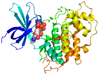
The catalytic subunit α of protein kinase A is a key regulatory enzyme that in humans is encoded by the PRKACA gene. This enzyme is responsible for phosphorylating other proteins and substrates, changing their activity. Protein kinase A catalytic subunit is a member of the AGC kinase family, and contributes to the control of cellular processes that include glucose metabolism, cell division, and contextual memory. PKA Cα is part of a larger protein complex that is responsible for controlling when and where proteins are phosphorylated. Defective regulation of PKA holoenzyme activity has been linked to the progression of cardiovascular disease, certain endocrine disorders and cancers.

<span class="mw-page-title-main">PPP2CA</span> Enzyme

Serine/threonine-protein phosphatase 2A catalytic subunit alpha isoform is an enzyme that is encoded by the PPP2CA gene.

<span class="mw-page-title-main">Myosin-light-chain phosphatase</span>

Myosin light-chain phosphatase, also called myosin phosphatase (EC 3.1.3.53; systematic name [myosin-light-chain]-phosphate phosphohydrolase), is an enzyme (specifically a serine/threonine-specific protein phosphatase) that dephosphorylates the regulatory light chain of myosin II:

<span class="mw-page-title-main">PPP1R3A</span> Protein-coding gene in the species Homo sapiens

Protein phosphatase 1 regulatory subunit 3A is an enzyme that in humans is encoded by the PPP1R3A gene.

<span class="mw-page-title-main">PPP2R5E</span> Protein-coding gene in the species Homo sapiens

Serine/threonine-protein phosphatase 2A 56 kDa regulatory subunit epsilon isoform is an enzyme that in humans is encoded by the PPP2R5E gene.

<span class="mw-page-title-main">Protein phosphorylation</span> Process of introducing a phosphate group on to a protein

Protein phosphorylation is a reversible post-translational modification of proteins in which an amino acid residue is phosphorylated by a protein kinase by the addition of a covalently bound phosphate group. Phosphorylation alters the structural conformation of a protein, causing it to become activated, deactivated, or otherwise modifying its function. Approximately 13,000 human proteins have sites that are phosphorylated.

The Akt signaling pathway or PI3K-Akt signaling pathway is a signal transduction pathway that promotes survival and growth in response to extracellular signals. Key proteins involved are PI3K and Akt.

The insulin transduction pathway is a biochemical pathway by which insulin increases the uptake of glucose into fat and muscle cells and reduces the synthesis of glucose in the liver and hence is involved in maintaining glucose homeostasis. This pathway is also influenced by fed versus fasting states, stress levels, and a variety of other hormones.

The integrated stress response is a cellular stress response conserved in eukaryotic cells that downregulates protein synthesis and upregulates specific genes in response to internal or environmental stresses.

Ceramide-activated protein phosphatases (CAPPs) are a group of enzymes that are activated by the lipid second messenger ceramide. Known CAPPs include members of the protein phosphatase 1 (PP1) and protein phosphatase 2A (PP2A) families. CAPPs are a subset of intracellular serine/threonine phosphatases. Each CAPP consists of a catalytic subunit which confers phosphatase activity and a regulatory subunit which confers substrate specificity. CAPP involvement has been implicated in glycogen metabolism, apoptotic pathways related to cancer and other cellular pathways related to Alzheimer’s disease.

References

  1. Tournebize R, Andersen SS, Verde F, Dorée M, Karsenti E, Hyman AA (September 1997). "Distinct roles of PP1 and PP2A-like phosphatases in control of microtubule dynamics during mitosis". The EMBO Journal. 16 (18): 5537–49. doi:10.1093/emboj/16.18.5537. PMC   1170186 . PMID   9312013.
  2. Fong NM, Jensen TC, Shah AS, Parekh NN, Saltiel AR, Brady MJ (November 2000). "Identification of binding sites on protein targeting to glycogen for enzymes of glycogen metabolism". The Journal of Biological Chemistry. 275 (45): 35034–9. doi: 10.1074/jbc.M005541200 . PMID   10938087.
  3. 1 2 Goldberg J, Huang HB, Kwon YG, Greengard P, Nairn AC, Kuriyan J (August 1995). "Three-dimensional structure of the catalytic subunit of protein serine/threonine phosphatase-1". Nature. 376 (6543): 745–53. Bibcode:1995Natur.376..745G. doi:10.1038/376745a0. PMID   7651533. S2CID   4256743.
  4. 1 2 3 4 Virshup DM, Shenolikar S (March 2009). "From promiscuity to precision: protein phosphatases get a makeover". Molecular Cell. 33 (5): 537–45. doi: 10.1016/j.molcel.2009.02.015 . PMID   19285938.
  5. Armstrong CG, Browne GJ, Cohen P, Cohen PT (November 1997). "PPP1R6, a novel member of the family of glycogen-targetting subunits of protein phosphatase 1". FEBS Letters. 418 (1–2): 210–4. doi:10.1016/S0014-5793(97)01385-9. PMID   9414128. S2CID   21169749.
  6. Egloff MP, Johnson DF, Moorhead G, Cohen PT, Cohen P, Barford D (April 1997). "Structural basis for the recognition of regulatory subunits by the catalytic subunit of protein phosphatase 1". The EMBO Journal. 16 (8): 1876–87. doi:10.1093/emboj/16.8.1876. PMC   1169791 . PMID   9155014.
  7. 1 2 Barford D, Das AK, Egloff MP (1998). "The structure and mechanism of protein phosphatases: insights into catalysis and regulation". Annual Review of Biophysics and Biomolecular Structure. 27: 133–64. doi:10.1146/annurev.biophys.27.1.133. PMID   9646865.
  8. Zhang Y, Zhang M, Zhang Y (March 2011). "Crystal structure of Ssu72, an essential eukaryotic phosphatase specific for the C-terminal domain of RNA polymerase II, in complex with a transition state analogue". The Biochemical Journal. 434 (3): 435–44. doi:10.1042/BJ20101471. PMID   21204787.
  9. Wera S, Hemmings BA (October 1995). "Serine/threonine protein phosphatases". The Biochemical Journal. 311 ( Pt 1) (1): 17–29. doi:10.1042/bj3110017. PMC   1136113 . PMID   7575450.
  10. MacKintosh C, Beattie KA, Klumpp S, Cohen P, Codd GA (May 1990). "Cyanobacterial microcystin-LR is a potent and specific inhibitor of protein phosphatases 1 and 2A from both mammals and higher plants". FEBS Letters. 264 (2): 187–92. doi: 10.1016/0014-5793(90)80245-E . PMID   2162782. S2CID   27643473.
  11. "Cantharidic Acid | CAS 28874-45-5".
  12. 1 2 3 4 5 Berg JM, Stryer L, Tymoczko JL (2010-12-24). Biochemistry (7th ed.). New York: W.H. Freeman. ISBN   978-1-4292-2936-4.
  13. Brady M, Saltiel A (January 1, 2001). "The role of protein phosphatase-1 in insulin action" (PDF). Recent Prog Horm Res . 56: 157–173. doi:10.1210/rp.56.1.157. PMID   11237211 . Retrieved December 15, 2023.
  14. Li Q, Zhao Q, Zhang J, Linkang L, Wenhao W, Chua B, Chen Y, Xu L, Li P (September 24, 2019). "The Protein Phosphatase 1 Complex Is a Direct Target of AKT that Links Insulin Signaling to Hepatic Glycogen Deposition". Cell Reports. 28 (13): 3406–3422. doi: 10.1016/j.celrep.2019.08.066 . PMID   31553910.
  15. Gong CX, Singh TJ, Grundke-Iqbal I, Iqbal K (September 1993). "Phosphoprotein phosphatase activities in Alzheimer disease brain". Journal of Neurochemistry. 61 (3): 921–7. doi:10.1111/j.1471-4159.1993.tb03603.x. PMID   8395566. S2CID   30225343.
  16. Nekhai S, Jerebtsova M, Jackson A, Southerland W (January 2007). "Regulation of HIV-1 transcription by protein phosphatase 1". Current HIV Research. 5 (1): 3–9. doi:10.2174/157016207779316279. PMID   17266553. S2CID   12105058.
  17. Ilinykh PA, Tigabu B, Ivanov A, Ammosova T, Obukhov Y, Garron T, Kumari N, Kovalskyy D, Platonov MO, Naumchik VS, Freiberg AN, Nekhai S, Bukreyev A (August 2014). "Role of protein phosphatase 1 in dephosphorylation of Ebola virus VP30 protein and its targeting for the inhibition of viral transcription". The Journal of Biological Chemistry. 289 (33): 22723–38. doi: 10.1074/jbc.M114.575050 . PMC   4132779 . PMID   24936058.
  18. Agarwalla PK, Aghi MK (2012). "Oncolytic herpes simplex virus engineering and preparation". Oncolytic Viruses. Methods in Molecular Biology. Vol. 797. pp. 1–19. doi:10.1007/978-1-61779-340-0_1. ISBN   978-1-61779-339-4. PMID   21948465.
  19. Cohen PT (January 2002). "Protein phosphatase 1--targeted in many directions". Journal of Cell Science. 115 (Pt 2): 241–56. doi:10.1242/jcs.115.2.241. PMID   11839776.