Ronald Vale

Last updated

Ronald David Vale [1]
Ronald Vale in September 2018.jpg
Vale in September 2018
Born (1959-01-11) January 11, 1959 (age 65)[ citation needed ]
NationalityAmerican
Alma mater Marine Biological Laboratory
Stanford University
University of California, Santa Barbara
Known forResearch in molecular motors, particularly kinesin and dynein
Awards Shaw Prize in Life Science and Medicine (2017)
Massry Prize (2013)
Albert Lasker Award for Basic Medical Research (2012)
Wiley Prize in Biomedical Sciences (2012)
Scientific career
Fields Biochemistry
Cell Biology
Institutions University of California, San Francisco
Howard Hughes Medical Institute
Thesis Nerve growth factor receptors and axonal transport  (1985)
Doctoral advisor Eric Shooter [2]

Ronald David Vale ForMemRS (born 1959) is an American biochemist and cell biologist. He is a professor at the Department of Cellular and Molecular Pharmacology, University of California, San Francisco. [3] His research is focused on motor proteins, particularly kinesin and dynein. [4] He was awarded the Canada Gairdner International Award for Biomedical Research in 2019, the Shaw Prize in Life Science and Medicine in 2017 together with Ian Gibbons, and the Albert Lasker Award for Basic Medical Research in 2012 alongside Michael Sheetz and James Spudich. He is a fellow of the American Academy of Arts and Sciences and a member of the National Academy of Sciences. He was the president of the American Society for Cell Biology in 2012. [5] He has also been an investigator at the Howard Hughes Medical Institute since 1995. [3] In 2019, Vale was named executive director of the Janelia Research Campus and a vice president of HHMI; his appointment began in early 2020. [6]

Contents

Early life and education

Vale was born in Hollywood, California. His mother, Evelyn, was a former actress; his father, Eugene, was a novelist and screenwriter. He finished high school at Hollywood High School. For his grade 10 science project, he set up a laboratory at the basement of his home to investigate the circadian rhythm of bean plants. His guidance counselor contacted Karl Hammer at the University of California, Los Angeles, who allowed Vale to continue his experiments at his laboratory. His guidance counselor also encouraged Vale to submit his work to the Westinghouse Science Talent Search (now the Regeneron Science Talent Search), where he was selected as one of the top forty students in the US. [2]

Vale is a first-generation university student. [2] He entered the College of Creative Studies, University of California, Santa Barbara, and earned a bachelor's degree in chemistry and biology in 1980. [7] During his study, he first worked at the laboratory of C. Fred Fox at UCLA, then at Robert Lefkowitz's group at Duke University, earning him two articles published in 1984 [8] and 1982, [9] respectively.

In 1980, Vale entered an MD/PhD program at Stanford University, supervised by Eric Shooter, [2] where he studied the nerve growth factor receptor (also known as the neurotrophic factor receptor). [10]

Vale obtained his PhD in neuroscience in 1985. He then spent one year as an NIH staff scientist in Tom Reese's laboratory at the Marine Biological Laboratory at Woods Hole, MA. [7]

Scientific career

While working on Nerve growth factor (NGF) receptors as a graduate student, Vale became interested in exploring the mechanism of how receptors and other molecules are transported in nerve axons. He then heard of the research of Michael Sheetz and James Spudich, who used a video camera on a microscope to film myosin-coated beads moving along actin filaments. In 1983, Vale and Sheetz decided to test whether the movement of myosin on actin was the source for organelle transport in axons, using the squid giant axon as a model. However, since no squid were caught that year at Stanford's Hopkins Marine Station, following Shooter's approval, they went to the Marine Biological Laboratory instead. [11]

At the Marine Biological Laboratory, Vale and Sheetz teamed up with Bruce Schnapp and Thomas J. Reese. [11] They found that membrane organelle transport occurred bidirectionally on a microtubule, and not actin filament as Vale had originally thought. [12] Vale further demonstrated that purified organelles by themselves rarely moved on microtubules, but movement was observed after adding the cytosol of the axon. He then discovered serendipitously that cytosol caused microtubules to translocate along a glass surface. Similarly, he found that cytosol-coated beads moved along microtubules. These two phenomena provided assays to study microtubule-based motility assay in vitro. [13] In 1985, Vale, Sheetz and Reese isolated the dominant motor protein in the cytosol, naming it "kinesin." They showed that kinesin only moved in one direction towards the plus ends of microtubules [14] and a second motor (later shown to be dynein by Richard Vallee) moved in the opposite direction. The results of Vale and colleagues on axonal transport were published in five papers in 1985.

Vale did not finish his MD, and joined the University of California, San Francisco as an assistant professor in 1986. [15] He was promoted to associate professor in 1992 and then to full professor in 1994. [7] In 1989, Vale, with Jonathan Howard and A. James Hudspeth developed a single molecular assay for kinesin. In 1991, he discovered the first protein that severed microtubules and later purified and named it katanin. [16] [17] In 1996, Vale and colleagues solved the crystal structure of the kinesin motor domain. [18] and discovered unexpectedly that it is structurally similar to myosin. [19] In that same year, working with Toshio Yanagida, Vale developed a single-molecule fluorescence assay for kinesin. In 1999, using various techniques, Vale and co-workers developed a mechanical model for how the two motor domains of the kinesin dimer walk in a “hand-over-hand” model along a microtubule.

Since 2003, Vale has focused on dynein, a motor protein discovered by Ian R. Gibbons in 1965. Although its discovery occurred 20 years before kinesin, its large size hampered its investigation. In 2006, Vale's laboratory prepared recombinant dynein from yeast, and elucidated how it walked on microtubules using single-molecule microscopy. [20] He then worked with Gibbons to determine the structure of the dynein microtubule-binding domain. [21] His team also solved the structure of the dynein motor domain. [22] Vale has extended his research to other fields, including T-cell signalling [23] and RNA biology. [24]

Outreach

Vale founded iBiology in 2006, a non-profit organization that produces and disseminates free online videos by leading biologists, speaking about biological principles and their research, and scientific training and professional development for practicing scientists. Vale recently [25] [26] founded and produced The Explorer's Guide to Biology (XBio), a free online undergraduate "textbook" that provides a storytelling and discover-focused approach to learning biology.

Between 2004 and 2008, Vale and Tim Mitchison co-directed the Physiology Course at the Marine Biological Laboratory in Woods Hole, transforming it into an interdisciplinary training environment that brings together biologists, physicists and computational scientists.

In 2009, Vale established the Young Investigators' Meeting in India, which provides a mentoring and networking workshop for postdocs and junior faculty in India. He founded ASAPbio (Accelerating Science and Publication in Biology) in 2015, promoting the use of preprints and an open and transparent peer-review process. [11] Also in 2009, Vale founded the Bangalore Microscopy Course, held at the National Centre for Biological Research, which provides international training in light microscopy. He also organized an online microscopy course through iBiology.

Nico Stuurman and Vale also conceived of and developed Micro-Manager, a free and open-source microscopy software [27] that was supported for many years through the Vale laboratory and now operates through the University of Wisconsin.

Awards and honours

Related Research Articles

<span class="mw-page-title-main">Microtubule</span> Polymer of tubulin that forms part of the cytoskeleton

Microtubules are polymers of tubulin that form part of the cytoskeleton and provide structure and shape to eukaryotic cells. Microtubules can be as long as 50 micrometres, as wide as 23 to 27 nm and have an inner diameter between 11 and 15 nm. They are formed by the polymerization of a dimer of two globular proteins, alpha and beta tubulin into protofilaments that can then associate laterally to form a hollow tube, the microtubule. The most common form of a microtubule consists of 13 protofilaments in the tubular arrangement.

<span class="mw-page-title-main">Cytoskeleton</span> Network of filamentous proteins that forms the internal framework of cells

The cytoskeleton is a complex, dynamic network of interlinking protein filaments present in the cytoplasm of all cells, including those of bacteria and archaea. In eukaryotes, it extends from the cell nucleus to the cell membrane and is composed of similar proteins in the various organisms. It is composed of three main components: microfilaments, intermediate filaments, and microtubules, and these are all capable of rapid growth or disassembly depending on the cell's requirements.
<span class="mw-page-title-main">Kinesin</span> Eukaryotic motor protein

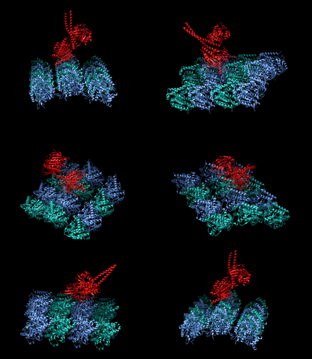
A kinesin is a protein belonging to a class of motor proteins found in eukaryotic cells. Kinesins move along microtubule (MT) filaments and are powered by the hydrolysis of adenosine triphosphate (ATP). The active movement of kinesins supports several cellular functions including mitosis, meiosis and transport of cellular cargo, such as in axonal transport, and intraflagellar transport. Most kinesins walk towards the plus end of a microtubule, which, in most cells, entails transporting cargo such as protein and membrane components from the center of the cell towards the periphery. This form of transport is known as anterograde transport. In contrast, dyneins are motor proteins that move toward the minus end of a microtubule in retrograde transport.

<span class="mw-page-title-main">Dynein</span> Class of enzymes

Dyneins are a family of cytoskeletal motor proteins that move along microtubules in cells. They convert the chemical energy stored in ATP to mechanical work. Dynein transports various cellular cargos, provides forces and displacements important in mitosis, and drives the beat of eukaryotic cilia and flagella. All of these functions rely on dynein's ability to move towards the minus-end of the microtubules, known as retrograde transport; thus, they are called "minus-end directed motors". In contrast, most kinesin motor proteins move toward the microtubules' plus-end, in what is called anterograde transport.
<span class="mw-page-title-main">Molecular motor</span> Biological molecular machines

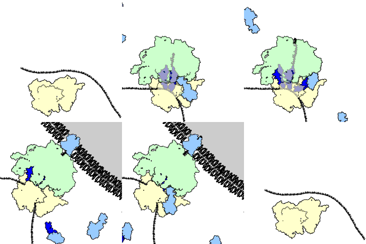
Molecular motors are natural (biological) or artificial molecular machines that are the essential agents of movement in living organisms. In general terms, a motor is a device that consumes energy in one form and converts it into motion or mechanical work; for example, many protein-based molecular motors harness the chemical free energy released by the hydrolysis of ATP in order to perform mechanical work. In terms of energetic efficiency, this type of motor can be superior to currently available man-made motors. One important difference between molecular motors and macroscopic motors is that molecular motors operate in the thermal bath, an environment in which the fluctuations due to thermal noise are significant.

<span class="mw-page-title-main">Axonal transport</span> Movement of organelles

Axonal transport, also called axoplasmic transport or axoplasmic flow, is a cellular process responsible for movement of mitochondria, lipids, synaptic vesicles, proteins, and other organelles to and from a neuron's cell body, through the cytoplasm of its axon called the axoplasm. Since some axons are on the order of meters long, neurons cannot rely on diffusion to carry products of the nucleus and organelles to the end of their axons. Axonal transport is also responsible for moving molecules destined for degradation from the axon back to the cell body, where they are broken down by lysosomes.
<span class="mw-page-title-main">Motor protein</span> Class of molecular proteins

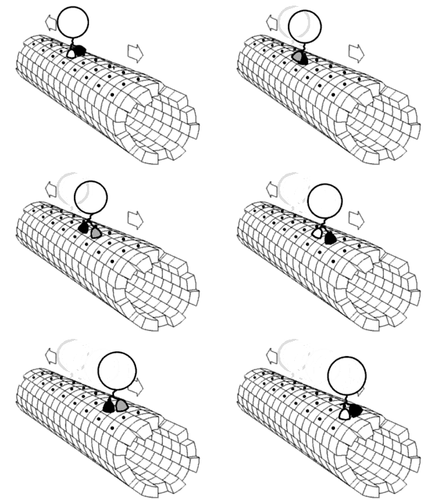
Motor proteins are a class of molecular motors that can move along the cytoplasm of cells. They convert chemical energy into mechanical work by the hydrolysis of ATP. Flagellar rotation, however, is powered by a proton pump.

Plus-end-directed kinesin ATPase (EC 3.6.4.4, kinesin) is an enzyme with systematic name kinesin ATP phosphohydrolase (plus-end-directed). This enzyme catalyses the following chemical reaction

<span class="mw-page-title-main">Dynactin</span>

Dynactin is a 23 subunit protein complex that acts as a co-factor for the microtubule motor cytoplasmic dynein-1. It is built around a short filament of actin related protein-1 (Arp1).

<span class="mw-page-title-main">KIF5B</span> Protein-coding gene in the species Homo sapiens

Kinesin family member 5B (KIF5B) is a protein that in humans is encoded by the KIF5B gene. It is part of the kinesin family of motor proteins.

<span class="mw-page-title-main">KIF3B</span> Protein-coding gene in the species Homo sapiens

Kinesin-like protein KIF3B is a protein that in humans is encoded by the KIF3B gene. KIF3B is an N-type protein that complexes with two other kinesin proteins to form two-headed anterograde motors. First, KIF3B forms a heterodimer with KIF3A ; (KIF3A/3B), that is membrane-bound and has ATPase activity. Then KIFAP3 binds to the tail domain to form a heterotrimeric motor. This motor has a plus end-directed microtubule sliding activity that exhibits a velocity of ~0.3 μm/s a. There are 14 kinesin protein families in the kinesin superfamily and KIF3B is part of the Kinesin-2 family, of kinesins that can all form heterotrimeric complexes. Expression of the three motor subunits is ubiquitous. The KIG3A/3B/KAP3 motors can transport 90 to 160 nm in diameter organelles.

<span class="mw-page-title-main">Kinesin-like protein KIF11</span> Protein-coding gene in the species Homo sapiens

Kinesin-like protein KIF11 is a molecular motor protein that is essential in mitosis. In humans it is coded for by the gene KIF11. Kinesin-like protein KIF11 is a member of the kinesin superfamily, which are nanomotors that move along microtubule tracks in the cell. Named from studies in the early days of discovery, it is also known as Kinesin-5, or as BimC, Eg5 or N-2, based on the founding members of this kinesin family.

<span class="mw-page-title-main">Intracellular transport</span> Directed movement of vesicles and substances within a cell

Intracellular transport is the movement of vesicles and substances within a cell. Intracellular transport is required for maintaining homeostasis within the cell by responding to physiological signals. Proteins synthesized in the cytosol are distributed to their respective organelles, according to their specific amino acid’s sorting sequence. Eukaryotic cells transport packets of components to particular intracellular locations by attaching them to molecular motors that haul them along microtubules and actin filaments. Since intracellular transport heavily relies on microtubules for movement, the components of the cytoskeleton play a vital role in trafficking vesicles between organelles and the plasma membrane by providing mechanical support. Through this pathway, it is possible to facilitate the movement of essential molecules such as membrane‐bounded vesicles and organelles, mRNA, and chromosomes.

<span class="mw-page-title-main">Anna Akhmanova</span> Russian cell biologist

Anna Sergeevna Akhmanova is a Russian-born professor of Cell Biology at Utrecht University in the Netherlands. She is best known for her research regarding microtubules and the proteins, called TIPs, that stabilize one specific end of the tubules. Among the awards she has won, she was one of the recipients of the 2018 Spinoza Prize, the highest honor for Dutch scientists.
<span class="mw-page-title-main">Ian R. Gibbons</span> English biophysicist and cell biologist

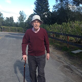
Ian Read Gibbons, was a biophysicist and cell biologist. He discovered and named dynein, and demonstrated energy source as ATP is sufficient for dynein to walk on microtubules. In 2017, he and Ronald Vale received the Shaw Prize for their research on microtubule motor proteins.
<span class="mw-page-title-main">Andrew P. Carter</span> British structural biologist

Andrew P. Carter is a British structural biologist who works at the Medical Research Council (MRC) Laboratory of Molecular Biology (LMB) in Cambridge, UK. He is known for his work on the microtubule motor dynein.

Samara Reck-Peterson is an American cell biologist and biophysicist. She is a Professor of Cellular and Molecular Medicine and Cell and Developmental Biology at the University of California, San Diego and an Investigator of the Howard Hughes Medical Institute. She is known for her contributions to our understanding of how dynein, an exceptionally large motor protein that moves many intracellular cargos, works and is regulated. She developed one of the first systems to produce recombinant dynein and discovered that, unlike other cytoskeletal motors, dynein can take a wide variety of step sizes, forward and back and even sideways. She lives in San Diego, California.

Edwin W. Taylor is an adjunct professor of cell and developmental biology at Northwestern University. He was elected to the National Academy of Sciences in 2001. Taylor received a BA in physics and chemistry from the University of Toronto in 1952; an MSc in physical chemistry from McMaster University in 1955, and a PhD in biophysics from the University of Chicago in 1957. In 2001 Taylor was elected to the National Academy of Scineces in Cellular and Developmental Biology and Biochemistry.

<span class="mw-page-title-main">Erika Holzbaur</span> American biologist

Erika L F. Holzbaur is an American biologist who is the William Maul Measey Professor of Physiology at University of Pennsylvania Perelman School of Medicine. Her research considers the dynamics of organelle motility along cytoskeleton of cells. She is particularly interested in the molecular mechanisms that underpin neurodegenerative diseases.

J. Richard McIntosh is a Distinguished Professor Emeritus in Molecular, Cellular, and Developmental Biology at the University of Colorado Boulder. McIntosh first graduated from Harvard with a BA in Physics in 1961, and again with a Ph.D. in Biophysics in 1968. He began his teaching career at Harvard but has spent most of his career at the University of Colorado Boulder. At the University of Colorado Boulder, McIntosh taught biology courses at both the undergraduate and graduate levels. Additionally, he created an undergraduate course in the biology of cancer towards the last several years of his teaching career. McIntosh's research career looks at a variety of things, including different parts of mitosis, microtubules, and motor proteins.
References

  1. "Ron Vale". American Society for Cell Biology . Retrieved November 11, 2018.
  2. 1 2 3 4 5 "Autobiography of Ronald D Vale". Shaw Prize Foundation. June 26, 2017. Archived from the original on November 6, 2018. Retrieved November 11, 2018.
  3. 1 2 "Ron Vale". University of California, San Francisco . Retrieved November 11, 2018.
  4. "Vale Lab". University of California, San Francisco. Retrieved November 11, 2018.
  5. "ASCB Presidents". American Society for Cell Biology. Retrieved November 11, 2018.
  6. "Ron Vale Named Next Executive Director of Janelia Research Campus and HHMI Vice President". Howard Hughes Medical Institute. Retrieved February 22, 2019.
  7. 1 2 3 "Biographical Notes of Laureates". Shaw Prize Foundation. Archived from the original on November 3, 2018. Retrieved November 11, 2018.
  8. Vale, Ronald D.; Peterson, Scott W.; Matiuk, Nicholas V.; Fox, C. Fred (March 1984). "Purified plasma membranes inhibit polypeptide growth factor-induced DNA synthesis in subconfluent 3T3 cells". Journal of Cell Biology . 98 (3): 1129–1132. doi:10.1083/jcb.98.3.1129. PMC   2113147 . PMID   6607925.
  9. Vale, Ronald D.; De Lean, Andre; Lefkowitz, Robert J.; Stadel, Jeffrey M. (November 1982). "Regulation of insulin receptors in frog erythrocytes by insulin and concanavalin A. Evidence for discrete classes of insulin binding sites". Molecular Pharmacology. 22 (3): 619–626. PMID   6759916.
  10. Vale, Ronald D. (October 5, 2012). "How lucky can one be? A perspective from a young sci entist at the right place at the right time". Nature Medicine . 18 (10): 1486–1488. doi:10.1038/nm.2925. PMID   23042358. S2CID   205390017.
  11. 1 2 3 Azvolinsky, Anna (September 2017). "Motor Man". The Scientist. Profile: 30988.
  12. Schnapp, Bruce J.; Vale, Ronald D.; Sheetz, Michael P.; Reese, Thomas S. (February 1985). "Single microtubules from squid axoplasm support bidirectional movement of organelles". Cell . 40 (2): 455–462. doi:10.1016/0092-8674(85)90160-6. PMID   2578325. S2CID   503297.
  13. Vale, Ronald D.; Schnapp, Bruce J.; Reese, Thomas S.; Sheetz, Michael P. (March 1985). "Organelle, bead, and microtubule translocations promoted by soluble factors from the squid giant axon". Cell. 40 (3): 559–569. doi:10.1016/0092-8674(85)90204-1. PMID   2578887. S2CID   23387327.
  14. Vale, Ronald D.; Reese, Thomas S.; Sheetz, Michael P. (August 1985). "Identification of a Novel Force-Generating Protein, Kinesin, Involved in Microtubule-Based Motility". Cell. 42 (1): 39–50. doi:10.1016/S0092-8674(85)80099-4. PMC   2851632 . PMID   3926325.
  15. Vale, Ron. "Autobiography of Ronald D. Vale". The Shaw Prize. Archived from the original on November 6, 2018. Retrieved November 18, 2019.
  16. Vale, Ronald D. (February 22, 1991). "Severing of stable microtubules by a mitotically activated protein in Xenopus egg extracts". Cell. 64 (4): 827–839. doi:10.1016/0092-8674(91)90511-V. PMID   1671762. S2CID   17047599.
  17. McNally, Francis J.; Vale, Ronald D. (November 3, 1993). "Identification of katanin, an ATPase that severs and disassembles stable microtubules". Cell. 75 (3): 419–429. doi:10.1016/0092-8674(93)90377-3. PMID   8221885. S2CID   10264319.
  18. Kull, F. Jon; Sablin, Elena P.; Lau, Rebecca; Fletterick, Robert J.; Vale, Ronald D. (April 11, 1996). "Crystal structure of the kinesin motor domain reveals a structural similarity to myosin". Nature . 380 (6574): 550–555. Bibcode:1996Natur.380..550J. doi:10.1038/380550a0. PMC   2851642 . PMID   8606779.
  19. Kull, F. Jon; Sablin, Elena; Lau, Rebecca; et al. (April 11, 1996). "Crystal structure of the kinesin motor domain reveals a structural similarity to myosin". Nature. 380 (6574): 550–555. Bibcode:1996Natur.380..550J. doi:10.1038/380550a0. PMC   2851642 . PMID   8606779.
  20. Reck-Peterson, Samara L.; Yildiz, Ahmet; Carter, Andrew P.; Gennerich, Arne; Zhang, Nan; Vale, Ronald D. (July 28, 2006). "Single-molecule analysis of dynein processivity and stepping behavior". Cell. 126 (2): 335–348. doi:10.1016/j.cell.2006.05.046. PMC   2851639 . PMID   16873064.
  21. Carter, Andrew P.; Garbarino, Joan E.; Wilson-Kubalek, Elizabeth M.; Shipley, Wesley E.; Cho, Carol; Milligan, Ronald A.; Vale, Ronald D.; Gibbons, Ian R. (December 12, 2008). "Structure and Functional Role of Dynein's Microtubule-Binding Domain". Science . 322 (5908): 1691–1695. Bibcode:2008Sci...322.1691C. doi:10.1126/science.1164424. PMC   2663340 . PMID   19074350.
  22. Carter, Andrew P.; Cho, Carol; Jin, Lan; Vale, Ronald D. (March 4, 2011). "Crystal Structure of the Dynein Motor Domain". Science. 331 (6021): 1159–1165. Bibcode:2011Sci...331.1159C. doi:10.1126/science.1202393. PMC   3169322 . PMID   21330489.
  23. James, John R.; Vale, Ronald D. (July 5, 2012). "Biophysical Mechanism of T Cell Receptor Triggering in a Reconstituted System". Nature. 487 (7405): 64–69. Bibcode:2012Natur.487...64J. doi:10.1038/nature11220. PMC   3393772 . PMID   22763440.
  24. Jain, Ankur; Vale, Ronald D. (June 8, 2017). "RNA Phase Transitions in Repeat Expansion Disorders". Nature. 546 (7657): 243–247. Bibcode:2017Natur.546..243J. doi:10.1038/nature22386. PMC   5555642 . PMID   28562589.
  25. @explorebiology (September 30, 2019). "The Explorer's Guide to Biology" (Tweet) via Twitter.
  26. "The Explorer's Guide to Biology". will rutter design. Retrieved June 4, 2021.
  27. "Micro-Manager Open Source Microscopy Software". Micro-Manager. Retrieved November 12, 2018.
  28. "Pfizer Award in Enzyme Chemistry" (PDF). Division of Biological Chemistry, American Chemical Society. Archived from the original (PDF) on January 1, 2017. Retrieved November 13, 2018.
  29. "Ronald D. Vale". National Academy of Sciences . Retrieved November 13, 2018.
  30. "Professor Ronald D. Vale". American Academy of Arts and Sciences. Archived from the original on October 3, 2018. Retrieved November 13, 2018.
  31. "Keith R. Porter Lecture Award". American Society for Cell Biology. Retrieved November 13, 2018.
  32. "Past Winners of the Wiley Prize in Biomedical Sciences". Wiley Foundation . Retrieved November 13, 2018.
  33. "2012 Albert Lasker Basic Medical Research Award". Lasker Foundation . Retrieved November 13, 2018.
  34. "Ronald D. Vale". European Molecular Biology Organization . Retrieved November 13, 2018.
  35. "Massry Prize Winners ( 1996 – Present )". Keck School of Medicine of USC . Retrieved November 13, 2018.
  36. "Ronald D. Vale, Ph.D." National Academy of Medicine . Retrieved November 13, 2018.
  37. "Foreign Fellow Details". Indian National Science Academy . Retrieved November 18, 2018.
  38. "Announcement of The Shaw Laureates 2017" (Press release). Shaw Prize Foundation. June 17, 2017. Archived from the original on August 13, 2017. Retrieved November 13, 2018.
  39. 2019 Canada Gairdner International Award