Causes of schizophrenia

Last updated

The causes of schizophrenia that underlie the development of schizophrenia, a psychiatric disorder, are complex and not clearly understood. A number of hypotheses including the dopamine hypothesis, and the glutamate hypothesis have been put forward in an attempt to explain the link between altered brain function and the symptoms and development of schizophrenia. [1]

Contents

Pathophysiology

The exact pathophysiology of schizophrenia remains poorly understood. The most commonly supported theories are the dopamine hypothesis and the glutamate hypothesis. [1] [2] [3] Other theories include the specific dysfunction of interneurons, abnormalities in the immune system, abnormalities in myelination, and oxidative stress. [4] [5] [6] [7] [8] [9]

Dopamine dysfunction

The first formulations of the dopamine hypothesis of schizophrenia came from post-mortem studies finding increased numbers of D2/D3 receptors in the striatum, and elevated cerebrospinal fluid levels of dopamine metabolites. Subsequently, most antipsychotics were found to have affinity for D2 receptors. Later investigations have suggested a link between striatal dopamine synthesis and positive symptoms, as well as increased dopamine transmission in subcortical regions, and decreased transmission in cortical regions.

A meta-analysis of molecular imaging studies observed increased presynaptic indicators of dopamine function, but no difference in the availability of dopamine transporters or dopamine D2/D3 receptors. Both studies using radio labeled L-DOPA, an indicator of dopamine synthesis, and studies using amphetamine release challenges observed significant differences between those with schizophrenia and control. These findings were interpreted as increased synthesis of dopamine, and increased release of dopamine respectively. These findings were localized to the striatum, and were noted to be limited by the quality of studies used. [10] A large degree of inconsistency has been observed in D2/D3 receptor binding, although a small but nonsignificant reduction in thalamic availability has been found. [11] The inconsistent findings with respect to receptor expression has been emphasized as not precluding dysfunction in dopamine receptors, as many factors such as regional heterogeneity and medication status may lead to variable findings. When combined with findings in presynaptic dopamine function, most evidence suggests dysregulation of dopamine in schizophrenia. [12]

Exactly how dopamine dysregulation can contribute to schizophrenia symptoms remains unclear. Some studies have suggested that disruption of the auditory thalamocortical projections give rise to hallucinations, [13] while dysregulated corticostriatal circuitry and reward circuitry in the form of aberrant salience can give rise to delusions. [14] Decreased inhibitory dopamine signals in the thalamus have been hypothesized to result in reduced sensory gating, and excessive activity in excitatory inputs into the cortex. [15]

One hypothesis linking delusions in schizophrenia to dopamine suggests that unstable representation of expectations in prefrontal neurons occurs in psychotic states due to insufficient D1 and NMDA receptor stimulation. This, when combined with hyperactivity of expectations to modification by salient stimuli is thought to lead to improper formation of beliefs. [16]

Glutamate dysfunction

Beside the dopamine hypothesis, interest has also focused on the neurotransmitter glutamate and the reduced function of the NMDA glutamate receptor in the pathophysiology of schizophrenia. This has largely been suggested by lower levels of glutamate receptors found in postmortem brains of people previously diagnosed with schizophrenia [17] and the discovery that glutamate blocking drugs such as phencyclidine and ketamine can mimic the symptoms and cognitive problems associated with the condition. [18]

The fact that reduced glutamate function is linked to poor performance on tests requiring frontal lobe and hippocampal function and that glutamate can affect dopamine function, all of which have been implicated in schizophrenia, have suggested an important mediating (and possibly causal) role of glutamate pathways in schizophrenia. [19] Positive symptoms fail however to respond to glutamatergic medication. [20]

Reduced mRNA and protein expression of several NMDA receptor subunits has also been reported in postmortem brains from people with schizophrenia. [21] In particular, the expression of mRNA for the NR1 receptor subunit, as well as the protein itself is reduced in the prefrontal cortex in post-mortem studies of those with schizophrenia. Fewer studies have examined other subunits, and results have been equivocal, except for a reduction in prefrontal NRC2. [22]

The large genome-wide association study mentioned above has supported glutamate abnormalities for schizophrenia, reporting several mutations in genes related to glutamatergic neurotransmission, such as GRIN2A, GRIA1, SRR, and GRM3. [23]

Interneuron dysfunction

Another hypothesis is closely related to the glutamate hypothesis, and involves the dysfunction of inhibitory GABAergic interneurons in the brain. [4] [5] [6] They are local, and one type, the fast-spiking parvalbumin-positive interneuron, has been suggested to play a key role in schizophrenia pathophysiology.

Early studies have identified decreases in GAD67 mRNA and protein in post-mortem brains from those with schizophrenia compared to controls. [24] These reductions were found in only a subset of cortical interneurons. Furthermore, GAD67 mRNA was completely undetectable in a subset of interneurons also expressing parvalbumin. Levels of parvalbumin protein and mRNA were also found to be lower in various regions in the brain. Actual numbers of parvalbumin interneurons have been found to be unchanged in these studies, however, except for a single study showing a decrease in parvalbumin interneurons in the hippocampus. [25] Finally, excitatory synapse density is lower selectively on parvalbumin interneurons in schizophrenia and predicts the activity-dependent down-regulation of parvalbumin and GAD67. [26] Together, this suggests that parvalbumin interneurons are somehow specifically affected in the disease.

Several studies have tried to assess levels in GABA in vivo in those with schizophrenia, but these findings have remained inconclusive.

Other studies have implicated that a loss-of-function translocation mutation in the DISC1 gene is a major risk factor in developing schizophrenia. The DISC1 gene codes for a scaffold protein that assists with neurite outgrowth and development of the cortex, operating at several intersections of neurodevelopmental pathways. [27]

EEG studies have indirectly also pointed to interneuron dysfunction in schizophrenia (see below). [28] These studies have pointed to abnormalities in oscillatory activity in schizophrenia, particularly in the gamma band (30–80 Hz). Gamma band activity appears to originate from intact functioning parvalbumin-positive interneuron. [29] Together with the post-mortem findings, these EEG abnormalities point to a role for dysfunctional parvalbumin interneurons in schizophrenia.

The largest meta-analysis on copy-number variations (CNVs), structural abnormalities in the form of genetic deletions or duplications, to date for schizophrenia, published in 2015, was the first genetic evidence for the broad involvement of GABAergic neurotransmission. [30]

Myelination abnormalities

Another hypothesis states that abnormalities in myelination are a core pathophysiology of schizophrenia. [31] [32] [33] This theory originated from structural imaging studies, which found that white matter regions, in addition to grey matter regions, showed volumetric reductions in people with schizophrenia. In addition, gene expression studies have shown abnormalities in myelination and oligodendrocytes in the post-mortem brains. Furthermore, oligodendrocyte numbers appear to be reduced in several post-mortem studies. [34]

It has been suggested that myelination abnormalities could originate from impaired maturation of oligodendrocyte precursor cells, [35] as these have been found to be intact in schizophrenia brains.

Immune system abnormalities

Inflammation and immune system abnormalities are seen to be key mechanisms for the development of schizophrenia. [9] A number of causes and consequences of inflammation have been implicated many of which are stress-related. Evidence suggests that early stress may contribute to the development of schizophrenia through alterations in the functioning of the immune system. [36] Adverse childhood experiences (ACEs) for example can give toxic stress. [37] ACEs and trauma can disrupt the control of immune responses and give rise to lasting inflammatory dysregulation throughout the nervous system. [36] Chronic trauma can promote chronic immune system activation. [38] Persistent systemic inflammation may lead to damage of the peripheral tissue and a subsequent breach of the blood-brain-barrier. If this happens microglia can be activated and cause neuroinflammation. [36] Inflammation can result in oxidative stress in schizophrenia which has damaging consequences for brain cells. [9]

The immune hypothesis is supported by findings of high levels of immune markers in the blood of people with schizophrenia. [39] High levels of immune markers have also been associated with having more severe psychotic symptoms. [40] [41] Furthermore, a meta-analysis of genome-wide association studies discovered that 129 out of 136 single-nucleotide polymorphisms significantly associated with schizophrenia were located in the major histocompatibility complex region of the genome. [42]

A systematic review investigating neuroinflammatory markers in post-mortem schizophrenia brains has shown quite some variability, with some studies showing alterations in various markers but others failing to find any differences. [43]

Oxidative stress

Another theory that has gained support is that a large role is played in the disease by oxidative stress. [8] [44] [45] Redox dysregulation in early development can potentially influence development of different cell types that have been shown to be impaired in the disease.

Oxidative stress has also been indicated through genetic studies into schizophrenia. [46]

Oxidative stress has been shown to affect maturation of oligodendrocytes, [47] the myelinating cell types in the brain, potentially underlying the white matter abnormalities found in the brain (see below).

Furthermore, oxidative stress could also influence the development of GABAergic interneurons, [48] which have also been found to be dysregulated in schizophrenia (see above).

Evidence that oxidative stress and oxidative DNA damage are increased in various tissues of people with schizophrenia has been reviewed by Markkanen et al. [49] The presence of increased oxidative DNA damage may be due, in part, to insufficient repair of such damages. Several studies have linked polymorphisms in DNA repair genes to the development of schizophrenia. [49] In particular, the base excision repair protein XRCC1 has been implicated. [49]

Neuropathology

The most consistent finding in post-mortem examinations of brain tissue is a lack of neurodegenerative lesions or gliosis. Abnormal neuronal organization and orientation (dysplasia) has been observed in the entorhinal cortex, hippocampus, and subcortical white matter, although results are not entirely consistent. A more consistent cytoarchitectural finding is reduced volume of purkinje cells and pyramidal cells in the hippocampus. This is consistent with the observation of decreased presynaptic terminals in the hippocampus, and a reduction in dendritic spines in the prefrontal cortex. [50] The reductions in prefrontal and increase in striatal spine densities seem to be independent of antipsychotic drug use. [51]

GI tract dysfunction

It has been hypothesized that in some people, development of schizophrenia is related to intestinal tract dysfunction such as seen with non-celiac gluten sensitivity or abnormalities in the gut microbiota. [52] A subgroup of persons with schizophrenia present an immune response to gluten differently from that found in people with celiac, with elevated levels of certain serum biomarkers of gluten sensitivity such as anti-gliadin IgG or anti-gliadin IgA antibodies. [53]

A link has been made between the gut microbiota and the development of TRS. The most prevalent cause put forward for TRS is that of mutation in the genes responsible for drug effectiveness. These include liver enzyme genes that control the availability of a drug to brain targets, and genes responsible for the structure and function of these targets. In the colon the bacteria encode a hundred times more genes than exist in the human genome. Only a fraction of ingested drugs reach the colon, having been already exposed to small intestinal bacteria, and absorbed in the portal circulation. This small fraction is then subject to the metabolic action of many communities of bacteria. Activation of the drug depends on the composition and enzymes of the bacteria and of the specifics of the drug, and therefore a great deal of individual variation can affect both the usefulness of the drug and its tolerability. It is suggested that parenteral administration of antipsychotics would bypass the gut and be more successful in overcoming TRS. The composition of gut microbiota is variable between individuals, but they are seen to remain stable. However, phyla can change in response to many factors including ageing, diet, substance use, and medications – especially antibiotics, laxatives, and antipsychotics. In FEP, schizophrenia has been linked to significant changes in the gut microbiota that can predict response to treatment. [54]

Sleep disorders

It has been suggested that sleep problems may be a core component of the pathophysiology of schizophrenia. [55]

Structural abnormalities

Beside theories concerning the functional mechanism underlying the disease, structural findings have been identified as well using a wide range of imaging techniques. Studies have tended to show various subtle average differences in the volume of certain areas of brain structure between people with and without diagnoses of schizophrenia, although it has become increasingly clear that no single pathological neuropsychological or structural neuroanatomic profile exists. [56]

Morphometry

Structural imaging studies have consistently reported differences in the size and structure of certain brain areas in schizophrenia.

The largest combined neuroimaging study with over 2000 subjects and 2500 controls focused on subcortical changes. [57] Volumetric increases were found in the lateral ventricles (+18%), caudate nucleus and pallidum, and extensive decreases in the hippocampus (-4%), thalamus, amygdala and nucleus accumbens. Together, this indicates that extensive changes do occur in the brains of people with schizophrenia.

A 2006 meta-analysis of MRI studies found that whole brain and hippocampal volume are reduced and that ventricular volume is increased in those with a first psychotic episode relative to healthy controls. The average volumetric changes in these studies are however close to the limit of detection by MRI methods, so it remains to be determined whether schizophrenia is a neurodegenerative process that begins at about the time of symptom onset, or whether it is better characterised as a neurodevelopmental process that produces abnormal brain volumes at an early age. [58] In first episode psychosis typical antipsychotics like haloperidol were associated with significant reductions in gray matter volume, whereas atypical antipsychotics like olanzapine were not. [59] Studies in non-human primates found gray and white matter reductions for both typical and atypical antipsychotics. [60]

Abnormal findings in the prefrontal cortex, temporal cortex and anterior cingulate cortex are found before the first onset of schizophrenia symptoms. These regions are the regions of structural deficits found in schizophrenia and first-episode subjects. [61] Positive symptoms, such as thoughts of being persecuted, were found to be related to the medial prefrontal cortex, amygdala, and hippocampus region. Negative symptoms were found to be related to the ventrolateral prefrontal cortex and ventral striatum. [61]

Ventricular and third ventricle enlargement, abnormal functioning of the amygdala, hippocampus, parahippocampal gyrus, neocortical temporal lobe regions, frontal lobe, prefrontal gray matter, orbitofrontal areas, parietal lobs abnormalities and subcortical abnormalities including the cavum septi pellucidi, basal ganglia, corpus callosum, thalamus and cerebellar abnormalities. Such abnormalities usually present in the form of loss of volume. [62]

Most schizophrenia studies have found average reduced volume of the left medial temporal lobe and left superior temporal gyrus, and half of studies have revealed deficits in certain areas of the frontal gyrus, parahippocampal gyrus and temporal gyrus. [63] However, at variance with some findings in individuals with chronic schizophrenia significant group differences of temporal lobe and amygdala volumes are not shown in first-episode people on average. [64]

Finally, MRI studies utilizing modern cortical surface reconstruction techniques have shown widespread reduction in cerebral cortical thickness (i.e., "cortical thinning") in frontal and temporal regions [65] [66] and somewhat less widespread cortical thinning in occipital and parietal regions [66] in people with schizophrenia, relative to healthy control subjects. Moreover, one study decomposed cortical volume into its constituent parts, cortical surface area and cortical thickness, and reported widespread cortical volume reduction in schizophrenia, mainly driven by cortical thinning, but also reduced cortical surface area in smaller frontal, temporal, parietal and occipital cortical regions. [67]

CT scans of the brains of people with schizophrenia show several pathologies. The brain ventricles are enlarged as compared to normal brains. The ventricles hold cerebrospinal fluid (CSF) and enlarged ventricles indicate a loss of brain volume. Additionally, the brains have widened sulci as compared to normal brains, also with increased CSF volumes and reduced brain volume. [62] [68]

Using machine learning, two neuroanatomical subtypes of schizophrenia have been described. Subtype 1 shows widespread low grey matter volumes, particularly in the thalamus, nucleus accumbens, medial temporal, medial prefrontal, frontal, and insular cortices. Subtype 2 shows increased volume in the basal ganglia and internal capsule, with otherwise normal brain volume. [69]

White matter

Diffusion tensor imaging (DTI) allows for the investigation of white matter more closely than traditional MRI. [62] Over 300 DTI imaging studies have been published examining white matter abnormalities in schizophrenia. [70] [71] Although quite some variation has been found pertaining to the specific regions affected, the general consensus states a reduced fractional anisotropy in brains from people with schizophrenia versus controls. Importantly, these differences between subjects and controls could potentially be attributed to lifestyle effects, medication effects etc. Other studies have looked at people with first-episode schizophrenia that have never received any medication, so-called medication-naive subjects. These studies, although few in number, also found reduced fractional anisotropy in subject brains compared to control brains. As with earlier findings, abnormalities can be found throughout the brain, although the corpus callosum seemed to be most commonly effected.

Functional abnormalities

During executive function tasks in people with schizophrenia, studies using functional magnetic resonance imaging (fMRI) demonstrated decreased activity relative to controls in the bilateral dorsolateral prefrontal cortex (dlPFC), right anterior cingulate cortex (ACC), and left mediodorsal nucleus of the thalamus. Increased activation was observed in the left ACC and left inferior parietal lobe. [72] During emotional processing tasks, reduced activations have been observed in the medial prefrontal cortex, ACC, dlPFC and amygdala. [73] A meta-analysis of facial emotional processing observed decreased activation in the amygdala, parahippocampus, lentiform nuclei, fusiform gyrus and right superior frontal gyrus, as well as increased activation in the left insula. [74]

One meta-analysis of fMRI during acute auditory verbal hallucinations has reported increased activations in areas implicated in language, including the bilateral inferior frontal and post central gyri, as well as the left parietal operculum. [75] Another meta analysis during both visual and auditory verbal hallucinations, replicated the findings in the inferior frontal and postcentral gyri during auditory verbal hallucinations, and also observed hippocampal, superior temporal, insular and medial prefrontal activations. Visual hallucinations were reported to be associated with increased activations in the secondary and associate visual cortices. [76]

PET

Data from a PET study suggests that the less the frontal lobes are activated (red) during a working memory task, the greater the increase in abnormal dopamine activity in the striatum (green), thought to be related to the neurocognitive deficits in schizophrenia. Schizophrenia PET scan.jpg
Data from a PET study suggests that the less the frontal lobes are activated (red) during a working memory task, the greater the increase in abnormal dopamine activity in the striatum (green), thought to be related to the neurocognitive deficits in schizophrenia.

PET scan findings in people with schizophrenia indicate cerebral blood flow decreases in the left parahippocampal region. A reduced ability to metabolize glucose in the thalamus and frontal cortex is also shown. PET scans show developmental abnormality in the medial part of the left temporal lobe, and the limbic, and frontal systems. PET scans show that thought disorders stem from increased blood flow in the frontal and temporal regions while delusions and hallucinations were associated with reduced flow in the cingulate, left frontal, and temporal areas. PET scans carried out during active auditory hallucinations revealed increased blood flow in the thalamus, left hippocampus, right striatum, parahippocampus, orbitofrontal, and cingulate areas. [62]

In addition, a decrease in NAA uptake has been reported in the hippocampus and both the grey and white matter of the prefrontal cortex. NAA may be an indicator of neural activity of number of viable neurons. However, given methodological limitations and variance it is not possible to use this as a diagnostic method. [78] Decreased prefrontal cortex connectivity has also been observed. [79] DOPA PET studies have confirmed an altered synthesis capacity of dopamine in the nigrostriatal system demonstrating a dopaminergic dysregulation. [80] [12]

Related Research Articles

<span class="mw-page-title-main">Schizophrenia</span> Mental disorder with psychotic symptoms

Schizophrenia is a mental disorder characterized by reoccurring episodes of psychosis that are correlated with a general misperception of reality. Other common signs include hallucinations, delusions, disorganized thinking, social withdrawal, and flat affect. Symptoms develop gradually and typically begin during young adulthood and are never resolved. There is no objective diagnostic test; diagnosis is based on observed behavior, a psychiatric history that includes the person's reported experiences, and reports of others familiar with the person. For a diagnosis of schizophrenia, the described symptoms need to have been present for at least six months or one month. Many people with schizophrenia have other mental disorders, especially substance use disorders, depressive disorders, anxiety disorders, and obsessive–compulsive disorder.

<span class="mw-page-title-main">Reelin</span> Large secreted extracellular matrix glycoprotein involved in neuronal migration

Reelin, encoded by the RELN gene, is a large secreted extracellular matrix glycoprotein that helps regulate processes of neuronal migration and positioning in the developing brain by controlling cell–cell interactions. Besides this important role in early development, reelin continues to work in the adult brain. It modulates synaptic plasticity by enhancing the induction and maintenance of long-term potentiation. It also stimulates dendrite and dendritic spine development and regulates the continuing migration of neuroblasts generated in adult neurogenesis sites like the subventricular and subgranular zones. It is found not only in the brain but also in the liver, thyroid gland, adrenal gland, fallopian tube, breast and in comparatively lower levels across a range of anatomical regions.

<span class="mw-page-title-main">Claustrum</span> Structure in the brain

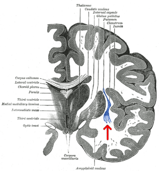
The claustrum is a thin sheet of neurons and supporting glial cells, that connects to the cerebral cortex and subcortical regions including the amygdala, hippocampus and thalamus of the brain. It is located between the insular cortex laterally and the putamen medially, encased by the extreme and external capsules respectively. Blood to the claustrum is supplied by the middle cerebral artery. It is considered to be the most densely connected structure in the brain, and thus hypothesized to allow for the integration of various cortical inputs such as vision, sound and touch, into one experience. Other hypotheses suggest that the claustrum plays a role in salience processing, to direct attention towards the most behaviorally relevant stimuli amongst the background noise. The claustrum is difficult to study given the limited number of individuals with claustral lesions and the poor resolution of neuroimaging.

The dopamine hypothesis of schizophrenia or the dopamine hypothesis of psychosis is a model that attributes the positive symptoms of schizophrenia to a disturbed and hyperactive dopaminergic signal transduction. The model draws evidence from the observation that a large number of antipsychotics have dopamine-receptor antagonistic effects. The theory, however, does not posit dopamine overabundance as a complete explanation for schizophrenia. Rather, the overactivation of D2 receptors, specifically, is one effect of the global chemical synaptic dysregulation observed in this disorder.

<span class="mw-page-title-main">Dopaminergic pathways</span> Projection neurons in the brain that synthesize and release dopamine

Dopaminergic pathways in the human brain are involved in both physiological and behavioral processes including movement, cognition, executive functions, reward, motivation, and neuroendocrine control. Each pathway is a set of projection neurons, consisting of individual dopaminergic neurons.

<span class="mw-page-title-main">Dizocilpine</span> Chemical compound

Dizocilpine (INN), also known as MK-801, is a pore blocker of the NMDA receptor, a glutamate receptor, discovered by a team at Merck in 1982. Glutamate is the brain's primary excitatory neurotransmitter. The channel is normally blocked with a magnesium ion and requires depolarization of the neuron to remove the magnesium and allow the glutamate to open the channel, causing an influx of calcium, which then leads to subsequent depolarization. Dizocilpine binds inside the ion channel of the receptor at several of PCP's binding sites thus preventing the flow of ions, including calcium (Ca2+), through the channel. Dizocilpine blocks NMDA receptors in a use- and voltage-dependent manner, since the channel must open for the drug to bind inside it. The drug acts as a potent anti-convulsant and probably has dissociative anesthetic properties, but it is not used clinically for this purpose because of the discovery of brain lesions, called Olney's lesions (see below), in laboratory rats. Dizocilpine is also associated with a number of negative side effects, including cognitive disruption and psychotic-spectrum reactions. It inhibits the induction of long term potentiation and has been found to impair the acquisition of difficult, but not easy, learning tasks in rats and primates. Because of these effects of dizocilpine, the NMDA receptor pore blocker ketamine is used instead as a dissociative anesthetic in human medical procedures. While ketamine may also trigger temporary psychosis in certain individuals, its short half-life and lower potency make it a much safer clinical option. However, dizocilpine is the most frequently used uncompetitive NMDA receptor antagonist in animal models to mimic psychosis for experimental purposes.

<span class="mw-page-title-main">Metabotropic glutamate receptor</span> Type of glutamate receptor

The metabotropic glutamate receptors, or mGluRs, are a type of glutamate receptor that are active through an indirect metabotropic process. They are members of the group C family of G-protein-coupled receptors, or GPCRs. Like all glutamate receptors, mGluRs bind with glutamate, an amino acid that functions as an excitatory neurotransmitter.

<span class="mw-page-title-main">Glutamate receptor</span> Cell-surface proteins that bind glutamate and trigger changes which influence the behavior of cells

Glutamate receptors are synaptic and non synaptic receptors located primarily on the membranes of neuronal and glial cells. Glutamate is abundant in the human body, but particularly in the nervous system and especially prominent in the human brain where it is the body's most prominent neurotransmitter, the brain's main excitatory neurotransmitter, and also the precursor for GABA, the brain's main inhibitory neurotransmitter. Glutamate receptors are responsible for the glutamate-mediated postsynaptic excitation of neural cells, and are important for neural communication, memory formation, learning, and regulation.

<span class="mw-page-title-main">Kynurenic acid</span> Chemical compound

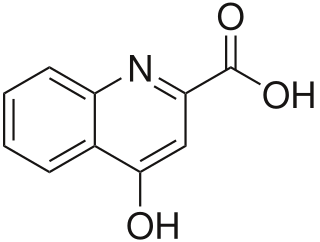
Kynurenic acid is a product of the normal metabolism of amino acid L-tryptophan. It has been shown that kynurenic acid possesses neuroactive activity. It acts as an antiexcitotoxic and anticonvulsant, most likely through acting as an antagonist at excitatory amino acid receptors. Because of this activity, it may influence important neurophysiological and neuropathological processes. As a result, kynurenic acid has been considered for use in therapy in certain neurobiological disorders. Conversely, increased levels of kynurenic acid have also been linked to certain pathological conditions.

<span class="mw-page-title-main">Chandelier cell</span>

Chandelier cells or chandelier neurons are a subset of GABAergic cortical interneurons. They are described as parvalbumin-containing and fast-spiking to distinguish them from other subtypes of GABAergic neurons, although some studies have suggested that only a subset of chandelier cells test positive for parvalbumin by immunostaining. The name comes from the specific shape of their axon arbors, with the terminals forming distinct arrays called "cartridges". The cartridges are immunoreactive to an isoform of the GABA membrane transporter, GAT-1, and this serves as their identifying feature. GAT-1 is involved in the process of GABA reuptake into nerve terminals, thus helping to terminate its synaptic activity. Chandelier neurons synapse exclusively to the axonal initial segment of pyramidal neurons, near the site where action potential is generated. It is believed that they provide inhibitory input to the pyramidal neurons, but there is data showing that in some circumstances the GABA from chandelier neurons could be excitatory.

<span class="mw-page-title-main">Synaptic gating</span>

Synaptic gating is the ability of neural circuits to gate inputs by either suppressing or facilitating specific synaptic activity. Selective inhibition of certain synapses has been studied thoroughly, and recent studies have supported the existence of permissively gated synaptic transmission. In general, synaptic gating involves a mechanism of central control over neuronal output. It includes a sort of gatekeeper neuron, which has the ability to influence transmission of information to selected targets independently of the parts of the synapse upon which it exerts its action.

<span class="mw-page-title-main">PPP1R1B</span> Protein

Protein phosphatase 1 regulatory subunit 1B (PPP1R1B), also known as dopamine- and cAMP-regulated neuronal phosphoprotein (DARPP-32), is a protein that in humans is encoded by the PPP1R1B gene.

The glutamate hypothesis of schizophrenia models the subset of pathologic mechanisms of schizophrenia linked to glutamatergic signaling. The hypothesis was initially based on a set of clinical, neuropathological, and, later, genetic findings pointing at a hypofunction of glutamatergic signaling via NMDA receptors. While thought to be more proximal to the root causes of schizophrenia, it does not negate the dopamine hypothesis, and the two may be ultimately brought together by circuit-based models. The development of the hypothesis allowed for the integration of the GABAergic and oscillatory abnormalities into the converging disease model and made it possible to discover the causes of some disruptions.

Scientific studies have found that different brain areas show altered activity in humans with major depressive disorder (MDD), and this has encouraged advocates of various theories that seek to identify a biochemical origin of the disease, as opposed to theories that emphasize psychological or situational causes. Factors spanning these causative groups include nutritional deficiencies in magnesium, vitamin D, and tryptophan with situational origin but biological impact. Several theories concerning the biologically based cause of depression have been suggested over the years, including theories revolving around monoamine neurotransmitters, neuroplasticity, neurogenesis, inflammation and the circadian rhythm. Physical illnesses, including hypothyroidism and mitochondrial disease, can also trigger depressive symptoms.

The biology of obsessive–compulsive disorder (OCD) refers biologically based theories about the mechanism of OCD. Cognitive models generally fall into the category of executive dysfunction or modulatory control. Neuroanatomically, functional and structural neuroimaging studies implicate the prefrontal cortex (PFC), basal ganglia (BG), insula, and posterior cingulate cortex (PCC). Genetic and neurochemical studies implicate glutamate and monoamine neurotransmitters, especially serotonin and dopamine.

<span class="mw-page-title-main">Visual processing abnormalities in schizophrenia</span>

Visual processing abnormalities in schizophrenia are commonly found, and contribute to poor social function.

<span class="mw-page-title-main">Hypofrontality</span>

Hypofrontality is a state of decreased cerebral blood flow (CBF) in the prefrontal cortex of the brain. Hypofrontality is symptomatic of several neurological medical conditions, such as schizophrenia, attention deficit hyperactivity disorder (ADHD), bipolar disorder, and major depressive disorder. This condition was initially described by Ingvar and Franzén in 1974, through the use of xenon blood flow technique with 32 detectors to image the brains of patients with schizophrenia. This finding was confirmed in subsequent studies using the improved spatial resolution of positron emission tomography with the fluorodeoxyglucose (18F-FDG) tracer. Subsequent neuroimaging work has shown that the decreases in prefrontal CBF are localized to the medial, lateral, and orbital portions of the prefrontal cortex. Hypofrontality is thought to contribute to the negative symptoms of schizophrenia.

<span class="mw-page-title-main">Biology of bipolar disorder</span> Biological Study Of Bipolar Disorder

Bipolar disorder is an affective disorder characterized by periods of elevated and depressed mood. The cause and mechanism of bipolar disorder is not yet known, and the study of its biological origins is ongoing. Although no single gene causes the disorder, a number of genes are linked to increase risk of the disorder, and various gene environment interactions may play a role in predisposing individuals to developing bipolar disorder. Neuroimaging and postmortem studies have found abnormalities in a variety of brain regions, and most commonly implicated regions include the ventral prefrontal cortex and amygdala. Dysfunction in emotional circuits located in these regions have been hypothesized as a mechanism for bipolar disorder. A number of lines of evidence suggests abnormalities in neurotransmission, intracellular signalling, and cellular functioning as possibly playing a role in bipolar disorder.

<span class="mw-page-title-main">Margarita Behrens</span> Neuroscientist and biochemist

Margarita Behrens is a neuroscientist and biochemist. She is currently an associate professor at the Salk Institute for Biological Studies where her lab studies the impact of oxidative stress on the post-natal brain through probing the biology of fast-spiking parvalbumin interneurons in models of schizophrenia.

<span class="mw-page-title-main">Bita Moghaddam</span> Iranian-American neuroscientist

Bita Moghaddam is an Iranian-American neuroscientist and author. She is currently the Ruth Matarazzo Professor of Behavioral Neuroscience at Oregon Health & Science University. Moghaddam investigates the neuronal processes underlying emotion and cognition as a first step to designing strategies to treat and prevent brain illnesses.

References

  1. 1 2 van Os J, Kapur S (August 2009). "Schizophrenia". Lancet. 374 (9690): 635–45. doi:10.1016/S0140-6736(09)60995-8. PMID   19700006. S2CID   208792724.
  2. Insel TR (November 2010). "Rethinking schizophrenia". Nature. 468 (7321): 187–93. Bibcode:2010Natur.468..187I. doi: 10.1038/nature09552 . PMID   21068826. S2CID   4416517.
  3. Elert E (April 2014). "Aetiology: Searching for schizophrenia's roots". Nature. 508 (7494): S2–3. Bibcode:2014Natur.508S...2E. doi: 10.1038/508S2a . PMID   24695332.
  4. 1 2 Marín O (January 2012). "Interneuron dysfunction in psychiatric disorders". Nature Reviews. Neuroscience. 13 (2): 107–20. doi:10.1038/nrn3155. PMID   22251963. S2CID   205507186.
  5. 1 2 Gonzalez-Burgos G, Cho RY, Lewis DA (June 2015). "Alterations in cortical network oscillations and parvalbumin neurons in schizophrenia". Biological Psychiatry. 77 (12): 1031–40. doi:10.1016/j.biopsych.2015.03.010. PMC   4444373 . PMID   25863358.
  6. 1 2 Pittman-Polletta BR, Kocsis B, Vijayan S, Whittington MA, Kopell NJ (June 2015). "Brain rhythms connect impaired inhibition to altered cognition in schizophrenia". Biological Psychiatry. 77 (12): 1020–30. doi:10.1016/j.biopsych.2015.02.005. PMC   4444389 . PMID   25850619.
  7. Feigenson KA, Kusnecov AW, Silverstein SM (January 2014). "Inflammation and the two-hit hypothesis of schizophrenia". Neuroscience and Biobehavioral Reviews. 38: 72–93. doi:10.1016/j.neubiorev.2013.11.006. PMC   3896922 . PMID   24247023.
  8. 1 2 Steullet P, Cabungcal JH, Monin A, Dwir D, O'Donnell P, Cuenod M, Do KQ (September 2016). "Redox dysregulation, neuroinflammation, and NMDA receptor hypofunction: A "central hub" in schizophrenia pathophysiology?". Schizophrenia Research. 176 (1): 41–51. doi:10.1016/j.schres.2014.06.021. PMC   4282982 . PMID   25000913.
  9. 1 2 3 Leza JC, García-Bueno B, Bioque M, Arango C, Parellada M, Do K, O'Donnell P, Bernardo M (August 2015). "Inflammation in schizophrenia: A question of balance". Neuroscience and Biobehavioral Reviews. 55: 612–26. doi:10.1016/j.neubiorev.2015.05.014. PMID   26092265. S2CID   35480356.
  10. Howes OD, Kambeitz J, Kim E, Stahl D, Slifstein M, Abi-Dargham A, Kapur S (August 2012). "The nature of dopamine dysfunction in schizophrenia and what this means for treatment". Archives of General Psychiatry. 69 (8): 776–86. doi:10.1001/archgenpsychiatry.2012.169. PMC   3730746 . PMID   22474070.
  11. Kambeitz J, Abi-Dargham A, Kapur S, Howes OD (June 2014). "Alterations in cortical and extrastriatal subcortical dopamine function in schizophrenia: systematic review and meta-analysis of imaging studies". The British Journal of Psychiatry. 204 (6): 420–9. doi: 10.1192/bjp.bp.113.132308 . PMID   25029687.
  12. 1 2 Weinstein JJ, Chohan MO, Slifstein M, Kegeles LS, Moore H, Abi-Dargham A (January 2017). "Pathway-Specific Dopamine Abnormalities in Schizophrenia". Biological Psychiatry. 81 (1): 31–42. doi:10.1016/j.biopsych.2016.03.2104. PMC   5177794 . PMID   27206569.
  13. Chun S, Westmoreland JJ, Bayazitov IT, Eddins D, Pani AK, Smeyne RJ, Yu J, Blundon JA, Zakharenko SS (June 2014). "Specific disruption of thalamic inputs to the auditory cortex in schizophrenia models". Science. 344 (6188): 1178–82. Bibcode:2014Sci...344.1178C. doi:10.1126/science.1253895. PMC   4349506 . PMID   24904170.
  14. Kapur S (January 2003). "Psychosis as a state of aberrant salience: a framework linking biology, phenomenology, and pharmacology in schizophrenia". The American Journal of Psychiatry. 160 (1): 13–23. doi:10.1176/appi.ajp.160.1.13. PMID   12505794.
  15. Takahashi H, Higuchi M, Suhara T (May 2006). "The role of extrastriatal dopamine D2 receptors in schizophrenia". Biological Psychiatry. 59 (10): 919–28. doi:10.1016/j.biopsych.2006.01.022. PMID   16682269. S2CID   20866362.
  16. Corlett PR, Taylor JR, Wang XJ, Fletcher PC, Krystal JH (November 2010). "Toward a neurobiology of delusions". Progress in Neurobiology. 92 (3): 345–69. doi:10.1016/j.pneurobio.2010.06.007. PMC   3676875 . PMID   20558235.
  17. Konradi C, Heckers S (February 2003). "Molecular aspects of glutamate dysregulation: implications for schizophrenia and its treatment". Pharmacology & Therapeutics. 97 (2): 153–79. doi:10.1016/S0163-7258(02)00328-5. PMC   4203361 . PMID   12559388.
  18. Lahti AC, Weiler MA, Tamara Michaelidis BA, Parwani A, Tamminga CA (October 2001). "Effects of ketamine in normal and schizophrenic volunteers". Neuropsychopharmacology. 25 (4): 455–67. doi: 10.1016/S0893-133X(01)00243-3 . PMID   11557159.
  19. Coyle JT, Tsai G, Goff D (November 2003). "Converging evidence of NMDA receptor hypofunction in the pathophysiology of schizophrenia". Annals of the New York Academy of Sciences. 1003 (1): 318–27. Bibcode:2003NYASA1003..318C. doi:10.1196/annals.1300.020. PMID   14684455. S2CID   1358620.
  20. Tuominen HJ, Tiihonen J, Wahlbeck K (January 2005). "Glutamatergic drugs for schizophrenia: a systematic review and meta-analysis". Schizophrenia Research. 72 (2–3): 225–34. doi:10.1016/j.schres.2004.05.005. PMID   15560967. S2CID   22407838.
  21. Weickert CS, Fung SJ, Catts VS, Schofield PR, Allen KM, Moore LT, Newell KA, Pellen D, Huang XF, Catts SV, Weickert TW (November 2013). "Molecular evidence of N-methyl-D-aspartate receptor hypofunction in schizophrenia". Molecular Psychiatry. 18 (11): 1185–92. doi:10.1038/mp.2012.137. PMC   3807670 . PMID   23070074.
  22. Catts VS, Lai YL, Weickert CS, Weickert TW, Catts SV (April 2016). "A quantitative review of the postmortem evidence for decreased cortical N-methyl-D-aspartate receptor expression levels in schizophrenia: How can we link molecular abnormalities to mismatch negativity deficits?". Biological Psychology. 116: 57–67. doi: 10.1016/j.biopsycho.2015.10.013 . PMID   26549579.
  23. Schizophrenia Working Group of the Psychiatric Genomics Consortium (July 2014). "Biological insights from 108 schizophrenia-associated genetic loci". Nature. 511 (7510): 421–7. Bibcode:2014Natur.511..421S. doi:10.1038/nature13595. PMC   4112379 . PMID   25056061.
  24. Gonzalez-Burgos G, Hashimoto T, Lewis DA (August 2010). "Alterations of cortical GABA neurons and network oscillations in schizophrenia". Current Psychiatry Reports. 12 (4): 335–44. doi:10.1007/s11920-010-0124-8. PMC   2919752 . PMID   20556669.
  25. Konradi C, Yang CK, Zimmerman EI, Lohmann KM, Gresch P, Pantazopoulos H, Berretta S, Heckers S (September 2011). "Hippocampal interneurons are abnormal in schizophrenia". Schizophrenia Research. 131 (1–3): 165–73. doi:10.1016/j.schres.2011.06.007. PMC   3159834 . PMID   21745723.
  26. Chung DW, Fish KN, Lewis DA (November 2016). "Pathological Basis for Deficient Excitatory Drive to Cortical Parvalbumin Interneurons in Schizophrenia". The American Journal of Psychiatry. 173 (11): 1131–1139. doi:10.1176/appi.ajp.2016.16010025. PMC   5089927 . PMID   27444795.
  27. Soares, Dinesh; et al. "DISC1: Structure, Function, and Therapeutic Potential for Major Mental Illness". PubMed Central. National Institutes of Health. Retrieved 12 March 2024.
  28. Senkowski D, Gallinat J (June 2015). "Dysfunctional prefrontal gamma-band oscillations reflect working memory and other cognitive deficits in schizophrenia". Biological Psychiatry. 77 (12): 1010–9. doi:10.1016/j.biopsych.2015.02.034. PMID   25847179. S2CID   206104940.
  29. Sohal VS, Zhang F, Yizhar O, Deisseroth K (June 2009). "Parvalbumin neurons and gamma rhythms enhance cortical circuit performance". Nature. 459 (7247): 698–702. Bibcode:2009Natur.459..698S. doi:10.1038/nature07991. PMC   3969859 . PMID   19396159.
  30. Pocklington AJ, Rees E, Walters JT, Han J, Kavanagh DH, Chambert KD, Holmans P, Moran JL, McCarroll SA, Kirov G, O'Donovan MC, Owen MJ (June 2015). "Novel Findings from CNVs Implicate Inhibitory and Excitatory Signaling Complexes in Schizophrenia". Neuron. 86 (5): 1203–14. doi:10.1016/j.neuron.2015.04.022. PMC   4460187 . PMID   26050040.
  31. Cassoli JS, Guest PC, Malchow B, Schmitt A, Falkai P, Martins-de-Souza D (2015-01-01). "Disturbed macro-connectivity in schizophrenia linked to oligodendrocyte dysfunction: from structural findings to molecules". npj Schizophrenia. 1: 15034. doi:10.1038/npjschz.2015.34. PMC   4849457 . PMID   27336040.
  32. Mighdoll MI, Tao R, Kleinman JE, Hyde TM (January 2015). "Myelin, myelin-related disorders, and psychosis". Schizophrenia Research. 161 (1): 85–93. doi:10.1016/j.schres.2014.09.040. PMID   25449713. S2CID   23435472.
  33. Haroutunian V, Katsel P, Roussos P, Davis KL, Altshuler LL, Bartzokis G (November 2014). "Myelination, oligodendrocytes, and serious mental illness". Glia. 62 (11): 1856–77. doi:10.1002/glia.22716. PMID   25056210. S2CID   571262.
  34. Stedehouder J, Kushner SA (January 2017). "Myelination of parvalbumin interneurons: a parsimonious locus of pathophysiological convergence in schizophrenia". Molecular Psychiatry. 22 (1): 4–12. doi:10.1038/mp.2016.147. PMC   5414080 . PMID   27646261.
  35. Mauney SA, Pietersen CY, Sonntag KC, Woo TW (December 2015). "Differentiation of oligodendrocyte precursors is impaired in the prefrontal cortex in schizophrenia". Schizophrenia Research. 169 (1–3): 374–380. doi:10.1016/j.schres.2015.10.042. PMC   4681621 . PMID   26585218.
  36. 1 2 3 Nettis MA, Pariante CM, Mondelli V (2020). "Early-Life Adversity, Systemic Inflammation and Comorbid Physical and Psychiatric Illnesses of Adult Life". Current Topics in Behavioral Neurosciences. 44: 207–225. doi:10.1007/7854_2019_89. ISBN   978-3-030-39140-9. PMID   30895531.
  37. Pearce J, Murray C, Larkin W (July 2019). "Childhood adversity and trauma: experiences of professionals trained to routinely enquire about childhood adversity". Heliyon. 5 (7): e01900. doi: 10.1016/j.heliyon.2019.e01900 . PMC   6658729 . PMID   31372522.
  38. Chase KA, Melbourne JK, Rosen C (March 2019). "Traumagenics: At the intersect of childhood trauma, immunity and psychosis". Psychiatry Research. 273: 369–377. doi:10.1016/j.psychres.2018.12.097. PMID   30682559. S2CID   56177674.
  39. Hope S, Melle I, Aukrust P, Steen NE, Birkenaes AB, Lorentzen S, Agartz I, Ueland T, Andreassen OA (November 2009). "Similar immune profile in bipolar disorder and schizophrenia: selective increase in soluble tumor necrosis factor receptor I and von Willebrand factor" (PDF). Bipolar Disorders. 11 (7): 726–34. doi:10.1111/j.1399-5618.2009.00757.x. hdl: 10852/34620 . PMID   19839997.
  40. Drexhage RC, Knijff EM, Padmos RC, Heul-Nieuwenhuijzen L, Beumer W, Versnel MA, Drexhage HA (January 2010). "The mononuclear phagocyte system and its cytokine inflammatory networks in schizophrenia and bipolar disorder". Expert Review of Neurotherapeutics. 10 (1): 59–76. doi:10.1586/ern.09.144. PMID   20021321. S2CID   207220109.
  41. Hope S, Ueland T, Steen NE, Dieset I, Lorentzen S, Berg AO, Agartz I, Aukrust P, Andreassen OA (April 2013). "Interleukin 1 receptor antagonist and soluble tumor necrosis factor receptor 1 are associated with general severity and psychotic symptoms in schizophrenia and bipolar disorder". Schizophrenia Research. 145 (1–3): 36–42. doi: 10.1016/j.schres.2012.12.023 . PMID   23403415.
  42. Schizophrenia Psychiatric Genome-Wide Association Study (GWAS) Consortium (September 2011). "Genome-wide association study identifies five new schizophrenia loci". Nature Genetics. 43 (10): 969–76. doi:10.1038/ng.940. PMC   3303194 . PMID   21926974.
  43. Trépanier MO, Hopperton KE, Mizrahi R, Mechawar N, Bazinet RP (August 2016). "Postmortem evidence of cerebral inflammation in schizophrenia: a systematic review". Molecular Psychiatry. 21 (8): 1009–26. doi:10.1038/mp.2016.90. PMC   4960446 . PMID   27271499.
  44. Emiliani FE, Sedlak TW, Sawa A (May 2014). "Oxidative stress and schizophrenia: recent breakthroughs from an old story". Current Opinion in Psychiatry. 27 (3): 185–90. doi:10.1097/YCO.0000000000000054. PMC   4054867 . PMID   24613987.
  45. Hardingham GE, Do KQ (February 2016). "Linking early-life NMDAR hypofunction and oxidative stress in schizophrenia pathogenesis" (PDF). Nature Reviews. Neuroscience. 17 (2): 125–34. doi:10.1038/nrn.2015.19. PMID   26763624. S2CID   27164937.
  46. Kushima I, Aleksic B, Nakatochi M, Shimamura T, Shiino T, Yoshimi A, et al. (March 2017). "High-resolution copy number variation analysis of schizophrenia in Japan". Molecular Psychiatry. 22 (3): 430–440. doi:10.1038/mp.2016.88. PMID   27240532. S2CID   3605506.
  47. Monin A, Baumann PS, Griffa A, Xin L, Mekle R, Fournier M, et al. (July 2015). "Glutathione deficit impairs myelin maturation: relevance for white matter integrity in schizophrenia patients". Molecular Psychiatry. 20 (7): 827–38. doi: 10.1038/mp.2014.88 . PMID   25155877. S2CID   15364819.
  48. Cabungcal JH, Steullet P, Kraftsik R, Cuenod M, Do KQ (March 2013). "Early-life insults impair parvalbumin interneurons via oxidative stress: reversal by N-acetylcysteine". Biological Psychiatry. 73 (6): 574–82. doi:10.1016/j.biopsych.2012.09.020. PMID   23140664. S2CID   24651769.
  49. 1 2 3 Markkanen E, Meyer U, Dianov GL (June 2016). "DNA Damage and Repair in Schizophrenia and Autism: Implications for Cancer Comorbidity and Beyond". Int J Mol Sci. 17 (6): 856. doi: 10.3390/ijms17060856 . PMC   4926390 . PMID   27258260.
  50. Harrison PJ (December 2000). "Postmortem studies in schizophrenia". Dialogues in Clinical Neuroscience. 2 (4): 349–57. doi:10.31887/DCNS.2000.2.4/pharrison. PMC   3181616 . PMID   22033474.
  51. Glausier JR, Lewis DA (October 2013). "Dendritic spine pathology in schizophrenia". Neuroscience. 251: 90–107. doi:10.1016/j.neuroscience.2012.04.044. PMC   3413758 . PMID   22546337.
  52. Nemani K, Hosseini Ghomi R, McCormick B, Fan X (January 2015). "Schizophrenia and the gut-brain axis". Progress in Neuro-Psychopharmacology & Biological Psychiatry. 56: 155–60. doi:10.1016/j.pnpbp.2014.08.018. PMID   25240858. S2CID   32315340.
  53. Lachance LR, McKenzie K (February 2014). "Biomarkers of gluten sensitivity in patients with non-affective psychosis: a meta-analysis". Schizophrenia Research (Review). 152 (2–3): 521–7. doi:10.1016/j.schres.2013.12.001. PMID   24368154. S2CID   26792210.
  54. Seeman MV (March 2020). "The Gut Microbiome and Treatment-Resistance in Schizophrenia". The Psychiatric Quarterly. 91 (1): 127–136. doi:10.1007/s11126-019-09695-4. PMID   31781943. S2CID   208329435.
  55. Pocivavsek, A; Rowland, LM (13 January 2018). "Basic Neuroscience Illuminates Causal Relationship Between Sleep and Memory: Translating to Schizophrenia". Schizophrenia Bulletin. 44 (1): 7–14. doi:10.1093/schbul/sbx151. PMC   5768044 . PMID   29136236.
  56. Flashman LA, Green MF (March 2004). "Review of cognition and brain structure in schizophrenia: profiles, longitudinal course, and effects of treatment". The Psychiatric Clinics of North America. 27 (1): 1–18, vii. doi:10.1016/S0193-953X(03)00105-9. PMID   15062627.
  57. van Erp TG, Hibar DP, Rasmussen JM, Glahn DC, Pearlson GD, Andreassen OA, et al. (April 2016). "Subcortical brain volume abnormalities in 2028 individuals with schizophrenia and 2540 healthy controls via the ENIGMA consortium". Molecular Psychiatry. 21 (4): 547–53. doi:10.1038/mp.2015.63. PMC   4668237 . PMID   26033243.
  58. Steen RG, Mull C, McClure R, Hamer RM, Lieberman JA (June 2006). "Brain volume in first-episode schizophrenia: systematic review and meta-analysis of magnetic resonance imaging studies". The British Journal of Psychiatry. 188 (6): 510–8. doi: 10.1192/bjp.188.6.510 . PMID   16738340.
  59. Lieberman JA, Bymaster FP, Meltzer HY, Deutch AY, Duncan GE, Marx CE, Aprille JR, Dwyer DS, Li XM, Mahadik SP, Duman RS, Porter JH, Modica-Napolitano JS, Newton SS, Csernansky JG (September 2008). "Antipsychotic drugs: comparison in animal models of efficacy, neurotransmitter regulation, and neuroprotection". Pharmacological Reviews. 60 (3): 358–403. doi:10.1124/pr.107.00107. PMC   4821196 . PMID   18922967.
  60. DeLisi LE (March 2008). "The concept of progressive brain change in schizophrenia: implications for understanding schizophrenia". Schizophrenia Bulletin. 34 (2): 312–21. doi:10.1093/schbul/sbm164. PMC   2632405 . PMID   18263882.
  61. 1 2 Jung WH, Jang JH, Byun MS, An SK, Kwon JS (December 2010). "Structural brain alterations in individuals at ultra-high risk for psychosis: a review of magnetic resonance imaging studies and future directions". Journal of Korean Medical Science. 25 (12): 1700–9. doi:10.3346/jkms.2010.25.12.1700. PMC   2995221 . PMID   21165282.
  62. 1 2 3 4 Shenton ME, Dickey CC, Frumin M, McCarley RW (April 2001). "A review of MRI findings in schizophrenia". Schizophrenia Research. 49 (1–2): 1–52. doi:10.1016/s0920-9964(01)00163-3. PMC   2812015 . PMID   11343862.
  63. Honea R, Crow TJ, Passingham D, Mackay CE (December 2005). "Regional deficits in brain volume in schizophrenia: a meta-analysis of voxel-based morphometry studies". The American Journal of Psychiatry. 162 (12): 2233–45. doi:10.1176/appi.ajp.162.12.2233. PMID   16330585.
  64. Vita A, De Peri L, Silenzi C, Dieci M (February 2006). "Brain morphology in first-episode schizophrenia: a meta-analysis of quantitative magnetic resonance imaging studies". Schizophrenia Research. 82 (1): 75–88. doi:10.1016/j.schres.2005.11.004. PMID   16377156. S2CID   28037606.
  65. Kuperberg GR, Broome MR, McGuire PK, David AS, Eddy M, Ozawa F, Goff D, West WC, Williams SC, van der Kouwe AJ, Salat DH, Dale AM, Fischl B (September 2003). "Regionally localized thinning of the cerebral cortex in schizophrenia". Archives of General Psychiatry. 60 (9): 878–88. doi: 10.1001/archpsyc.60.9.878 . PMID   12963669.
  66. 1 2 Rimol LM, Hartberg CB, Nesvåg R, Fennema-Notestine C, Hagler DJ, Pung CJ, Jennings RG, Haukvik UK, Lange E, Nakstad PH, Melle I, Andreassen OA, Dale AM, Agartz I (July 2010). "Cortical thickness and subcortical volumes in schizophrenia and bipolar disorder". Biological Psychiatry. 68 (1): 41–50. doi:10.1016/j.biopsych.2010.03.036. PMID   20609836. S2CID   7718134.
  67. Rimol LM, Nesvåg R, Hagler DJ, Bergmann O, Fennema-Notestine C, Hartberg CB, Haukvik UK, Lange E, Pung CJ, Server A, Melle I, Andreassen OA, Agartz I, Dale AM (March 2012). "Cortical volume, surface area, and thickness in schizophrenia and bipolar disorder". Biological Psychiatry. 71 (6): 552–60. doi:10.1016/j.biopsych.2011.11.026. PMID   22281121. S2CID   27413169.
  68. Ropper AH, Brown RH (2005). Adams and Victor's Principles of Neurology (8th ed.). New York: McGraw-Hill. p. 1324. ISBN   0-07-141620-X.
  69. Chand GB, Dwyer DB, Erus G, et al. (27 February 2020). "Two distinct neuroanatomical subtypes of schizophrenia revealed using machine learning". Brain: A Journal of Neurology. 143 (3): 1027–1038. doi:10.1093/brain/awaa025. PMC   7089665 . PMID   32103250.
  70. Fitzsimmons J, Kubicki M, Shenton ME (March 2013). "Review of functional and anatomical brain connectivity findings in schizophrenia". Current Opinion in Psychiatry. 26 (2): 172–87. doi:10.1097/YCO.0b013e32835d9e6a. PMID   23324948. S2CID   24317327.
  71. Ellison-Wright I, Bullmore E (March 2009). "Meta-analysis of diffusion tensor imaging studies in schizophrenia". Schizophrenia Research. 108 (1–3): 3–10. doi:10.1016/j.schres.2008.11.021. PMID   19128945. S2CID   39073686.
  72. Minzenberg MJ, Laird AR, Thelen S, Carter CS, Glahn DC (August 2009). "Meta-analysis of 41 functional neuroimaging studies of executive function in schizophrenia". Archives of General Psychiatry. 66 (8): 811–22. doi:10.1001/archgenpsychiatry.2009.91. PMC   2888482 . PMID   19652121.
  73. Taylor SF, Kang J, Brege IS, Tso IF, Hosanagar A, Johnson TD (January 2012). "Meta-analysis of functional neuroimaging studies of emotion perception and experience in schizophrenia". Biological Psychiatry. 71 (2): 136–45. doi:10.1016/j.biopsych.2011.09.007. PMC   3237865 . PMID   21993193.
  74. Li H, Chan RC, McAlonan GM, Gong QY (September 2010). "Facial emotion processing in schizophrenia: a meta-analysis of functional neuroimaging data". Schizophrenia Bulletin. 36 (5): 1029–39. doi:10.1093/schbul/sbn190. PMC   2930350 . PMID   19336391.
  75. Kühn S, Gallinat J (June 2012). "Quantitative meta-analysis on state and trait aspects of auditory verbal hallucinations in schizophrenia". Schizophrenia Bulletin. 38 (4): 779–86. doi:10.1093/schbul/sbq152. PMC   3406531 . PMID   21177743.
  76. Zmigrod L, Garrison JR, Carr J, Simons JS (October 2016). "The neural mechanisms of hallucinations: A quantitative meta-analysis of neuroimaging studies". Neuroscience and Biobehavioral Reviews. 69: 113–23. doi: 10.1016/j.neubiorev.2016.05.037 . PMID   27473935.
  77. Meyer-Lindenberg A, Miletich RS, Kohn PD, Esposito G, Carson RE, Quarantelli M, Weinberger DR, Berman KF (March 2002). "Reduced prefrontal activity predicts exaggerated striatal dopaminergic function in schizophrenia". Nature Neuroscience. 5 (3): 267–71. doi:10.1038/nn804. PMID   11865311. S2CID   26098214.
  78. Steen RG, Hamer RM, Lieberman JA (November 2005). "Measurement of brain metabolites by 1H magnetic resonance spectroscopy in patients with schizophrenia: a systematic review and meta-analysis". Neuropsychopharmacology. 30 (11): 1949–62. doi: 10.1038/sj.npp.1300850 . PMID   16123764.
  79. Davis KL, American College of Neuropsychopharmacology (2002). "Chapter 53: Neural Circuitry and the Pathophysiology of Schizophrenia". Neuropsychopharmacology: The Fifth Generation of Progress : an Official Publication of the American College of Neuropsychopharmacology. Lippincott Williams & Wilkins. pp. 731–743. ISBN   9780781728379.
  80. Fusar-Poli P, Meyer-Lindenberg A (January 2013). "Striatal presynaptic dopamine in schizophrenia, part II: meta-analysis of [(18)F/(11)C]-DOPA PET studies". Schizophrenia Bulletin. 39 (1): 33–42. doi:10.1093/schbul/sbr180. PMC   3523905 . PMID   22282454.