Eosinophilic gastroenteritis

Last updated
Eosinophilic gastroenteritis
Eos gastroen.jpg
H&E Stain: Dense Eosinophilic infiltration of gastro-duodenal wall
Specialty Immunology, Gastroenterology

Eosinophilic gastroenteritis (EG or EGE), also known as eosinophilic enteritis, [1] is a rare and heterogeneous condition characterized by patchy or diffuse eosinophilic infiltration of gastrointestinal (GI) tissue, first described by Kaijser in 1937. [2] [3] Presentation may vary depending on location as well as depth and extent of bowel wall involvement and usually runs a chronic relapsing course. It can be classified into mucosal, muscular and serosal types based on the depth of involvement. [4] [5] Any part of the GI tract can be affected, and isolated biliary tract involvement has also been reported. [6] [7] The stomach is the organ most commonly affected, followed by the small intestine and the colon. [8] [9]

Contents

Signs and symptoms

EG typically presents with a combination of chronic nonspecific GI symptoms which include abdominal pain, diarrhea, occasional nausea and vomiting, weight loss and abdominal distension. Approximately 80% have symptoms for several years; [7] a high degree of clinical suspicion is often required to establish the diagnosis, as the disease is extremely rare. It doesn't come all of a sudden but takes about 3–4 years to develop depending upon the age of the patient. Occasionally, the disease may manifest itself as an acute abdomen or bowel obstruction. [10] [11]

Pathophysiology

Peripheral blood eosinophilia and elevated serum IgE are usual but not universal. The damage to the gastrointestinal tract wall is caused by eosinophilic infiltration and degranulation. [15]

As a part of host defense mechanism, eosinophils are normally present in gastrointestinal mucosa, though the finding in deeper tissue is almost always pathologic. [16] What triggers such dense infiltration in EG is not clear. It is possible that different pathogenetic mechanisms of disease is involved in several subgroups of patients. Food allergy and variable IgE response to food substances has been observed in some patients which implies role of hypersensitive response in pathogenesis. Many patients indeed have history of other atopic conditions like eczema, asthma, etc.[ citation needed ]

Eosinophil recruitment into inflammatory tissue is a complex process, regulated by a number of inflammatory cytokines. In EG cytokines IL-3, IL-5 and granulocyte macrophage colony stimulating factor (GM-CSF) may be behind the recruitment and activation. They have been observed immunohistochemically in diseased intestinal wall. [17] In addition eotaxin has been shown to have an integral role in regulating the homing of eosinophils into the lamina propria of stomach and small intestine. [18] In the allergic subtype of disease, it is thought that food allergens cross the intestinal mucosa and trigger an inflammatory response that includes mast cell degranulation and recruitment of eosinophils. [18] [19]

Diagnosis

Spiral CT showing ascites and concentric thickening of colon and ileum in EG Eosinophilic gastroenteritis CT.jpg
Spiral CT showing ascites and concentric thickening of colon and ileum in EG

Talley et al. [20] suggested 3 diagnostic criteria which are still widely used:

  1. the presence of gastrointestinal symptoms,
  2. histological demonstration of eosinophilic infiltration in one or more areas of the gastrointestinal tract or presence of high eosinophil count in ascitic fluid (latter usually indicates subserosal variety),
  3. no evidence of parasitic or extraintestinal disease.

Hypereosinophilia, the hallmark of allergic response, may be absent in up to 20% of patients, but hypoalbuminaemia and other abnormalities suggestive of malabsorption may be present. CT scans may show nodular and irregular thickening of the folds in the distal stomach and proximal small bowel, but these findings can also be present in other conditions like Crohn's disease and lymphoma.[ citation needed ]

The endoscopic appearance in eosinophilic gastroenteritis is nonspecific; it includes erythematous, friable, nodular, and occasional ulcerative changes. [21] Sometimes diffuse inflammation results in complete loss of villi, involvement of multiple layers, submucosal oedema and fibrosis. [22] [23]

Definitive diagnosis involves histological evidence of eosinophilic infiltration in biopsy slides. Microscopy reveals >20 eosinophils per high power field. [12] [20] Infiltration is often patchy, can be missed and laparoscopic full thickness biopsy may be required.

Radio isotope scan using technetium (99mTc) exametazime-labeled leukocyte SPECT may be useful in assessing the extent of disease and response to treatment but has little value in diagnosis, as the scan does not help differentiating EG from other causes of inflammation. [24] [25]

When eosinophilic gastroenteritis is observed in association with eosinophilic infiltration of other organ systems, the diagnosis of idiopathic hypereosinophilic syndrome should be considered. [26]

Management

Corticosteroids are the mainstay of therapy with a 90% response rate in some studies. Appropriate duration of steroid treatment is unknown and relapse often necessitates long term treatment. Various steroid sparing agents e.g. sodium cromoglycate (a stabilizer of mast cell membranes), ketotifen (an antihistamine), and montelukast (a selective, competitive leukotriene receptor antagonist) have been proposed, centering on an allergic hypothesis, with mixed results. [19] [27] Oral budesonide (an oral steroid) can be useful in treatment, as well. [28] An elimination diet may be successful if a limited number of food allergies are identified. [21] [29] An elemental diet may also be successful in the treatment of children. [30]

In a randomized clinical trial, lirentelimab was found to improve eosinophil counts and symptoms in individuals with eosinophilic gastritis and duodenitis. [31] [32]

Epidemiology

Epidemiology may differ between studies, as number of cases are small, with approximately 300 EG cases reported in published literature.

EG can present at any age and across all races, with a slightly higher incidence in males. [33] Earlier studies showed higher incidence in the third to fifth decades of life. [2] [4]

Other gastrointestinal conditions associated with allergy

See also

Related Research Articles

<span class="mw-page-title-main">Eosinophil</span> Variety of white blood cells

Eosinophils, sometimes called eosinophiles or, less commonly, acidophils, are a variety of white blood cells and one of the immune system components responsible for combating multicellular parasites and certain infections in vertebrates. Along with mast cells and basophils, they also control mechanisms associated with allergy and asthma. They are granulocytes that develop during hematopoiesis in the bone marrow before migrating into blood, after which they are terminally differentiated and do not multiply. They form about 2 to 3% of white blood cells in the body.

<span class="mw-page-title-main">Eosinophilia</span> Blood condition

Eosinophilia is a condition in which the eosinophil count in the peripheral blood exceeds 5×10⁸/L (500/μL). Hypereosinophilia is an elevation in an individual's circulating blood eosinophil count above 1.5 × 109/L (i.e. 1,500/μL). The hypereosinophilic syndrome is a sustained elevation in this count above 1.5 × 109/L (i.e. 1,500/μL) that is also associated with evidence of eosinophil-based tissue injury.

<span class="mw-page-title-main">Enteritis</span> Intestinal inflammatory disease

Enteritis is inflammation of the small intestine. It is most commonly caused by food or drink contaminated with pathogenic microbes, such as Serratia, but may have other causes such as NSAIDs, radiation therapy as well as autoimmune conditions like Crohn's disease and celiac disease. Symptoms include abdominal pain, cramping, diarrhea, dehydration, and fever. Related diseases of the gastrointestinal system involve inflammation of the stomach and large intestine.

Functional gastrointestinal disorders (FGID), also known as disorders of gut–brain interaction, include a number of separate idiopathic disorders which affect different parts of the gastrointestinal tract and involve visceral hypersensitivity and motility disturbances.

<span class="mw-page-title-main">Eosinophilic granulomatosis with polyangiitis</span> Medical condition

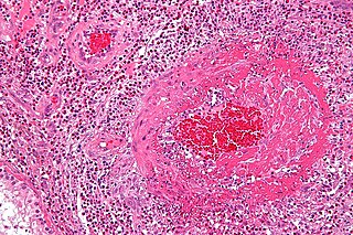
Eosinophilic granulomatosis with polyangiitis (EGPA), formerly known as allergic granulomatosis, is an extremely rare autoimmune condition that causes inflammation of small and medium-sized blood vessels (vasculitis) in persons with a history of airway allergic hypersensitivity (atopy).

Food intolerance is a detrimental reaction, often delayed, to a food, beverage, food additive, or compound found in foods that produces symptoms in one or more body organs and systems, but generally refers to reactions other than food allergy. Food hypersensitivity is used to refer broadly to both food intolerances and food allergies.

<span class="mw-page-title-main">Eosinophilic esophagitis</span> Allergic inflammatory condition of the esophagus

Eosinophilic esophagitis (EoE) is an allergic inflammatory condition of the esophagus that involves eosinophils, a type of white blood cell. In healthy individuals, the esophagus is typically devoid of eosinophils. In EoE, eosinophils migrate to the esophagus in large numbers. When a trigger food is eaten, the eosinophils contribute to tissue damage and inflammation. Symptoms include swallowing difficulty, food impaction, vomiting, and heartburn.

<span class="mw-page-title-main">Intestinal pseudo-obstruction</span> Medical condition

Intestinal pseudo-obstruction (IPO) is a clinical syndrome caused by severe impairment in the ability of the intestines to push food through. It is characterized by the signs and symptoms of intestinal obstruction without any lesion in the intestinal lumen. Clinical features mimic those seen with mechanical intestinal obstructions and can include abdominal pain, nausea, abdominal distension, vomiting, dysphagia and constipation depending upon the part of the gastrointestinal tract involved.

Loeffler endocarditis is a form of heart disease characterized by a stiffened, poorly-functioning heart caused by infiltration of the heart by white blood cells known as eosinophils. Restrictive cardiomyopathy is a disease of the heart muscle which results in impaired diastolic filling of the heart ventricles, i.e. the large heart chambers which pump blood into the pulmonary or systemic circulation. Diastole is the part of the cardiac contraction-relaxation cycle in which the heart fills with venous blood after the emptying done during its previous systole.

<span class="mw-page-title-main">Sucrose intolerance</span> Medical condition

Sucrose intolerance or genetic sucrase-isomaltase deficiency (GSID) is the condition in which sucrase-isomaltase, an enzyme needed for proper metabolism of sucrose (sugar) and starch, is not produced or the enzyme produced is either partially functional or non-functional in the small intestine. All GSID patients lack fully functional sucrase, while the isomaltase activity can vary from minimal functionality to almost normal activity. The presence of residual isomaltase activity may explain why some GSID patients are better able to tolerate starch in their diet than others with GSID.

Gluten-sensitive enteropathy–associated conditions are comorbidities or complications of gluten-related gastrointestinal distress. GSE has key symptoms typically restricted to the bowel and associated tissues; however, there are a wide variety of associated conditions. These include bowel disorders, eosinophilic gastroenteritis and increase with coeliac disease (CD) severity. With some early onset and a large percentage of late onset disease, other disorders appear prior to the coeliac diagnosis or allergic-like responses markedly increased in GSE. Many of these disorders persist on a strict gluten-free diet, and are thus independent of coeliac disease after triggering. For example, autoimmune thyroiditis is a common finding with GSE.

<span class="mw-page-title-main">Esophageal food bolus obstruction</span> Medical condition

An esophageal food bolus obstruction is a medical emergency caused by the obstruction of the esophagus by an ingested foreign body.

An aeroallergen is any airborne substance, such as pollen or spores, which triggers an allergic reaction.

<span class="mw-page-title-main">Eosinophilic bronchitis</span> Medical condition

Eosinophilic bronchitis (EB) is a type of airway inflammation due to excessive mast cell recruitment and activation in the superficial airways as opposed to the smooth muscles of the airways as seen in asthma. It often results in a chronic cough. Lung function tests are usually normal. Inhaled corticosteroids are often an effective treatment.

<span class="mw-page-title-main">Eosinophilic cystitis</span> Medical condition

Eosinophilic cystitis is a rare type of interstitial cystitis first reported in 1960 by Edwin Brown. Eosinophilic cystitis has been linked to a number of etiological factors, including allergies, bladder tumors, trauma to the bladder, parasitic infections, and chemotherapy drugs, though the exact cause of the condition is still unknown. The antigen-antibody response is most likely the cause of eosinophilic cystitis. This results in the generation of different immunoglobulins, which activate eosinophils and start the inflammatory process.

Non-celiac gluten sensitivity (NCGS) or gluten sensitivity is a controversial disorder which can cause both gastrointestinal and other problems.

Eosinophilic myocarditis is inflammation in the heart muscle that is caused by the infiltration and destructive activity of a type of white blood cell, the eosinophil. Typically, the disorder is associated with hypereosinophilia, i.e. an eosinophil blood cell count greater than 1,500 per microliter. It is distinguished from non-eosinophilic myocarditis, which is heart inflammation caused by other types of white blood cells, i.e. lymphocytes and monocytes, as well as the respective descendants of these cells, NK cells and macrophages. This distinction is important because the eosinophil-based disorder is due to a particular set of underlying diseases and its preferred treatments differ from those for non-eosinophilic myocarditis.

Lymphocyte-variant hypereosinophilia is a rare disorder in which eosinophilia or hypereosinophilia is caused by an aberrant population of lymphocytes. These aberrant lymphocytes function abnormally by stimulating the proliferation and maturation of bone marrow eosinophil-precursor cells termed colony forming unit-eosinophils or CFU-Eos.

Familial eosinophilia is a rare congenital disorder characterized by the presence of sustained elevations in blood eosinophil levels that reach ranges diagnostic of eosinophilia or, far more commonly, hypereosinophilia. Although high eosinophil levels are associated with certain diseases and thought to contribute to the tissue destruction found in many other eosinophilia-related diseases, clinical manifestations and tissue destruction related to the eosinophilia in familial eosinophilia is uncommon: this genetic disease typically has a benign phenotype and course compared to other congenital and acquired eosinophilic diseases.

<span class="mw-page-title-main">Lymphocytic esophagitis</span> Medical condition

Lymphocytic esophagitis is a rare and poorly understood medical disorder involving inflammation in the esophagus. The disease is named from the primary inflammatory process, wherein lymphocytes are seen within the esophageal mucosa. Symptoms of the condition include difficulty swallowing, heartburn and food bolus obstruction. The condition was first described in 2006 by Rubio and colleagues. Initial reports questioned whether this was a true medical disorder, or whether the inflammation was secondary to another condition, such as gastroesophageal reflux disease.

References

  1. Pineton de Chambrun, Guillaume; Dufour, Gaspard; Tassy, Barbara; Rivière, Benjamin; Bouta, Najima; Bismuth, Michael; Panaro, Fabrizio; Funakoshi, Natalie; Ramos, Jeanne; Valats, Jean-Christophe; Blanc, Pierre (2018-07-02). "Diagnosis, Natural History and Treatment of Eosinophilic Enteritis: a Review". Current Gastroenterology Reports. 20 (8): 37. doi:10.1007/s11894-018-0645-6. ISSN   1534-312X.
  2. 1 2 Kaijser R. Zur Kenntnis der allergischen Affektionen des Verdauugskanals vom Standpunkt des Chirurgen aus. Arch Klin Chir 1937; 188:36–64.
  3. Whitaker I, Gulati A, McDaid J, Bugajska-Carr U, Arends M (2004). "Eosinophilic gastroenteritis presenting as obstructive jaundice". European Journal of Gastroenterology & Hepatology. 16 (4): 407–9. doi:10.1097/00042737-200404000-00007. PMID   15028974.
  4. 1 2 Klein N, Hargrove R, Sleisenger M, Jeffries G (1970). "Eosinophilic gastroenteritis". Medicine (Baltimore). 49 (4): 299–319. doi: 10.1097/00005792-197007000-00003 . PMID   5426746. S2CID   45969740.
  5. Treiber, Treiber; Weidner, S (2007). "Eosinophilic Gastroenteritis". Clinical Gastroenterology and Hepatology. 5 (5): e16. doi:10.1016/j.cgh.2007.01.011. PMID   17428742.
  6. Polyak S, Smith T, Mertz H (2002). "Eosinophilic gastroenteritis causing pancreatitis and pancreaticobiliary ductal dilation". Dig. Dis. Sci. 47 (5): 1091–5. doi:10.1023/A:1015046309132. PMID   12018905. S2CID   24453648.
  7. 1 2 Christopher V, Thompson M, Hughes S (2002). "Eosinophilic gastroenteritis mimicking pancreatic cancer". Postgraduate Medical Journal. 78 (922): 498–9. doi:10.1136/pmj.78.922.498. PMC   1742453 . PMID   12185230.
  8. Naylor A (1990). "Eosinophilic gastroenteritis". Scottish Medical Journal. 35 (6): 163–5. doi:10.1177/003693309003500601. PMID   2077646. S2CID   43539786.
  9. Jimenez-Saenz M, Villar-Rodriguez J, Torres Y, Carmona I, Salas-Herrero E, Gonzalez-Vilches J, Herrerias-Gutierrez J (2003). "Biliary tract disease: a rare manifestation of eosinophilic gastroenteritis". Dig. Dis. Sci. 48 (3): 624–7. doi:10.1023/A:1022521707420. PMID   12757181. S2CID   23627059.
  10. Shweiki E, West J, Klena J, Kelley S, Colley A, Bross R, Tyler W (1999). "Eosinophilic gastroenteritis presenting as an obstructing cecal mass--a case report and review of the literature". Am. J. Gastroenterol. 94 (12): 3644–5. doi:10.1111/j.1572-0241.1999.01625.x. PMID   10606337. S2CID   21217147.
  11. Tran D, Salloum L, Tshibaka C, Moser R (2000). "Eosinophilic gastroenteritis mimicking acute appendicitis". The American Surgeon. 66 (10): 990–2. doi:10.1177/000313480006601018. PMID   11261632. S2CID   37194432.
  12. 1 2 Baig M, Qadir A, Rasheed J (2006). "A review of eosinophilic gastroenteritis". Journal of the National Medical Association. 98 (10): 1616–9. PMC   2569760 . PMID   17052051.
  13. Lee C, Changchien C, Chen P, Lin D, Sheen I, Wang C, Tai D, Sheen-Chen S, Chen W, Wu C (1993). "Eosinophilic gastroenteritis: 10 years experience". Am. J. Gastroenterol. 88 (1): 70–4. PMID   8420276.
  14. Miyamoto T, Shibata T, Matsuura S, Kagesawa M, Ishizawa Y, Tamiya K (1996). "Eosinophilic gastroenteritis with ileus and ascites". Intern. Med. 35 (10): 779–82. doi: 10.2169/internalmedicine.35.779 . PMID   8933185.)
  15. Tan A, Kruimel J, Naber T (2001). "Eosinophilic gastroenteritis treated with non-enteric-coated budesonide tablets". European Journal of Gastroenterology & Hepatology. 13 (4): 425–7. doi:10.1097/00042737-200104000-00021. PMID   11338074.
  16. Blackshaw A, Levison D (1986). "Eosinophilic infiltrates of the gastrointestinal tract". J. Clin. Pathol. 39 (1): 1–7. doi:10.1136/jcp.39.1.1. PMC   499605 . PMID   2869055.
  17. Desreumaux P, Bloget F, Seguy D, Capron M, Cortot A, Colombel J, Janin A (1996). "Interleukin 3, granulocyte-macrophage colony-stimulating factor, and interleukin 5 in eosinophilic gastroenteritis". Gastroenterology. 110 (3): 768–74. doi: 10.1053/gast.1996.v110.pm8608886 . PMID   8608886.
  18. 1 2 Mishra A, Hogan S, Brandt E, Rothenberg M (2001). "An etiological role for aeroallergens and eosinophils in experimental esophagitis". J. Clin. Invest. 107 (1): 83–90. doi:10.1172/JCI10224. PMC   198543 . PMID   11134183.
  19. 1 2 Pérez-Millán A, Martín-Lorente J, López-Morante A, Yuguero L, Sáez-Royuela F (1997). "Subserosal eosinophilic gastroenteritis treated efficaciously with sodium cromoglycate". Dig. Dis. Sci. 42 (2): 342–4. doi:10.1023/A:1018818003002. PMID   9052516. S2CID   19266537.
  20. 1 2 Talley N, Shorter R, Phillips S, Zinsmeister A (1990). "Eosinophilic gastroenteritis: a clinicopathological study of patients with disease of the mucosa, muscle layer, and subserosal tissues". Gut. 31 (1): 54–8. doi:10.1136/gut.31.1.54. PMC   1378340 . PMID   2318432.
  21. 1 2 Chen M, Chu C, Lin S, Shih S, Wang T (2003). "Eosinophilic gastroenteritis: clinical experience with 15 patients". World J. Gastroenterol. 9 (12): 2813–6. doi: 10.3748/wjg.v9.i12.2813 . PMC   4612059 . PMID   14669340.
  22. Johnstone J, Morson B (1978). "Eosinophilic gastroenteritis". Histopathology. 2 (5): 335–48. doi:10.1111/j.1365-2559.1978.tb01726.x. PMID   363591. S2CID   43074488.
  23. Katz A, Goldman H, Grand R (1977). "Gastric mucosal biopsy in eosinophilic (allergic) gastroenteritis". Gastroenterology. 73 (4 Pt 1): 705–9. doi: 10.1016/S0016-5085(19)31769-X . PMID   892374.
  24. Lee K, Hahm K, Kim Y, Kim J, Cho S, Jie H, Park C, Yim H (1997). "The usefulness of Tc-99m HMPAO labeled WBC SPECT in eosinophilic gastroenteritis". Clinical Nuclear Medicine. 22 (8): 536–41. doi:10.1097/00003072-199708000-00005. PMID   9262899.
  25. Imai E, Kaminaga T, Kawasugi K, Yokokawa T, Furui S (2003). "The usefulness of 99mTc-hexamethylpropyleneamineoxime white blood cell scintigraphy in a patient with eosinophilic gastroenteritis". Annals of Nuclear Medicine. 17 (7): 601–3. doi:10.1007/BF03006675. PMID   14651361. S2CID   32498521.
  26. Matsushita M, Hajiro K, Morita Y, Takakuwa H, Suzaki T (1995). "Eosinophilic gastroenteritis involving the entire digestive tract". Am. J. Gastroenterol. 90 (10): 1868–70. PMID   7572911.
  27. Barbie D, Mangi A, Lauwers G (2004). "Eosinophilic gastroenteritis associated with systemic lupus erythematosus". J. Clin. Gastroenterol. 38 (10): 883–6. doi:10.1097/00004836-200411000-00010. PMID   15492606.
  28. Alsayegh, Mohammad; Mack, Douglas (2012-11-02). "Eosinophilic gastroenteritis with gastric and small bowel involvement: successful treatment with oral budesonide". Allergy, Asthma, and Clinical Immunology. 8 (Suppl 1): A6. doi: 10.1186/1710-1492-8-S1-A6 . ISSN   1710-1484. PMC   3487875 .
  29. Katz A, Twarog F, Zeiger R, Falchuk Z (1984). "Milk-sensitive and eosinophilic gastroenteropathy: similar clinical features with contrasting mechanisms and clinical course". The Journal of Allergy and Clinical Immunology. 74 (1): 72–8. doi: 10.1016/0091-6749(84)90090-3 . PMID   6547462.
  30. Lucendo, Alfredo J.; Serrano-Montalbán, Beatriz; Arias, Ángel; Redondo, Olga; Tenias, José M. (July 2015). "Efficacy of Dietary Treatment for Inducing Disease Remission in Eosinophilic Gastroenteritis". Journal of Pediatric Gastroenterology and Nutrition. 61 (1): 56–64. doi: 10.1097/MPG.0000000000000766 . ISSN   0277-2116. PMID   25699593. S2CID   26195645.
  31. Dellon, ES; Peterson, KA; Murray, JA; Falk, GW; Gonsalves, N; Chehade, M; Genta, RM; Leung, J; Khoury, P; Klion, AD; Hazan, S; Vaezi, M; Bledsoe, AC; Durrani, SR; Wang, C; Shaw, C; Chang, AT; Singh, B; Kamboj, AP; Rasmussen, HS; Rothenberg, ME; Hirano, I (22 October 2020). "Anti-Siglec-8 Antibody for Eosinophilic Gastritis and Duodenitis". The New England Journal of Medicine. 383 (17): 1624–1634. doi:10.1056/NEJMoa2012047. PMC   7600443 . PMID   33085861.
  32. Young, Alex (October 29, 2019). "Therapeutic antibody effective in eosinophilic gastritis". Healio. Retrieved 14 December 2020.
  33. Guandalini, Stefano (2004). Essential Pediatric Gastroenterology and Nutrition. City: McGraw-Hill Professional. ISBN   978-0-07-141630-6. Page 210.