Leukoencephalopathy with vanishing white matter

Last updated
Leukoencephalopathy with vanishing white matter
Other namesChildhood ataxia with central nervous system hypomyelinization, Vanishing white matter leukodystrophy, Cree leukoencephalopathy, Vanishing white matter leukodystrophy with ovarian failure, included, Myelinopathia centralis diffusa
Autosomal recessive - en.svg
This condition is inherited in an autosomal recessive manner

Leukoencephalopathy with vanishing white matter (VWM disease) is an autosomal recessive neurological disease. The cause of the disease are mutations in any of the 5 genes encoding subunits of the translation initiation factor eIF2B: EIF2B1, EIF2B2, EIF2B3, EIF2B4, or EIF2B5. The disease belongs to a family of conditions called the Leukodystrophies.[ citation needed ]

Contents

Symptoms and signs

Onset usually occurs in childhood, however some adult cases have been found. Generally, physicians look for the symptoms in children. Symptoms include cerebellar ataxia, spasticity, optic atrophy, epilepsy, [1] loss of motor functions, irritability, vomiting, coma, [2] and even fever has been tied to VWM. [3] The neurological disorders and symptoms which occur with VWM are not specific to countries; they are the same all over the world. [4] Neurological abnormalities may not always be present in those who experience onset as adults. Symptoms generally appear in young children or infants who were previously developing fairly normally.[ citation needed ]

Causes

VWM is a leukodystrophy which has unique biochemical abnormalities. [3] A unique characteristic of VWM is that only oligodendrocytes and astrocytes are negatively affected while other glial cells and neurons seem to be unaffected. This is the central question behind VWM. The real reasons behind this behavior are unknown since the cells are in the brain and have been rarely studied. However, there is a theory which is generally accepted by most experts in the field. The main characteristic of these cells is the fact that they synthesize a lot of proteins. These cells produce a large amount of proteins from a small amount of precursors and so are constantly working and under a reasonable amount of stress. So with a mutation in eIF2B, slight increases in the amount of stress these cells encounter occur, making them more susceptible to failure due to stress. [4] The large amount of oligodendrocytes which display apoptotic characteristics and express apoptotic proteins suggests cell number reduction in the early stages of the disease. [1] Premature ovarian failure has also been associated with diminishing white matter. However through an intensive survey, it was determined that even if an individual has premature ovarian failure, she does not necessarily have VWM. [5]

eIF2B's role

Overview of eIF2 and eIF2B's purpose in cells EIF2regulation.jpg
Overview of eIF2 and eIF2B's purpose in cells

eIF2B is the guanine nucleotide-exchange factor for eIF2, and is composed of 5 subunits. The largest subunit, eIF2B5 contains the most mutations for VWM. eIF2B is a complex which is very involved with the regulation of in the translation of mRNA into proteins. [6] eIF2B is essential for the exchange of guanosine diphosphate(GDP) for guanosine-5'-triphosphate(GTP) in the initiation of translation via eIF2, because eIF2 is regenerated through this exchange. A decrease in eIF2B activity has been correlated with the onset of VWM. [7] A common factor among VWM patients is mutations in the five subunits of eIF2B (21 discovered thus far), [6] expressed in over 60% of the patients. These mutations lead to the decreased activity of eIF2B. The most common mutation is R113H, which is the mutation of histidine to arginine. The homozygous form of the mutation is the least severe form. [4] This mutation has also been documented in rodents, but they do not acquire VWM, while humans do. [6] Another common mutation is G584A found in the eIF2B5 subunit. [8] A correlation with stress has also been made, as eIF2B plays a central role in stress management – it is essential in down regulation protein synthesis in different stress conditions – and VWM patients are highly sensitive to stress. [7] Protein eIF2B exists in all cells, and if this protein is reduced enough the cell will be negatively affected, and if it is reduced to zero, the cell will die. In affected cells, the protein is reduced to about 50%, which is acceptable for functionality in most cells, but not in glial cells since they synthesize a large amount of proteins constantly and need as many functioning proteins within them as possible. This would lower the baseline of the amount of stress a cell can handle, and thus in a stressed environment, it would have detrimental effects on these cells. [4] Mutations in three of the subunits of eIF2B (2,4,&5) has been seen in both VWM and premature ovarian failure. [5] The North American Cree population has also been found to have a distinctive mutation, R195H, which can lead to VWM. [6] All patients who have been studied only have one mutation present in the gene, causing the eIF2B to still be active, which leads to VWM. If two mutations occurred, then eIF2B activity would be stopped by the body. [9]

Neuropathology

Upon autopsy, the full effect of VWM has been documented. The gray matter remains normal in all characteristics while the white matter changes texture, becoming soft and gelatinous. Rarefaction of the white matter is seen through light microscopy and the small number of axons and U-fibers that were affected can also be seen. Numerous small cavities in the white matter are also apparent. The key characteristic that sets VWM apart from the other leukodystrophies is the presence of foamy oligodendrocytes. These foamy oligodendrocytes tend to have increased cytoplasmic structures, a greater number of irregular mitochondria and a higher rate of apoptosis. Abnormally shaped astrocytes with fibrile infections are very prevalent throughout the capillaries in the brain. Strangely, astrocytes are affected more than oligodendrocytes; there is even a reduction in the astrocyte progenitors, yet axons remain relatively unharmed. [10]

Diagnosis

Most diagnosis occurs in the early years of life around 2 to 6 years old. [2] There have been cases in which onset and diagnosis have occurred late into adulthood. Those with onset at this time have different signs, particularly the lack of cognitive deterioration. Overall, detection of adult forms of VWM is difficult as MRI was not a common tool when they were diagnosed. [3] Common signs to look for include chronic progressive neurological deterioration with cerebellar ataxia, spasticity, mental decline, decline of vision, mild epilepsy, hand tremor, the ability to chew and swallow food becomes difficult, rapid deterioration and fibrile infections following head trauma or fright, loss of motor functions, irritability, behavioural changes, vomiting, and even coma. Those who go into coma, if they do come out usually die within a few years. [2] The diagnosis can be difficult if the physician does not take an MRI. [4]

Case report on diagnosis of adult-onset VWM

The individual was examined at age 32, but he stated that he started noting differences 5 years before. He noticed sexual impotency, social isolation, unexplained aggression and sadness, loss of motivation, inert laughs, auditory hallucinations, thought insertion, delusions, and imperative commenting. He showed very minimal physical impairments, commonly seen in child-onsets. However, his MRI showed characteristic signs of VWM disease. [11]

MRI

The MRI of patients with VWM shows a well defined leukodystrophy. These MRIs display reversal of signal intensity of the white matter in the brain. Recovery sequences and holes in the white matter are also visible. [4] Over time, the MRI is excellent at showing rarefaction and cystic degeneration of the white matter as it is replaced by fluid. To show this change, displaying white matter as a high signal (T2-weighted), proton density, and Fluid attenuated inversion recovery (FLAIR) images are the best approach. T2-weighted images also displaying cerebrospinal fluid and rarefied/cystic white matter. To view the remaining tissue, and get perspective on the damage done (also helpful in determining the rate of deterioration) (T1-weighted), proton density, and FLAIR images are ideal as they show radiating stripe patterns in the degenerating white matter. A failure of MRI images is their ineffectiveness and difficulty in interpretation in infants since the brain has not fully developed yet. Though some patterns and signs may be visible, it is still difficult to conclusively diagnose. This often leads to misdiagnosis in infants particularly if the MRI results in equivocal patterns or because of the high water content in infants' brains. The easiest way to fix this problem is a follow-up MRI in the following weeks. A potentially similar appearance of MRI with white matter abnormalities and cystic changes may be seen in some patients with hypomelanosis of Ito, some forms of Lowe's (oculocerebrorenal) disease, or some of the mucopolysaccharidoses. [2]

Common misdiagnosis

Often with VWM, the lack of knowledge of the disease causes a misdiagnosis among physicians. As VWM is a member of the large group of leukodystrophy syndromes, it is often misdiagnosed as another type such as metachromatic leukodystrophy. More often than not, it is simply classified as a non-specific leukodystrophy. [4] The characteristics of the brain upon autopsy are often very similar to atypical diffuse sclerosis, such as the presence of fibrillary astrocytes and scant sudanophilic lipids. [2] Adult-onset VWM disease can present with psychosis and may be hard to differentiate from schizophrenia. [11] Common misdiagnosis from misinterpreting the MRI include asphyxia, congenital infections, metabolic diseases. [2]

Multiple sclerosis is often a misdiagnosis, but only in children due to its neurological characteristics, onset in early years, and MRI abnormalities. [1] However, there are many differences between the two diseases. The glial cells express a loss of myelin. This loss of myelin is different from that seen in other diseases where hypomyelination occurs. In VWM, the cells never produce the normal amounts, whereas with diseases like MS, the cells' normal amounts are deteriorated. Also, with MS, the demyelination occurs due to inflammation, which is not the case in VWM. [4] Cell differences include a lower penetration of the macrophages and microglia, as well as the lack of T cells and B cells in VWM. [1] Finally, patients with MS have widespread demyelination, but those with VWM only express demyelination in a localized area. [4]

Some atypical forms of multiple sclerosis (multiple sclerosis with cavitary lesions) can be specially difficult to differentiate but there are some clues in MRI imaging that can help. [12]

Treatment

There are no treatments, only precautions which can be taken, mainly to reduce trauma to the head and avoiding physiological stress. [2] Melatonin has been shown to provide cytoprotective traits to glial cells exposed to stressors such as excitotoxicity and oxidative stress. These stressors would be detrimental to cells with a genetically reduced activity of protein eIF2B. However, research connecting these ideas have not been conducted yet. [4]

Epidemiology

Extensive pathological and biochemical tests were performed, however the cause was found by studying a small population in which mutations in the eIF2B gene were found. [10] No effective systemic studies have been conducted to determine the incidence around the world, but through the studies conducted thus far, it appears to be more prevalent in the white populations. [2] VWM appears to have a lower number of cases in the Middle East, and Turkey has not yet had a reported case. Its prevalence is limited by the physician's ability to identify the disease. [4] As of 2006, more than 200 people have been identified with VWM, many of whom were originally diagnosed with an unclassified leukodystrophy. [10]

History

The first time this disease was documented was in 1962 when Eickle studied a 36-year-old woman. Her first symptoms, gait difficulties and secondary amenorrhoea, occurred when she was 31 years old. Throughout the duration of her life, she experienced chronic episodes with extensive deterioration of her brain following minor physical trauma. Upon death, autopsy was performed in which the cerebral white matter displayed dispersed cystic areas. These areas were surrounded by a dense net of oligodendrocytes in which only mild fibrillary astrocytes and scant sudanophilic lipids were found. [2]

As the years progressed, more accounts of similar patients with similar symptoms were documented; however no one classified all the accounts as the same disease. It was not until 1993-94 when Dr. Hanefeld and Dr. Schiffmann and their colleagues identified the disease as childhood-onset progressive leukoencephalopathy. They determined it was autosomal recessive. They too saw that head trauma was a trigger for the onset of VWM. The key factor which allowed them to connect these patients together was the results of the magnetic-resonance spectroscopy in which the normal white matter signals were gone and often replaced with resonances indicative of lactate and glucose. They determined the cause was hypomyelination. in 1997–98, Dr. Marjo S. van der Knaap and colleagues saw the same characteristics in another set of patients, but these patients also expressed febrile infections. Dr. van der Knaap used MRI as well as magnetic-resonance spectroscopy and determined that ongoing cystic degeneration of the cerebral white matter [2] and matter rarefaction [7] was more descriptive of the disease rather than hypomyelination and proposed the name vanishing white matter. [2] The name proposed by Dr. Schiffmann in 1994, childhood ataxia with central hypomyelination (CACH) is another commonly accepted name. [7]

See also

Related Research Articles

<span class="mw-page-title-main">Myelin</span> Fatty substance that surrounds nerve cell axons to insulate them and increase transmission speed

In vertebrates, most neuronal cell axons are encased in myelin. Simply put, myelin insulates axons and increases the rate at which electrical impulses are passed along the axon. The myelinated axon can be likened to an electrical wire with insulating material (myelin) around it. However, unlike the plastic covering on an electrical wire, myelin does not form a single long sheath over the entire length of the axon. Rather, myelin ensheaths the axon in segments: in general, each axon is encased in multiple long myelin sheaths separated by short gaps called nodes of Ranvier.

<span class="mw-page-title-main">Pelizaeus–Merzbacher disease</span> X-linked leukodystrophy

Pelizaeus–Merzbacher disease is an X-linked neurological disorder that damages oligodendrocytes in the central nervous system. It is caused by mutations in proteolipid protein 1 (PLP1), a major myelin protein. It is characterized by a decrease in the amount of insulating myelin surrounding the nerves (hypomyelination) and belongs to a group of genetic diseases referred to as leukodystrophies.

<span class="mw-page-title-main">Demyelinating disease</span> Any neurological disease in which the myelin sheath of neurons is damaged

A demyelinating disease refers to any disease affecting the nervous system where the myelin sheath surrounding neurons is damaged. This damage disrupts the transmission of signals through the affected nerves, resulting in a decrease in their conduction ability. Consequently, this reduction in conduction can lead to deficiencies in sensation, movement, cognition, or other functions depending on the nerves affected.

<span class="mw-page-title-main">Alexander disease</span> Rare genetic disorder of the white matter of the brain

Alexander disease is a very rare autosomal dominant leukodystrophy, which are neurological conditions caused by anomalies in the myelin which protects nerve fibers in the brain. The most common type is the infantile form that usually begins during the first two years of life. Symptoms include mental and physical developmental delays, followed by the loss of developmental milestones, an abnormal increase in head size and seizures. The juvenile form of Alexander disease has an onset between the ages of 2 and 13 years. These children may have excessive vomiting, difficulty swallowing and speaking, poor coordination, and loss of motor control. Adult-onset forms of Alexander disease are less common. The symptoms sometimes mimic those of Parkinson’s disease or multiple sclerosis, or may present primarily as a psychiatric disorder.

<span class="mw-page-title-main">Leukodystrophy</span> Group of disorders characterised by degeneration of white matter in the brain

Leukodystrophies are a group of, usually, inherited disorders, characterized by degeneration of the white matter in the brain. The word leukodystrophy comes from the Greek roots leuko, "white", dys, "abnormal" and troph, "growth". The leukodystrophies are caused by imperfect growth or development of the glial cells which produce the myelin sheath, the fatty insulating covering around nerve fibers. Leukodystrophies may be classified as hypomyelinating or demyelinating diseases, respectively, depending on whether the damage is present before birth or occurs after. Other demyelinating diseases are usually not congenital and have a toxic or autoimmune cause.

<span class="mw-page-title-main">Pathophysiology of multiple sclerosis</span>

Multiple sclerosis is an inflammatory demyelinating disease of the CNS in which activated immune cells invade the central nervous system and cause inflammation, neurodegeneration, and tissue damage. The underlying cause is currently unknown. Current research in neuropathology, neuroimmunology, neurobiology, and neuroimaging, together with clinical neurology, provide support for the notion that MS is not a single disease but rather a spectrum.

<span class="mw-page-title-main">Lesional demyelinations of the central nervous system</span>

Multiple sclerosis and other demyelinating diseases of the central nervous system (CNS) produce lesions and glial scars or scleroses. They present different shapes and histological findings according to the underlying condition that produces them.

<span class="mw-page-title-main">EIF2B5</span> Protein-coding gene in humans

Translation initiation factor eIF-2B subunit epsilon is a protein that in humans is encoded by the EIF2B5 gene.

<span class="mw-page-title-main">MLC1</span> Protein-coding gene in the species Homo sapiens

Membrane protein MLC1 is a protein that in humans is encoded by the MLC1 gene.

<span class="mw-page-title-main">EIF2B2</span> Protein-coding gene in the species Homo sapiens

Translation initiation factor eIF-2B subunit beta is a protein that in humans is encoded by the EIF2B2 gene.

<span class="mw-page-title-main">EIF2B1</span> Protein-coding gene in humans

Translation initiation factor eIF-2B subunit alpha is a protein that in humans is encoded by the EIF2B1 gene.

<span class="mw-page-title-main">EIF2B4</span> Protein-coding gene in the species Homo sapiens

Translation initiation factor eIF-2B subunit delta is a protein that in humans is encoded by the EIF2B4 gene.

<span class="mw-page-title-main">EIF2B3</span> Protein-coding gene in the species Homo sapiens

Translation initiation factor eIF-2B subunit gamma is a protein that in humans is encoded by the EIF2B3 gene.

Leukoencephalopathy with neuroaxonal spheroids (LENAS) is an extremely rare kind of leukoencephalopathy and is classified as a neurodegenerative disease. LENAS is a cause of severe and subacute dementia that results from damage to certain areas of the brain. This damage is to a type of brain tissue called white matter and axon damage due to swellings which are termed spheroids.

Eukaryotic Initiation Factor 2 (eIF2) is an eukaryotic initiation factor. It is required for most forms of eukaryotic translation initiation. eIF2 mediates the binding of tRNAiMet to the ribosome in a GTP-dependent manner. eIF2 is a heterotrimer consisting of an alpha, a beta, and a gamma subunit.

Megalencephalic leukoencephalopathy with subcortical cysts is a form of hereditary CNS demyelinating disease. It belongs to a group of disorders called leukodystrophies. It is characterized by early-onset enlargement of the head (macrocephaly) as well as delayed-onset neurological deterioration to include spasticity, epilepsy, and lack of muscular coordination. MLC does not appear to be a disease that is fatal at birth or early in life despite its symptoms, although the number of patients throughout history known to have the disease is fairly limited.

Ribose-5-phosphate isomerase deficiency is a human disorder caused by mutations in ribose-5-phosphate isomerase, an enzyme of the pentose phosphate pathway. With only four diagnosed patients over a 27-year period, RPI deficiency is the second rarest disease known as of now, being beaten only by Fields Condition affecting three individuals, Catherine and Kirstie Fields, and one unknown person.

<span class="mw-page-title-main">Hereditary diffuse leukoencephalopathy with spheroids</span> Medical condition

Hereditary diffuse leukoencephalopathy with spheroids (HDLS) is a rare adult onset autosomal dominant disorder characterized by cerebral white matter degeneration with demyelination and axonal spheroids leading to progressive cognitive and motor dysfunction. Spheroids are axonal swellings with discontinuous or absence of myelin sheaths. It is believed that the disease arises from primary microglial dysfunction that leads to secondary disruption of axonal integrity, neuroaxonal damage, and focal axonal spheroids leading to demyelination. Spheroids in HDLS resemble to some extent those produced by shear stress in a closed head injury with damage to axons, causing them to swell due to blockage of axoplasmic transport. In addition to trauma, axonal spheroids can be found in aged brain, stroke, and in other degenerative diseases. In HDLS, it is uncertain whether demyelination occurs prior to the axonal spheroids or what triggers neurodegeneration after apparently normal brain and white matter development, although genetic deficits suggest that demyelination and axonal pathology may be secondary to microglial dysfunction. The clinical syndrome in patients with HDLS is not specific and it can be mistaken for Alzheimer's disease, frontotemporal dementia, atypical Parkinsonism, multiple sclerosis, or corticobasal degeneration.

<span class="mw-page-title-main">Hypomyelination-congenital cataract syndrome</span> Medical condition

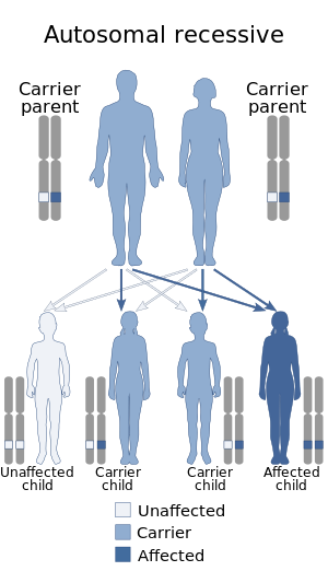
Hypomyelination-congenital cataract syndrome is a rare autosomal recessive hereditary disorder that affects the brain's white matter and is characterized by congenital cataract, psychomotor development delays, and moderate intellectual disabilities. It is a type of leukoencephalopathy.

<span class="mw-page-title-main">Autosomal dominant leukodystrophy with autonomic disease</span> Medical condition

Autosomal dominant leukodystrophy with autonomic disease is a rare neurological condition of genetic origin which is characterized by gradual demyelination of the central nervous system which results in various impairments, including ataxia, mild cognitive disability and autonomic dysfunction. It is part of a group of disorders called "leukodystrophies".

References

  1. 1 2 3 4 Kuhlmann, T.; Lassmann, H.; Bruck, W (2008). "Diagnosis of inflammatory demyelination in biopsy specimens: a practical approach". Acta Neuropathol. 115 (3): 275–287. doi:10.1007/s00401-007-0320-8. PMC   2668559 . PMID   18175128.
  2. 1 2 3 4 5 6 7 8 9 10 11 van der Knaap, M.S.; Pronk, J.C.; Scheper, G.C. (2006). "Vanishing White Matter Disease". Lancet Neurology. 5 (5): 413–423. doi:10.1016/S1474-4422(06)70440-9. PMID   16632312. S2CID   44301370.
  3. 1 2 3 Baumann, N.; Turpin, J-C (2000). "Adult-onset leukodystrophies". Journal of Neurology. 247 (10): 751–9. doi:10.1007/s004150070088. PMID   11127529. S2CID   6817046.
  4. 1 2 3 4 5 6 7 8 9 10 11 Dr. Raphael Schiffmann (Director of Baylor Research Institute at Dallas, Texas). Telephone Interview 2008-11-08.
  5. 1 2 Goswami, D.; Conway, G.S. (2005). "Premature ovarian failure". Human Reproduction Update. 11 (4): 391–410. doi: 10.1093/humupd/dmi012 . PMID   15919682. S2CID   23396413.
  6. 1 2 3 4 Abbott, C.M.; Proud, C.G. (2004). "Translation factors: in sickness and in health". Trends in Biochemical Sciences. 29 (1): 25–31. doi:10.1016/j.tibs.2003.11.006. PMID   14729329.
  7. 1 2 3 4 Pronk, J.C.; van Kollenburg, B.; Scheper, G.C.; van der Knaap, M.S. (2006). "Vanishing White Matter Disease: A Review with Focus on Its Genetics". Mental Retardation and Developmental Disabilities Research Reviews. 12 (2): 123–8. doi:10.1002/mrdd.20104. PMID   16807905.
  8. Laberge, A-M.; Michaud, J.; Richter, A.; Lambert, M.; Brais, B.; Mitchell G.A. (2005). "Population history and its impact on medical genetics in Quebec". Clinical Genetics. 68 (4): 287–301. doi: 10.1111/j.1399-0004.2005.00497.x . PMID   16143014. S2CID   26368522.
  9. Scheper, G.C.; Proud, C.G.; van der Knaap, M.S. (2006). "Defective translation initiation causes vanishing of cerebral white matter". Trends in Molecular Medicine. 12 (4): 159–166. doi:10.1016/j.molmed.2006.02.006. PMID   16545608.
  10. 1 2 3 Schiffmann, R.; Elroy-Stein, O. (2006). "Childhood ataxia with CNS hypomyelination/vanishing white matter disease – A common leukodystrophy caused by abnormal control of protein synthesis". Molecular Genetics and Metabolism. 88 (1): 7–15. doi:10.1016/j.ymgme.2005.10.019. PMID   16378743.
  11. 1 2 Denier C, Orgibet A, Roffi F, Jouvent E, Buhl C, Niel F, Boespflug-Tanguy O, Said G, Ducreux D (2007). "Adult-onset vanishing white matter leukoencephalopathy presenting as psychosis". Neurology. 68 (18): 1538–9. doi:10.1212/01.wnl.0000260701.76868.44. PMID   17470759. S2CID   28752485.
  12. Ayrignac et al. Brain magnetic resonance imaging helps to differentiate atypical multiple sclerosis with cavitary lesions and vanishing white matter disease, European Journal of Neurology Volume 23, Issue 6, 04 January 2016, doi : 10.1111/ene.12931