Pendred syndrome

Last updated
Pendred syndrome
Other namesGoiter-deafness syndrome [1]
Pendred syndrome diagram.jpg
The normal cochlea has 2 & a half turns, but, in Pendred Syndrome, there is abnormal partitioning (the central bony core is reduced in size and complexity)and a reduced number of turns leading to a Mondini cochlea which has a basal turn and a dilated apical turn (1 & a half turns). There is also a dilated endolymphatic duct and sac with a widened vestibular aqueduct
Specialty Endocrinology   OOjs UI icon edit-ltr-progressive.svg
Named after Vaughan Pendred

Pendred syndrome is a genetic disorder leading to congenital bilateral (both sides) sensorineural hearing loss and goitre with euthyroid or mild hypothyroidism (decreased thyroid gland function). There is no specific treatment, other than supportive measures for the hearing loss and thyroid hormone supplementation in case of hypothyroidism. It is named after Vaughan Pendred (1869–1946), the British doctor who first described the condition in an Irish family living in Durham in 1896. [2] [3] It accounts for 7.5% to 15% of all cases of congenital deafness. [4]

Contents

Signs and symptoms

The hearing loss of Pendred syndrome is often, although not always, present from birth, and language acquisition may be a significant problem if deafness is severe in childhood. The hearing loss typically worsens over the years, and progression can be step-wise and related to minor head trauma. In some cases, language development worsens after head injury, demonstrating that the inner ear is sensitive to trauma in Pendred syndrome; this is as a consequence of the widened vestibular aqueducts usual in this syndrome. [4] Vestibular function varies in Pendred syndrome and vertigo can be a feature of minor head trauma. A goitre is present in 75% of all cases. [4]

Genetics

Pendred syndrome has an autosomal recessive pattern of inheritance. Autorecessive.svg
Pendred syndrome has an autosomal recessive pattern of inheritance.

Pendred syndrome is inherited in an autosomal recessive manner, meaning that one would need to inherit an abnormal gene from each parent to develop the condition. This also means that a sibling of a patient with Pendred syndrome has a 25% chance of also having the condition if the parents are unaffected carriers. [5]

It has been linked to mutations in the PDS gene, which codes for the pendrin protein (solute carrier family 26, member 4, SLC26A4). The gene is located on the long arm of chromosome 7 (7q31). [6] [7] Mutations in the same gene also cause enlarged vestibular aqueduct syndrome (EVA or EVAS), another congenital cause of deafness; specific mutations are more likely to cause EVAS, while others are more linked with Pendred syndrome. [8]

Pathophysiology

SLC26A4 can be found in the cochlea (part of the inner ear), thyroid and the kidney. In the kidney, it participates in the secretion of bicarbonate. However, Pendred syndrome is not known to lead to kidney problems. [9] It functions as an iodide/chloride transporter. [10] In the thyroid, this leads to reduced organification of iodine (i.e. its incorporation into thyroid hormone). [6]

Diagnosis

People with Pendred syndrome present with a hearing loss either at birth or during childhood. The hearing loss is commonly progressive. In early stages it is usually a mixed hearing loss (both conductive and sensorineural hearing loss) because of a third window effect due to the inner ear malformation (widened vestibular aqueducts). A thyroid goitre may be present in the first decade and is usual towards the end of the second decade. MRI scanning of the inner ear usually shows widened or large vestibular aqueducts with enlarged endolymphatic sacs and may show abnormalities of the cochleae that are known as Mondini dysplasia. [4] Genetic testing to identify the pendrin gene usually establishes the diagnosis. If the condition is suspected, a "perchlorate discharge test" is sometimes performed. This test is highly sensitive, but may also be abnormal in other thyroid conditions. [4] If a goitre is present, thyroid function tests are performed to identify mild cases of thyroid dysfunction even if they are not yet causing symptoms. [11]

Treatment

No specific treatment exists for Pendred syndrome. If thyroid hormone levels are decreased, thyroid hormone supplements may be required. Patients are advised to take precautions against head injury. [11]

Related Research Articles

<span class="mw-page-title-main">Tietz syndrome</span> Congenital disorder

Tietz syndrome, also called Tietz albinism-deafness syndrome or albinism and deafness of Tietz, is an autosomal dominant congenital disorder characterized by deafness and leucism. It is caused by a mutation in the microphthalmia-associated transcription factor (MITF) gene. Tietz syndrome was first described in 1963 by Walter Tietz (1927–2003) a German Physician working in California.

<span class="mw-page-title-main">Conductive hearing loss</span> Medical condition

Conductive hearing loss (CHL) occurs when there is a problem transferring sound waves anywhere along the pathway through the outer ear, tympanic membrane (eardrum), or middle ear (ossicles). If a conductive hearing loss occurs in conjunction with a sensorineural hearing loss, it is referred to as a mixed hearing loss. Depending upon the severity and nature of the conductive loss, this type of hearing impairment can often be treated with surgical intervention or pharmaceuticals to partially or, in some cases, fully restore hearing acuity to within normal range. However, cases of permanent or chronic conductive hearing loss may require other treatment modalities such as hearing aid devices to improve detection of sound and speech perception.

<span class="mw-page-title-main">Sensorineural hearing loss</span> Hearing loss caused by an inner ear or vestibulocochlear nerve defect

Sensorineural hearing loss (SNHL) is a type of hearing loss in which the root cause lies in the inner ear or sensory organ or the vestibulocochlear nerve. SNHL accounts for about 90% of reported hearing loss. SNHL is usually permanent and can be mild, moderate, severe, profound, or total. Various other descriptors can be used depending on the shape of the audiogram, such as high frequency, low frequency, U-shaped, notched, peaked, or flat.

<span class="mw-page-title-main">Primrose syndrome</span> Medical condition

Primrose syndrome is a rare, slowly progressive genetic disorder that can vary symptomatically between individual cases, but is generally characterised by ossification of the external ears, learning difficulties, and facial abnormalities. It was first described in 1982 in Scotland's Royal National Larbert Institution by Dr D.A.A. Primrose.

<span class="mw-page-title-main">Jervell and Lange-Nielsen syndrome</span> Medical condition

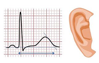
Jervell and Lange-Nielsen syndrome (JLNS) is a rare type of long QT syndrome associated with severe, bilateral sensorineural hearing loss. Those with JLNS are at risk of abnormal heart rhythms called arrhythmias, which can lead to fainting, seizures, or sudden death. JLNS, like other forms of long QT syndrome, causes the cardiac muscle to take longer than usual to recharge between beats. It is caused by genetic variants responsible for producing ion channels that carry transport potassium out of cells. The condition is usually diagnosed using an electrocardiogram, but genetic testing can also be used. Treatment includes lifestyle measures, beta blockers, and implantation of a defibrillator in some cases. It was first described by Anton Jervell and Fred Lange-Nielsen in 1957.

Mondini dysplasia, also known as Mondini malformation and Mondini defect, is an abnormality of the inner ear that is associated with sensorineural hearing loss.

Albinism-black lock-cell migration disorder is the initialism for the following terms and concepts that describe a condition affecting a person's physical appearance and physiology: (1) A – albinism, (2) B – black lock of hair, (3) C – cell migration disorder of the neurocytes of the gut, and (4) D – sensorineural deafness. The syndrome is caused by mutation in the endothelin B receptor gene (EDNRB).

Nonsyndromic deafness is hearing loss that is not associated with other signs and symptoms. In contrast, syndromic deafness involves hearing loss that occurs with abnormalities in other parts of the body. Genetic changes are related to the following types of nonsyndromic deafness.

Pendrin is an anion exchange protein that in humans is encoded by the SLC26A4 gene . Pendrin was initially identified as a sodium-independent chloride-iodide exchanger with subsequent studies showing that it also accepts formate and bicarbonate as substrates. Pendrin is similar to the Band 3 transport protein found in red blood cells. Pendrin is the protein which is mutated in Pendred syndrome, which is an autosomal recessive disorder characterized by sensorineural hearing loss, goiter and a partial organification problem detectable by a positive perchlorate test.

<span class="mw-page-title-main">Thyroid dyshormonogenesis</span> Medical condition

Thyroid dyshormonogenesis is a rare condition due to genetic defects in the synthesis of thyroid hormones.

<span class="mw-page-title-main">Cochlin</span> Protein highly abundant in the cochlea and vestibule of the inner ear

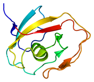
Cochlin is a protein that in humans is encoded by the COCH gene. It is an extracellular matrix (ECM) protein highly abundant in the cochlea and vestibule of the inner ear, constituting the major non-collagen component of the ECM of the inner ear. The protein is highly conserved in human, mouse, and chicken, showing 94% and 79% amino acid identity of human to mouse and chicken sequences, respectively.

<span class="mw-page-title-main">Björnstad syndrome</span> Medical condition

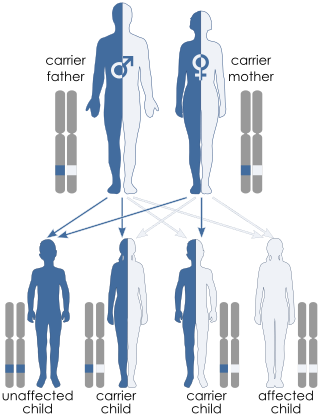
Björnstad syndrome is an autosomal recessive congenital condition involving pili torti, sensorineural deafness, and hair abnormalities. It was first characterized in 1965, in Oslo, by prof. Roar Theodor Bjørnstad after he observed an association between pili torti and hearing loss. The condition is extremely rare, with less than 50 cases documented in medical literature worldwide.

EAST syndrome is a syndrome consisting of epilepsy, ataxia, sensorineural deafness and salt-wasting renal tubulopathy. The tubulopathy in this condition predispose to hypokalemic metabolic alkalosis with normal blood pressure. Hypomagnesemia may also be present.

<span class="mw-page-title-main">Enlarged vestibular aqueduct</span> Human disease

Large vestibular aqueduct is a structural deformity of the inner ear. Enlargement of this duct is one of the most common inner ear deformities and is commonly associated with hearing loss during childhood. The term was first discovered in 1791 by Mondini when he was completing a temporal bone dissection. It was then defined by Valvassori and Clemis as a vestibular aqueduct that is greater than or equal to 2.0 mm at the operculum and/or greater than or equal to 1.0 mm at the midpoint. Some use the term enlarged vestibular aqueduct syndrome, but this is felt by others to be erroneous as it is a clinical finding which can occur in several syndromes.

<span class="mw-page-title-main">Mohr–Tranebjærg syndrome</span> Medical condition

Mohr–Tranebjærg syndrome (MTS) is a rare X-linked recessive syndrome also known as deafness–dystonia syndrome and caused by mutation in the TIMM8A gene. It is characterized by clinical manifestations commencing with early childhood onset hearing loss, followed by adolescent onset progressive dystonia or ataxia, visual impairment from early adulthood onwards and dementia from the 4th decade onwards. The severity of the symptoms may vary, but they progress usually to severe deafness and dystonia and sometimes are accompanied by cortical deterioration of vision and mental deterioration.

<span class="mw-page-title-main">Donnai–Barrow syndrome</span> Medical condition

Donnai–Barrow syndrome is a genetic disorder first described by Dian Donnai and Margaret Barrow in 1993. It is associated with LRP2. It is an inherited (genetic) disorder that affects many parts of the body.

<span class="mw-page-title-main">FOXI1</span> Protein-coding gene in the species Homo sapiens

Forkhead box I1 is a protein that in humans is encoded by the FOXI1 gene.

Nasodigitoacoustic syndrome, also called Keipert syndrome, is a rare congenital syndrome first described by J.A. Keipert and colleagues in 1973. The syndrome is characterized by a misshaped nose, broad thumbs and halluces, brachydactyly, sensorineural hearing loss, facial features such as hypertelorism, and developmental delay.

<span class="mw-page-title-main">Distal 18q-</span> Human disease

Distal 18q- is a genetic condition caused by a deletion of genetic material within one of the two copies of chromosome 18. The deletion involves the distal section of 18q and typically extends to the tip of the long arm of chromosome 18.

<span class="mw-page-title-main">Michel aplasia</span> Medical condition

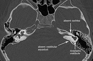
Michel aplasia, also known as complete labyrinthine aplasia (CLA), is a congenital abnormality of the inner ear. It is characterized by the bilateral absence of differentiated inner ear structures and results in complete deafness (anacusis). Michel aplasia should not be confused with michel dysplasia. It may affect one or both ears.

References

  1. RESERVED, INSERM US14-- ALL RIGHTS. "Orphanet: Pendred syndrome". www.orpha.net. Retrieved 29 September 2019.
  2. Pendred V (1896). "Deaf-mutism and goitre". Lancet. 2 (3808): 532. doi:10.1016/S0140-6736(01)74403-0.
  3. Pearce JM (2007). "Pendred's syndrome". Eur. Neurol. 58 (3): 189–90. doi: 10.1159/000104724 . PMID   17622729.
  4. 1 2 3 4 5 Reardon W, Coffey R, Phelps PD, et al. (July 1997). "Pendred syndrome--100 years of underascertainment?". QJM. 90 (7): 443–7. doi: 10.1093/qjmed/90.7.443 . PMID   9302427.
  5. Wémeau JL, Kopp P (March 2017). "Pendred syndrome". Best Practice & Research. Clinical Endocrinology & Metabolism. 31 (2): 213–224. doi:10.1016/j.beem.2017.04.011. PMID   28648509.
  6. 1 2 Sheffield VC, Kraiem Z, Beck JC, et al. (April 1996). "Pendred syndrome maps to chromosome 7q21-34 and is caused by an intrinsic defect in thyroid iodine organification". Nat. Genet. 12 (4): 424–6. doi:10.1038/ng0496-424. PMID   8630498. S2CID   25888014.
  7. Coyle B, Coffey R, Armour JA, Gausden E, Hochberg Z, Grossman A, et al. (April 1996). "Pendred syndrome (goitre and sensorineural hearing loss) maps to chromosome 7 in the region containing the nonsyndromic deafness gene DFNB4". Nature Genetics. 12 (4): 421–3. doi:10.1038/ng0496-421. PMID   8630497. S2CID   7166946.
  8. Azaiez H, Yang T, Prasad S, Sorensen JL, Nishimura CJ, Kimberling WJ, Smith RJ (December 2007). "Genotype-phenotype correlations for SLC26A4-related deafness". Human Genetics. 122 (5): 451–7. doi:10.1007/s00439-007-0415-2. PMC   10519375 . PMID   17690912. S2CID   19451595.
  9. Royaux IE, Wall SM, Karniski LP, et al. (March 2001). "Pendrin, encoded by the Pendred syndrome gene, resides in the apical region of renal intercalated cells and mediates bicarbonate secretion". Proc. Natl. Acad. Sci. U.S.A. 98 (7): 4221–6. Bibcode:2001PNAS...98.4221R. doi: 10.1073/pnas.071516798 . PMC   31206 . PMID   11274445.
  10. Scott DA, Wang R, Kreman TM, Sheffield VC, Karniski LP (April 1999). "The Pendred syndrome gene encodes a chloride-iodide transport protein". Nature Genetics. 21 (4): 440–3. doi:10.1038/7783. PMID   10192399. S2CID   23717390.
  11. 1 2 National Institute on Deafness and Other Communication Disorders (October 2006). "Pendred Syndrome". Archived from the original on 2008-05-09. Retrieved 2008-05-05.