Shiga toxin

Last updated
Ribbon diagram of Shiga toxin (Stx) from S. dysenteriae. From PDB: 1R4Q . Shiga toxin (Stx) PDB 1r4q.png
Ribbon diagram of Shiga toxin (Stx) from S. dysenteriae. From PDB: 1R4Q .
Shiga-like toxin beta subunit
Identifiers
SymbolSLT_beta
Pfam PF02258
InterPro IPR003189
SCOP2 2bos / SCOPe / SUPFAM
TCDB 1.C.54
Available protein structures:
Pfam   structures / ECOD  
PDB RCSB PDB; PDBe; PDBj
PDBsum structure summary
Shiga-like toxin subunit A
Identifiers
SymbolShiga-like_toxin_subunit_A
InterPro IPR016331
SCOP2 1r4q / SCOPe / SUPFAM

Shiga toxins are a family of related toxins with two major groups, Stx1 and Stx2, expressed by genes considered to be part of the genome of lambdoid prophages. [1] The toxins are named after Kiyoshi Shiga, who first described the bacterial origin of dysentery caused by Shigella dysenteriae . [2] Shiga-like toxin (SLT) is a historical term for similar or identical toxins produced by Escherichia coli . [3] The most common sources for Shiga toxin are the bacteria S. dysenteriae and some serotypes of Escherichia coli (STEC), which includes serotypes O157:H7, and O104:H4. [4] [5]

Contents

Nomenclature

Microbiologists use many terms to describe Shiga toxin and differentiate more than one unique form. Many of these terms are used interchangeably.

  1. Shiga toxin type 1 and type 2 (Stx-1 and 2) are the Shiga toxins produced by some E. coli strains. Stx-1 is identical to Stx of Shigella spp. or differs by only one amino acid. [6] Stx-2 shares 56% sequence identity with Stx-1.[ citation needed ]
  2. Cytotoxins – an archaic denotation for Stx – is used in a broad sense.
  3. Verocytotoxins/verotoxins – a seldom-used term for Stx – is from the hypersensitivity of Vero cells to Stx. [7] [8] [9]
  4. The term Shiga-like toxins is another antiquated term which arose prior to the understanding that Shiga and Shiga-like toxins were identical. [10]

History

The toxin is named after Kiyoshi Shiga, who discovered S. dysenteriae in 1897. [2] In 1977, researchers in Ottawa, Ontario discovered the Shiga toxin normally produced by Shigella dysenteriae in a line of E. coli. [11] The E. coli version of the toxin was named "verotoxin" because of its ability to kill Vero cells (African green monkey kidney cells) in culture. Shortly after, the verotoxin was referred to as Shiga-like toxin because of its similarities to Shiga toxin.

It has been suggested by some researchers that the gene coding for Shiga-like toxin comes from a toxin-converting lambdoid bacteriophage, such as H-19B or 933W, inserted into the bacteria's chromosome via transduction. [12] Phylogenetic studies of the diversity of E. coli suggest that it may have been relatively easy for Shiga toxin to transduce into certain strains of E. coli, because Shigella is itself a subgenus of Escherichia ; in fact, some strains traditionally considered E. coli (including those that produce this toxin) in fact belong to this lineage. Being closer relatives of Shigella dysenteriae than of the typical E. coli, it is not at all unusual that toxins similar to that of S. dysenteriae are produced by these strains. As microbiology advances, the historical variation in nomenclature (which arose because of gradually advancing science in multiple places) is increasingly giving way to recognizing all of these molecules as "versions of the same toxin" rather than "different toxins". [13] :2–3

Transmission

The toxin requires highly specific receptors on the cells' surface in order to attach and enter the cell; species such as cattle, swine, and deer which do not carry these receptors may harbor toxigenic bacteria without any ill effect, shedding them in their feces, from where they may be spread to humans. [14]

Clinical significance

Symptoms of Shiga toxin ingestion include abdominal pain as well as watery diarrhea. Severe life-threatening cases are characterized by hemorrhagic colitis (HC). [15]

The toxin is associated with hemolytic-uremic syndrome. In contrast, Shigella species may also produce shigella enterotoxins, which are the cause of dysentery.

The toxin is effective against small blood vessels, such as found in the digestive tract, the kidney, and lungs, but not against large vessels such as the arteries or major veins. A specific target for the toxin appears to be the vascular endothelium of the glomerulus. This is the filtering structure that is a key to the function of the kidney. Destroying these structures leads to kidney failure and the development of the often deadly and frequently debilitating hemolytic uremic syndrome. Food poisoning with Shiga toxin often also has effects on the lungs and the nervous system.

Structure and mechanism

SLT2 from Escherichia coli O157:H7. A-subunit is shown above (viridian), with B-subunit pentamer below (multicolored). From PDB: 1R4P . 1R4P Structure.png
SLT2 from Escherichia coli O157:H7. A-subunit is shown above (viridian), with B-subunit pentamer below (multicolored). From PDB: 1R4P .

Mechanism

The B subunits of the toxin bind to a component of the cell membrane known as glycolipid globotriaosylceramide (Gb3). Binding of the subunit B to Gb3 causes induction of narrow tubular membrane invaginations, which drives formation of inward membrane tubules for the bacterial uptake into the cell. These tubules are essential for uptake into the host cell. [16] The Shiga toxin (a non-pore forming toxin) is transferred to the cytosol via Golgi network and endoplasmic reticulum (ER). From the Golgi toxin is trafficked to the ER. Shiga toxins act to inhibit protein synthesis within target cells by a mechanism similar to that of the infamous plant toxin ricin. [17] [18] After entering a cell via a macropinosome, [19] the payload (A subunit) cleaves a specific adenine nucleobase from the 28S RNA of the 60S subunit of the ribosome, thereby halting protein synthesis. [20] As they mainly act on the lining of the blood vessels, the vascular endothelium, a breakdown of the lining and hemorrhage eventually occurs.[ clarification needed ] The first response is commonly a bloody diarrhea. This is because Shiga toxin is usually taken in with contaminated food or water.

The bacterial Shiga toxin can be used for targeted therapy of gastric cancer, because this tumor entity expresses the receptor of the Shiga toxin. For this purpose an unspecific chemotherapeutical is conjugated to the B-subunit to make it specific. In this way only the tumor cells, but not healthy cells, are destroyed during therapy. [21]

Structure

The toxin has two subunits—designated A (mol. wt. 32000 Da) and B (mol. wt. 7700 Da)—and is one of the AB5 toxins. The B subunit is a pentamer that binds to specific glycolipids on the host cell, specifically globotriaosylceramide (Gb3). [22] [23] Following this, the A subunit is internalised and cleaved into two parts. The A1 component then binds to the ribosome, disrupting protein synthesis. Stx-2 has been found to be about 400 times more toxic (as quantified by LD50 in mice) than Stx-1.

Gb3 is, for unknown reasons, present in greater amounts in renal epithelial tissues, to which the renal toxicity of Shiga toxin may be attributed. Gb3 is also found in central nervous system neurons and endothelium, which may lead to neurotoxicity. [24] Stx-2 is also known to increase the expression of its receptor GB3 and cause neuronal dysfunctions. [25]

See also

Related Research Articles

<i>Escherichia coli</i> Enteric, rod-shaped, gram-negative bacterium

Escherichia coli ( ESH-ə-RIK-ee-ə KOH-ly) is a gram-negative, facultative anaerobic, rod-shaped, coliform bacterium of the genus Escherichia that is commonly found in the lower intestine of warm-blooded organisms. Most E. coli strains are harmless, but some serotypes such as EPEC, and ETEC are pathogenic and can cause serious food poisoning in their hosts, and are occasionally responsible for food contamination incidents that prompt product recalls. Most strains are part of the normal microbiota of the gut and are harmless or even beneficial to humans (although these strains tend to be less studied than the pathogenic ones). For example, some strains of E. coli benefit their hosts by producing vitamin K2 or by preventing the colonization of the intestine by pathogenic bacteria. These mutually beneficial relationships between E. coli and humans are a type of mutualistic biological relationship — where both the humans and the E. coli are benefitting each other. E. coli is expelled into the environment within fecal matter. The bacterium grows massively in fresh fecal matter under aerobic conditions for three days, but its numbers decline slowly afterwards.

<i>Escherichia coli</i> O157:H7 Serotype of the bacteria Escherichia coli

Escherichia coli O157:H7 is a serotype of the bacterial species Escherichia coli and is one of the Shiga-like toxin–producing types of E. coli. It is a cause of disease, typically foodborne illness, through consumption of contaminated and raw food, including raw milk and undercooked ground beef. Infection with this type of pathogenic bacteria may lead to hemorrhagic diarrhea, and to kidney failure; these have been reported to cause the deaths of children younger than five years of age, of elderly patients, and of patients whose immune systems are otherwise compromised.

<span class="mw-page-title-main">Shigellosis</span> Medical condition

Shigellosis is an infection of the intestines caused by Shigella bacteria. Symptoms generally start one to two days after exposure and include diarrhea, fever, abdominal pain, and feeling the need to pass stools even when the bowels are empty. The diarrhea may be bloody. Symptoms typically last five to seven days and it may take several months before bowel habits return entirely to normal. Complications can include reactive arthritis, sepsis, seizures, and hemolytic uremic syndrome.

<i>Shigella</i> Genus of bacteria

Shigella is a genus of bacteria that is Gram-negative, facultatively anaerobic, non–spore-forming, nonmotile, rod-shaped, and is genetically closely related to Escherichia. The genus is named after Kiyoshi Shiga, who discovered it in 1897.

<span class="mw-page-title-main">Hemolytic–uremic syndrome</span> Group of blood disorders related to bacterial infection

Hemolytic–uremic syndrome (HUS) is a group of blood disorders characterized by low red blood cells, acute kidney injury, and low platelets. Initial symptoms typically include bloody diarrhea, fever, vomiting, and weakness. Kidney problems and low platelets then occur as the diarrhea progresses. Children are more commonly affected, but most children recover without permanent damage to their health, although some children may have serious and sometimes life-threatening complications. Adults, especially the elderly, may present a more complicated presentation. Complications may include neurological problems and heart failure.

<i>Shigella dysenteriae</i> Bacterial species

Shigella dysenteriae is a species of the rod-shaped bacterial genus Shigella. Shigella species can cause shigellosis. Shigellae are Gram-negative, non-spore-forming, facultatively anaerobic, nonmotile bacteria. S. dysenteriae has the ability to invade and replicate in various species of epithelial cells and enterocytes.

<i>Shigella flexneri</i> Species of bacterium

Shigella flexneri is a species of Gram-negative bacteria in the genus Shigella that can cause diarrhea in humans. Several different serogroups of Shigella are described; S. flexneri belongs to group B. S. flexneri infections can usually be treated with antibiotics, although some strains have become resistant. Less severe cases are not usually treated because they become more resistant in the future. Shigella are closely related to Escherichia coli, but can be differentiated from E.coli based on pathogenicity, physiology and serology.

<span class="mw-page-title-main">Diphtheria toxin</span> Exotoxin

Diphtheria toxin is an exotoxin secreted mainly by Corynebacterium diphtheriae but also by Corynebacterium ulcerans and Corynebacterium pseudotuberculosis, the pathogenic bacterium that causes diphtheria. The toxin gene is encoded by a prophage called corynephage β. The toxin causes the disease in humans by gaining entry into the cell cytoplasm and inhibiting protein synthesis.

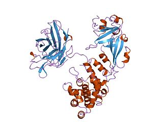
The AB5 toxins are six-component protein complexes secreted by certain pathogenic bacteria known to cause human diseases such as cholera, dysentery, and hemolytic–uremic syndrome. One component is known as the A subunit, and the remaining five components are B subunits. All of these toxins share a similar structure and mechanism for entering targeted host cells. The B subunit is responsible for binding to receptors to open up a pathway for the A subunit to enter the cell. The A subunit is then able to use its catalytic machinery to take over the host cell's regular functions.

<span class="mw-page-title-main">P1PK blood group system</span> Human blood group system

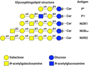
P1PK is a human blood group system based upon the A4GALT gene on chromosome 22. The P antigen was first described by Karl Landsteiner and Philip Levine in 1927. The P1PK blood group system consists of three glycosphingolipid antigens: Pk, P1 and NOR. In addition to glycosphingolipids, terminal Galα1→4Galβ structures are present on complex-type N-glycans. The GLOB antigen is now the member of the separate GLOB (globoside) blood group system.

Microbial toxins are toxins produced by micro-organisms, including bacteria, fungi, protozoa, dinoflagellates, and viruses. Many microbial toxins promote infection and disease by directly damaging host tissues and by disabling the immune system. Endotoxins most commonly refer to the lipopolysaccharide (LPS) or lipooligosaccharide (LOS) that are in the outer plasma membrane of Gram-negative bacteria. The botulinum toxin, which is primarily produced by Clostridium botulinum and less frequently by other Clostridium species, is the most toxic substance known in the world. However, microbial toxins also have important uses in medical science and research. Currently, new methods of detecting bacterial toxins are being developed to better isolate and understand these toxins. Potential applications of toxin research include combating microbial virulence, the development of novel anticancer drugs and other medicines, and the use of toxins as tools in neurobiology and cellular biology.

<span class="mw-page-title-main">Globotriaosylceramide</span>

Globotriaosylceramide is a globoside. It is also known as CD77, Gb3, GL3, and ceramide trihexoside. It is one of the few clusters of differentiation that is not a protein.

Enteroinvasive Escherichia coli (EIEC) is a type of pathogenic bacteria whose infection causes a syndrome that is identical to shigellosis, with profuse diarrhea and high fever. EIEC are highly invasive, and they use adhesin proteins to bind to and enter intestinal cells. They produce no toxins, but severely damage the intestinal wall through mechanical cell destruction.

Escherichia coli O104:H4 is an enteroaggregative Escherichia coli strain of the bacterium Escherichia coli, and the cause of the 2011 Escherichia coli O104:H4 outbreak. The "O" in the serological classification identifies the cell wall lipopolysaccharide antigen, and the "H" identifies the flagella antigen.

Shigatoxigenic Escherichia coli (STEC) and verotoxigenic E. coli (VTEC) are strains of the bacterium Escherichia coli that produce Shiga toxin. Only a minority of the strains cause illness in humans. The ones that do are collectively known as enterohemorrhagic E. coli (EHEC) and are major causes of foodborne illness. When infecting the large intestine of humans, they often cause gastroenteritis, enterocolitis, and bloody diarrhea and sometimes cause a severe complication called hemolytic-uremic syndrome (HUS). Cattle is an important natural reservoir for EHEC because the colonised adult ruminants are asymptomatic. This is because they lack vascular expression of the target receptor for Shiga toxins. The group and its subgroups are known by various names. They are distinguished from other strains of intestinal pathogenic E. coli including enterotoxigenic E. coli (ETEC), enteropathogenic E. coli (EPEC), enteroinvasive E. coli (EIEC), enteroaggregative E. coli (EAEC), and diffusely adherent E. coli (DAEC).

<span class="mw-page-title-main">Cytolethal distending toxin</span>

Cytolethal distending toxins are a class of heterotrimeric toxins produced by certain gram-negative bacteria that display DNase activity. These toxins trigger G2/M cell cycle arrest in specific mammalian cell lines, leading to the enlarged or distended cells for which these toxins are named. Affected cells die by apoptosis.

Pathogenic <i>Escherichia coli</i> Strains of E. coli that can cause disease

Escherichia coli is a gram-negative, rod-shaped bacterium that is commonly found in the lower intestine of warm-blooded organisms (endotherms). Most E. coli strains are harmless, but pathogenic varieties cause serious food poisoning, septic shock, meningitis, or urinary tract infections in humans. Unlike normal flora E. coli, the pathogenic varieties produce toxins and other virulence factors that enable them to reside in parts of the body normally not inhabited by E. coli, and to damage host cells. These pathogenic traits are encoded by virulence genes carried only by the pathogens.

<span class="mw-page-title-main">Heat-labile enterotoxin family</span> Family of toxic protein complexes

In molecular biology, the heat-labile enterotoxin family includes Escherichia coli heat-labile enterotoxin and cholera toxin (Ctx) secreted by Vibrio cholerae.

Enteroaggregative Escherichia coli are a pathotype of Escherichia coli which cause acute and chronic diarrhea in both the developed and developing world. They may also cause urinary tract infections. EAEC are defined by their "stacked-brick" pattern of adhesion to the human laryngeal epithelial cell line HEp-2. The pathogenesis of EAEC involves the aggregation of and adherence of the bacteria to the intestinal mucosa, where they elaborate enterotoxins and cytotoxins that damage host cells and induce inflammation that results in diarrhea.

Bacterial effectors are proteins secreted by pathogenic bacteria into the cells of their host, usually using a type 3 secretion system (TTSS/T3SS), a type 4 secretion system (TFSS/T4SS) or a Type VI secretion system (T6SS). Some bacteria inject only a few effectors into their host’s cells while others may inject dozens or even hundreds. Effector proteins may have many different activities, but usually help the pathogen to invade host tissue, suppress its immune system, or otherwise help the pathogen to survive. Effector proteins are usually critical for virulence. For instance, in the causative agent of plague, the loss of the T3SS is sufficient to render the bacteria completely avirulent, even when they are directly introduced into the bloodstream. Gram negative microbes are also suspected to deploy bacterial outer membrane vesicles to translocate effector proteins and virulence factors via a membrane vesicle trafficking secretory pathway, in order to modify their environment or attack/invade target cells, for example, at the host-pathogen interface.

References

  1. Friedman D; Court D (2001). "Bacteriophage lambda: alive and well and still doing its thing". Current Opinion in Microbiology. 4 (2): 201–7. doi:10.1016/S1369-5274(00)00189-2. PMID   11282477.
  2. 1 2 Trofa, Andrew F.; Ueno-Olsen, Hannah; Oiwa, Ruiko; Yoshikawa, Masanosuke (1999-11-01). "Dr. Kiyoshi Shiga: Discoverer of the Dysentery Bacillus". Clinical Infectious Diseases. 29 (5): 1303–1306. doi: 10.1086/313437 . ISSN   1058-4838. PMID   10524979.
  3. Zhu Q; Li L; Guo Z; Yang R (June 2002). "Identification of Shiga-like toxin Escherichia coli isolated from children with diarrhea by polymerase chain reaction". Chin. Med. J. 115 (6): 815–8. PMID   12123543.
  4. Beutin L (2006). "Emerging enterohaemorrhagic Escherichia coli, causes and effects of the rise of a human pathogen". Journal of Veterinary Medicine. B, Infectious Diseases and Veterinary Public Health. 53 (7): 299–305. doi:10.1111/j.1439-0450.2006.00968.x. PMID   16930272.
  5. Spears; et al. (2006). "A comparison of Enteropathogenic and enterohaemorragic E.coli pathogenesis". FEMS Microbiology Letters. 255 (2): 187–202. doi: 10.1111/j.1574-6968.2006.00119.x . PMID   16448495.
  6. Kaper JB, O'Brien AD (2014). Sperandio V, Hovde CJ (eds.). "Overview and Historical Perspectives". Microbiology Spectrum. 2 (6). doi:10.1128/microbiolspec.EHEC-0028-2014. PMC   4290666 . PMID   25590020.
  7. Beutin L; Geier D; Steinrück H; Zimmermann S; Scheutz F (September 1993). "Prevalence and some properties of verotoxin (Shiga-like toxin)-producing Escherichia coli in seven different species of healthy domestic animals". Journal of Clinical Microbiology. 31 (9): 2483–8. doi:10.1128/JCM.31.9.2483-2488.1993. PMC   265781 . PMID   8408571.
  8. Bitzan M; Richardson S; Huang C; Boyd B; Petric M; Karmali MA (August 1994). "Evidence that verotoxins (Shiga-like toxins) from Escherichia coli bind to P blood group antigens of human erythrocytes in vitro". Infection and Immunity. 62 (8): 3337–47. doi:10.1128/IAI.62.8.3337-3347.1994. PMC   302964 . PMID   8039905.
  9. Giraldi R; Guth BE; Trabulsi LR (June 1990). "Production of Shiga-like toxin among Escherichia coli strains and other bacteria isolated from diarrhea in São Paulo, Brazil". Journal of Clinical Microbiology. 28 (6): 1460–2. doi:10.1128/JCM.28.6.1460-1462.1990. PMC   267957 . PMID   2199511.
  10. Scheutz F, Teel LD, Beutin L, Piérard D, Buvens G, Karch H, Mellmann A, Caprioli A, Tozzoli R, Morabito S, Strockbine NA, Melton-Celsa AR, Sanchez M, Persson S, O'Brien AD (September 2012). "Multicenter evaluation of a sequence-based protocol for subtyping Shiga toxins and standardizing Stx nomenclature". Journal of Clinical Microbiology. 50 (9): 2951–63. doi:10.1128/JCM.00860-12. PMC   3421821 . PMID   22760050.
  11. Konowalchuk J, Speirs JI, Stavric S (December 1977). "Vero response to a cytotoxin of Escherichia coli". Infection and Immunity. 18 (3): 775–9. doi:10.1128/IAI.18.3.775-779.1977. PMC   421302 . PMID   338490.
  12. Mizutani S, Nakazono N, Sugino Y (April 1999). "The so-called chromosomal verotoxin genes are actually carried by defective prophages". DNA Research. 6 (2): 141–3. doi: 10.1093/dnares/6.2.141 . PMID   10382973.
  13. Silva CJ, Brandon DL, Skinner CB, He X, et al. (2017), "Chapter 3: Structure of Shiga toxins and other AB5 toxins", Shiga toxins: A Review of Structure, Mechanism, and Detection, Springer, ISBN   978-3319505800.
  14. Asakura H, Makino S, Kobori H, Watarai M, Shirahata T, Ikeda T, Takeshi K (August 2001). "Phylogenetic diversity and similarity of active sites of Shiga toxin (stx) in Shiga toxin-producing Escherichia coli (STEC) isolates from humans and animals". Epidemiology and Infection. 127 (1): 27–36. doi:10.1017/S0950268801005635. PMC   2869726 . PMID   11561972.
  15. Beutin, L; Miko, A; Krause, G; Pries, K; Haby, S; Steege, K; Albrecht, N (2007). "Identification of human-pathogenic strains of Shiga toxin-producing Escherichia coli from food by a combination of serotyping and molecular typing of Shiga toxin genes". Applied and Environmental Microbiology. 73 (15): 4769–75. doi:10.1128/AEM.00873-07. PMC   1951031 . PMID   17557838.
  16. Römer W, Berland L, Chambon V, Gaus K, Windschiegl B, Tenza D, Aly MR, Fraisier V, Florent JC, Perrais D, Lamaze C, Raposo G, Steinem C, Sens P, Bassereau P, Johannes L (November 2007). "Shiga toxin induces tubular membrane invaginations for its uptake into cells". Nature. 450 (7170): 670–5. Bibcode:2007Natur.450..670R. doi:10.1038/nature05996. PMID   18046403. S2CID   4410673.
  17. Sandvig K, van Deurs B (November 2000). "Entry of ricin and Shiga toxin into cells: molecular mechanisms and medical perspectives". The EMBO Journal. 19 (22): 5943–50. doi:10.1093/emboj/19.22.5943. PMC   305844 . PMID   11080141.
  18. Mercatelli D, Bortolotti M, Giorgi FM (August 2020). "Transcriptional network inference and master regulator analysis of the response to ribosome-inactivating proteins in leukemia cells". Toxicology. 441: 152531. doi:10.1016/j.tox.2020.152531. PMID   32593706. S2CID   220255474.
  19. Lukyanenko V, Malyukova I, Hubbard A, Delannoy M, Boedeker E, Zhu C, Cebotaru L, Kovbasnjuk O (November 2011). "Enterohemorrhagic Escherichia coli infection stimulates Shiga toxin 1 macropinocytosis and transcytosis across intestinal epithelial cells". American Journal of Physiology. Cell Physiology. 301 (5): C1140-9. doi:10.1152/ajpcell.00036.2011. PMC   3213915 . PMID   21832249.
  20. Donohue-Rolfe A, Acheson DW, Keusch GT (2010). "Shiga toxin: purification, structure, and function". Reviews of Infectious Diseases. 13 Suppl 4 (7): S293-7. doi:10.1016/j.toxicon.2009.11.021. PMID   2047652.
  21. Gastric adenocarcinomas express the glycosphingolipid Gb3/CD77: Targeting of gastric cancer cells with Shiga toxin B-subunit
  22. Stein PE, Boodhoo A, Tyrrell GJ, Brunton JL, Read RJ (February 1992). "Crystal structure of the cell-binding B oligomer of verotoxin-1 from E. coli". Nature. 355 (6362): 748–50. Bibcode:1992Natur.355..748S. doi:10.1038/355748a0. PMID   1741063. S2CID   4274763.
  23. Kaper JB, Nataro JP, Mobley HL (February 2004). "Pathogenic Escherichia coli". Nature Reviews. Microbiology. 2 (2): 123–40. doi:10.1038/nrmicro818. PMID   15040260. S2CID   3343088.
  24. Obata F, Tohyama K, Bonev AD, Kolling GL, Keepers TR, Gross LK, Nelson MT, Sato S, Obrig TG (November 2008). "Shiga toxin 2 affects the central nervous system through receptor globotriaosylceramide localized to neurons". The Journal of Infectious Diseases. 198 (9): 1398–406. doi:10.1086/591911. PMC   2684825 . PMID   18754742.
  25. Tironi-Farinati C, Loidl CF, Boccoli J, Parma Y, Fernandez-Miyakawa ME, Goldstein J (May 2010). "Intracerebroventricular Shiga toxin 2 increases the expression of its receptor globotriaosylceramide and causes dendritic abnormalities". Journal of Neuroimmunology. 222 (1–2): 48–61. doi:10.1016/j.jneuroim.2010.03.001. hdl: 11336/16303 . PMID   20347160. S2CID   11910897.