Solid pseudopapillary tumour

Last updated
Solid pseudopapillary tumour
Other namesSolid pseudopapillary neoplasm, solid pseudopapillary tumour/neoplasm of the pancreas, Frantz's tumour
Cytopathology of solid pseudopapillary neoplasm.png
Cytopathology of solid pseudopapillary tumour/neoplasm with main findings that distinguish it from pancreatic endocrine neoplasms and acinar cell carcinomas. [1] Diff-Quik stain.
Specialty Oncology

A solid pseudopapillary tumour is a low-grade malignant neoplasm of the pancreas of papillary architecture that typically afflicts young women. [2]

Contents

Signs and symptoms

Solid pseudopapillary tumours are often asymptomatic and are identified incidentally on imaging performed for unrelated reasons. Less often, they may cause abdominal pain. Solid pseudopapillary tumours tend to occur in women, and most often present in the third decade of life. [3]

Diagnosis

Papillae vs pseudopapillae: True papillae are outgrowths of epithelium, surrounding fibrovascular cores of stroma and at least one blood vessel. In contrast, pseudopapillae (such as in solid pseudopapillary tumours) are nests of proliferating cells that eventually grow to become almost back-to-back, with cells in the centers of nests disintegrating, leaving rims of cells lining the periphery of each nest. Discohesive cells and some formations lacking central blood vessels are visual clues. Papillae vs pseudopapillae.png
Papillae vs pseudopapillae: True papillae are outgrowths of epithelium, surrounding fibrovascular cores of stroma and at least one blood vessel. In contrast, pseudopapillae (such as in solid pseudopapillary tumours) are nests of proliferating cells that eventually grow to become almost back-to-back, with cells in the centers of nests disintegrating, leaving rims of cells lining the periphery of each nest. Discohesive cells and some formations lacking central blood vessels are visual clues.

The gold standard for diagnosing solid pseudopapillary tumour of the pancreas is cytopathology by endoscopic ultrasound (EUS) guided fine needle aspiration (FNA) of the lesion. [4] After surgical excision, the tumor can undergo histopathology evaluation for cancer staging.

Gross morphology

Solid pseudopapillary tumours are typically round, well-demarcated, measuring 2–17 cm in diameter (average 8 cm), with solid and cystic areas with hemorrhage on cut sections. [5]

Histomorphology

Solid pseudopapillary tumours consist of solid sheets of cells that are focally dyscohesive. The cells in the lesion usually have uniform nuclei with occasional nuclear grooves, eosinophilic or clear cytoplasm and PAS positive eosinophilic intracytoplasmic globules. [6] Necrosis is usually present and, as cell death preferentially occurs distant from blood vessels, lead to the formation of pseudopapillae.[ citation needed ]

Immunohistochemistry

Solid pseudopapillary tumours show positive nuclear staining for beta catenin, as well as positive immunostaining for CD10, CD56, vimentin, alpha 1-antitrypsin, and neuron specific enolase; they are negative for chromogranin and pancreatic enzymes. [7] [8]

Management

In most cases, solid pseudopapillary tumours should be resected surgically, as there is a risk of malignancy (cancer).[ citation needed ]

See also

Related Research Articles

<span class="mw-page-title-main">Thyroid neoplasm</span> Medical condition

Thyroid neoplasm is a neoplasm or tumor of the thyroid. It can be a benign tumor such as thyroid adenoma, or it can be a malignant neoplasm, such as papillary, follicular, medullary or anaplastic thyroid cancer. Most patients are 25 to 65 years of age when first diagnosed; women are more affected than men. The estimated number of new cases of thyroid cancer in the United States in 2023 is 43,720 compared to only 2,120 deaths. Of all thyroid nodules discovered, only about 5 percent are cancerous, and under 3 percent of those result in fatalities.

<span class="mw-page-title-main">Pancreatic cancer</span> Type of endocrine gland cancer

Pancreatic cancer arises when cells in the pancreas, a glandular organ behind the stomach, begin to multiply out of control and form a mass. These cancerous cells have the ability to invade other parts of the body. A number of types of pancreatic cancer are known.

<span class="mw-page-title-main">Keratin 20</span> Protein-coding gene in the species Homo sapiens

Keratin 20, often abbreviated CK20, is a protein that in humans is encoded by the KRT20 gene.

<span class="mw-page-title-main">Warthin's tumor</span> Medical condition

Warthin's tumor, also known as papillary cystadenoma lymphomatosum, is a benign cystic tumor of the salivary glands containing abundant lymphocytes and germinal centers. It is named for pathologist Aldred Scott Warthin, who described two cases in 1929.

<span class="mw-page-title-main">Cystadenoma</span> Medical condition

Cystadenoma is a type of cystic adenoma. When malignant, it is called cystadenocarcinoma.

Pancreatic diseases are diseases that affect the pancreas, an organ in most vertebrates and in humans and other mammals located in the abdomen. The pancreas plays a role in the digestive and endocrine system, producing enzymes which aid the digestion process and the hormone insulin, which regulates blood sugar levels. The most common pancreatic disease is pancreatitis, an inflammation of the pancreas which could come in acute or chronic form. Other pancreatic diseases include diabetes mellitus, exocrine pancreatic insufficiency, cystic fibrosis, pseudocysts, cysts, congenital malformations, tumors including pancreatic cancer, and hemosuccus pancreaticus.

<span class="mw-page-title-main">Acinic cell carcinoma</span> Medical condition

Acinic cell carcinoma is a malignant tumor representing 2% of all salivary tumors. 90% of the time found in the parotid gland, 10% intraorally on buccal mucosa or palate. The disease presents as a slow growing mass, associated with pain or tenderness in 50% of the cases. Often appears pseudoencapsulated.

<span class="mw-page-title-main">Pancreatoblastoma</span> Medical condition

Pancreatoblastoma is a rare type of pancreatic cancer. It occurs mainly in childhood and has a relatively good prognosis.

<span class="mw-page-title-main">Cystadenocarcinoma</span> Medical condition

Cystadenocarcinoma is a malignant form of a cystadenoma and is a cancer derived from glandular epithelium, in which cystic accumulations of retained secretions are formed. The neoplastic cells manifest varying degrees of anaplasia and invasiveness, and local extension and metastases occur. Cystadenocarcinomas develop frequently in the ovaries, where pseudomucinous and serous types are recognized. Similar tumor histology has also been reported in the pancreas, although it is a considerably rarer entity representing 1–1.5% of all Pancreatic cancer.

<span class="mw-page-title-main">Mucinous cystadenoma</span> Medical condition

Mucinous cystadenoma is a benign cystic tumor lined by a mucinous epithelium. It is a type of cystic adenoma (cystadenoma).

<span class="mw-page-title-main">Intraductal papillary mucinous neoplasm</span> Medical condition

Intraductal papillary mucinous neoplasm (IPMN) is a type of tumor that can occur within the cells of the pancreatic duct. IPMN tumors produce mucus, and this mucus can form pancreatic cysts. Although intraductal papillary mucinous neoplasms are benign tumors, they can progress to pancreatic cancer. As such IPMN is viewed as a precancerous condition. Once an intraductal papillary mucinous neoplasm has been found, the management options include close monitoring and pre-emptive surgery.

Mucinous cystadenocarcinoma of the lung (MCACL) is a very rare malignant mucus-producing neoplasm arising from the uncontrolled growth of transformed epithelial cells originating in lung tissue.

<span class="mw-page-title-main">Pancreatic serous cystadenoma</span> Medical condition

Pancreatic serous cystadenoma is a benign tumour of the pancreas. It is usually solitary and found in the body or tail of the pancreas, and may be associated with von Hippel–Lindau syndrome.

<span class="mw-page-title-main">Acinar cell carcinoma of the pancreas</span> Medical condition

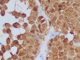
Acinar cell carcinoma of the pancreas, also acinar cell carcinoma, is a rare malignant exocrine tumour of the pancreas. It represents 5% of all exocrine tumours of the pancreas, making it the second most common type of pancreatic cancer. It is abbreviated ACC. It typically has a guarded prognosis.

<span class="mw-page-title-main">Pancreatic tumor</span> Medical condition

A pancreatic tumor is an abnormal growth in the pancreas. In adults, almost 90% are pancreatic cancer and a few are benign. Pancreatic tumors are rare in children.

<span class="mw-page-title-main">Cystic lesions of the pancreas</span> Medical condition

Cystic lesions of the pancreas are a group of pancreatic lesions characterized by a cystic appearance. They can be benign or malignant.

<span class="mw-page-title-main">Pancreatic cyst</span> Medical condition

A pancreatic cyst is a fluid filled sac within the pancreas.

A mucinous cystic neoplasm is an abnormal and excessive growth of tissue (neoplasm) that typically has elements of mucin and one or more cysts. By location, they include:

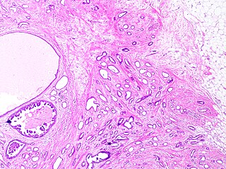
Pancreatic mucinous cystic neoplasm (MCN) is a type of cystic lesion that occurs in the pancreas. Amongst individuals undergoing surgical resection of a pancreatic cyst, about 23 percent were mucinous cystic neoplasms. These lesions are benign, though there is a high rate of progression to cancer. As such, surgery should be pursued when feasible. The rate of malignancy present in MCN is about 10 percent. If resection is performed before invasive malignancy develops, prognosis is excellent. The extent of invasion is the single most important prognostic factor in predicting survival.

<span class="mw-page-title-main">Tubular carcinoma</span> Type of breast cancer

Tubular carcinoma is a subtype of invasive ductal carcinoma of the breast. More rarely, tubular carcinomas may arise in the pancreas or kidney. Most tubular carcinomas begin in the milk duct of the breast and spread to healthy tissue around it.

References

  1. Source for mentioned findings: Pitman MB, Centeno BA, Daglilar ES, Brugge WR, Mino-Kenudson M (2014). "Cytological criteria of high-grade epithelial atypia in the cyst fluid of pancreatic intraductal papillary mucinous neoplasms". Cancer Cytopathol. 122 (1): 40–7. doi:10.1002/cncy.21344. PMID   23939829. S2CID   205677185.
    Images and annotations: Mikael Häggström, M.D.
  2. Patil TB, Shrikhande SV, Kanhere HA, Saoji RR, Ramadwar MR, Shukla PJ (2006). "Solid pseudopapillary neoplasm of the pancreas: a single institution experience of 14 cases". HPB. 8 (2): 148–50. doi:10.1080/13651820510035721. PMC   2131425 . PMID   18333264.
  3. Shuja, Asim (May 2014). "Solid pseudopapillary tumor: a rare neoplasm of the pancreas". Gastroenterol Rep (Oxf). 2 (2): 145–149. doi: 10.1093/gastro/gou006 . PMC   4020125 . PMID   24759340.
  4. De Moura, D.T.H. (2018). "The importance of endoscopic ultrasound fine-needle aspiration in the diagnosis of solid pseudopapillary tumor of the pancreas: two case reports". Journal of Medical Case Reports. 12 (107): 107. doi: 10.1186/s13256-018-1585-3 . PMC   5918898 . PMID   29695287.
  5. Fletcher CDM (2007). Diagnostic Histopathology of Tumors. Vol. I (3rd ed.). Elsevier. p. 478.
  6. Serra S, Chetty R (November 2008). "Revision 2: an immunohistochemical approach and evaluation of solid pseudopapillary tumour of the pancreas". J. Clin. Pathol. 61 (11): 1153–9. doi: 10.1136/jcp.2008.057828 . PMID   18708424.
  7. Stömmer P, Kraus J, Stolte M, Giedl J (March 1991). "Solid and cystic pancreatic tumors. Clinical, histochemical, and electron microscopic features in ten cases". Cancer. 67 (6): 1635–41. doi: 10.1002/1097-0142(19910315)67:6<1635::aid-cncr2820670627>3.0.co;2-m . PMID   1900454.
  8. Pettinato G, Manivel JC, Ravetto C, et al. (November 1992). "Papillary cystic tumor of the pancreas: A clinicopathologic study of 20 cases with cytologic, immunohistochemical, ultrastructural, and flow cytometric observations, and a review of the literature". American Journal of Clinical Pathology . 98 (5): 478–88. doi:10.1093/ajcp/98.5.478. PMID   1283055.
  9. Image by Mikael Häggström, MD.
    Reference for features: Pooja Navale, M.D., Omid Savari, M.D., Joseph F. Tomashefski, Jr., M.D., Monika Vyas, M.D. "Solid pseudopapillary neoplasm".{{cite web}}: CS1 maint: multiple names: authors list (link) Last author update: 4 March 2022
  10. Wang Y, Miller FH, Chen ZE, Merrick L, Mortele KJ, Hoff FL, et al. (2011). "Diffusion-weighted MR imaging of solid and cystic lesions of the pancreas". Radiographics. 31 (3): E47-64. doi:10.1148/rg.313105174. PMID   21721197.
    Diagram by Mikael Häggström, M.D.
  11. Pooja Navale, M.D., Omid Savari, M.D., Joseph F. Tomashefski, Jr., M.D., Monika Vyas, M.D. "Solid pseudopapillary neoplasm". Pathology Outlines.{{cite web}}: CS1 maint: multiple names: authors list (link) Last author update: 4 March 2022