Appendix cancer

Last updated
Appendix cancer
Other namesAppendiceal cancer
Appendiceal carcinoid 1.JPG
An excised biopsy of an appendiceal carcinoid tumor
Specialty Oncology, general surgery
Symptoms Bloating, discomfort in lower right abdomen, shortness of breath, loss of appetite [1]
Usual onset~50-55 years old [2]
TypesColonic-Type Adenocarcinoma, Non-carcinoid Appendix Tumors, Signet-Ring Cell Adenocarcinoma [1]
Risk factors Smoking, family history, Multiple endocrine neoplasia type 1 [3]
Diagnostic method Biopsy, CT Scan, MRI [1]
Differential diagnosis Acid reflux, Irritable bowel syndrome, Lactose intolerance, Stomach cancer [1]
Treatment Appendectomy, chemotherapy, radiation therapy [4]
Prognosis Five-year survival rate 25-88% (U.S.) [5]
Frequency~1,000 cases per year (U.S.) [6]
DeathsUnknown

Appendix cancer, also known as appendiceal cancer, is a very rare malignant tumor that forms in the vermiform appendix. [7]

Contents

Gastrointestinal stromal tumors are rare tumors with malignant potential. [8] Primary lymphomas can occur in the appendix. Breast cancer, colon cancer, and tumors of the female genital tract may metastasize to the appendix. [9]

Diagnosis

Appendix neoplasms by incidence and prognosis. Appendix neoplasms by incidence and prognosis.png
Appendix neoplasms by incidence and prognosis.

Carcinoid tumors are the most common tumors of the appendix. [10] Other common forms are mucinous adenocarcinomas, adenocarcinoma not otherwise specified (NOS), and signet ring cell adenocarcinoma listed from highest to lowest incidence. [11]

Carcinoid

Histopathology of an appendiceal carcinoid. The arrow points out a cluster of neuroendocrine cells. There are also inflammatory cells consistent with acute appendicitis. Histopathology of appendiceal carcinoid.jpg
Histopathology of an appendiceal carcinoid. The arrow points out a cluster of neuroendocrine cells. There are also inflammatory cells consistent with acute appendicitis.

A carcinoid is a neuroendocrine tumor (NET) of the intestines. [12] Incidence rates among carcinoids occur at about .15 per 100,000 per year. This subgroup makes up a large amount of neoplasias both malignant and benign. Almost 3 out of 4 of these tumors are associated with the region at the end of the appendix, and tend to be diagnosed in the 4th to 5th decades in life. Both women and Caucasian individuals show a minor prevalence regarding neuroendocrine tumor diagnosis without an explanation. [13] Prognosis of 5 year survival rates of carcinoids averages between 70 and 80% for typical cases. Advanced cases for 5 year survival range from 12 to 28%.

Mucinous neoplasm

Low-grade appendiceal mucinous neoplasm: Minimal cytological atypia of the epithelial cells. Histopathology of low-grade appendiceal mucinous neoplasm.jpg
Low-grade appendiceal mucinous neoplasm: Minimal cytological atypia of the epithelial cells.

Mucinous cystadenoma is an obsolete term for appendiceal mucinous neoplasm. [15]

Treatment

Small neuroendocrine tumors. [16] (<2 cm) without features of malignancy may be treated by appendectomy if complete removal is possible. Other neuroendocrine tumors and adenocarcinomas may require right hemicolectomy.

Pseudomyxoma peritonei treatment includes cytoreductive surgery which includes the removal of visible tumor and affected essential organs within the abdomen and pelvis. The peritoneal cavity is infused with heated chemotherapy known as HIPEC in an attempt to eradicate residual disease. The surgery may or may not be preceded or followed with intravenous chemotherapy or HIPEC. [17]

Epidemiology

A study of primary malignancies in the United States found a rate of 0.12 cases per 1,000,000 population per year. Carcinoids that were not identified as malignant were not included in this data. [18] Carcinoid is found in roughly 1 in 300-400 appendectomies for acute appendicitis. [19]

In a systematic literature review where 4765 appendiceal cancer patients were identified, the incidence of appendiceal cancer was shown to have increased regardless of the type of tumor, age, sex, and stage of appendiceal cancer. [11] Roughly 75% of appendiceal cases listed in the review had some form of metastases occurring. No observed trends have been noticed as to why this increase is occurring. One theory proposed is the increased use of computed tomography imaging in emergency departments since the early 1990s allowing for detection to occur before a surgery may be performed.

Notable cases

Related Research Articles

<span class="mw-page-title-main">Krukenberg tumor</span> Medical condition

A Krukenberg tumor refers to a malignancy in the ovary that metastasized from a primary site, classically the gastrointestinal tract, although it can arise in other tissues such as the breast. Gastric adenocarcinoma, especially at the pylorus, is the most common source. Krukenberg tumors are often found in both ovaries, consistent with its metastatic nature.

<span class="mw-page-title-main">Pancreatic cancer</span> Type of endocrine gland cancer

Pancreatic cancer arises when cells in the pancreas, a glandular organ behind the stomach, begin to multiply out of control and form a mass. These cancerous cells have the ability to invade other parts of the body. A number of types of pancreatic cancer are known.

<span class="mw-page-title-main">Ovarian cancer</span> Cancer originating in or on the ovary

Ovarian cancer is a cancerous tumor of an ovary. It may originate from the ovary itself or more commonly from communicating nearby structures such as fallopian tubes or the inner lining of the abdomen. The ovary is made up of three different cell types including epithelial cells, germ cells, and stromal cells. When these cells become abnormal, they have the ability to divide and form tumors. These cells can also invade or spread to other parts of the body. When this process begins, there may be no or only vague symptoms. Symptoms become more noticeable as the cancer progresses. These symptoms may include bloating, vaginal bleeding, pelvic pain, abdominal swelling, constipation, and loss of appetite, among others. Common areas to which the cancer may spread include the lining of the abdomen, lymph nodes, lungs, and liver.

<span class="mw-page-title-main">Malignancy</span> Tendency of a medical condition to become progressively worse

Malignancy is the tendency of a medical condition to become progressively worse; the term is most familiar as a characterization of cancer.

<span class="mw-page-title-main">Surface epithelial-stromal tumor</span> Medical condition

Surface epithelial-stromal tumors are a class of ovarian neoplasms that may be benign or malignant. Neoplasms in this group are thought to be derived from the ovarian surface epithelium or from ectopic endometrial or fallopian tube (tubal) tissue. Tumors of this type are also called ovarian adenocarcinoma. This group of tumors accounts for 90% to 95% of all cases of ovarian cancer; however is mainly only found in postmenopausal women with the exception of the United States where 7% of cases occur in women under the age of 40. Serum CA-125 is often elevated but is only 50% accurate so it is not a useful tumor marker to assess the progress of treatment. 75% of women with epithelial ovarian cancer are found within the advanced-stages; however younger patients are more likely to have better prognoses than older patients.

<span class="mw-page-title-main">Pseudomyxoma peritonei</span> Medical condition

Pseudomyxoma peritonei (PMP) is a clinical condition caused by cancerous cells that produce abundant mucin or gelatinous ascites. The tumors cause fibrosis of tissues and impede digestion or organ function, and if left untreated, the tumors and mucin they produce will fill the abdominal cavity. This will result in compression of organs and will destroy the function of the colon, small intestine, stomach, or other organs. Prognosis with treatment in many cases is optimistic, but the disease is lethal if untreated, with death occurring via cachexia, bowel obstruction, or other types of complications.

<span class="mw-page-title-main">Carcinoid</span> Slow-growing type of neuroendocrine tumor

A carcinoid is a slow-growing type of neuroendocrine tumor originating in the cells of the neuroendocrine system. In some cases, metastasis may occur. Carcinoid tumors of the midgut are associated with carcinoid syndrome.

The International Classification of Diseases for Oncology (ICD-O) is a domain-specific extension of the International Statistical Classification of Diseases and Related Health Problems for tumor diseases. This classification is widely used by cancer registries.

<span class="mw-page-title-main">Blastoma</span> Type of cancer arising from precursor cells

A blastoma is a type of cancer, more common in children, that is caused by malignancies in precursor cells, often called blasts. Examples are nephroblastoma, medulloblastoma, and retinoblastoma. The suffix -blastoma is used to imply a tumor of primitive, incompletely differentiated cells, e.g., chondroblastoma is composed of cells resembling the precursor of chondrocytes.

<span class="mw-page-title-main">Cystadenoma</span> Medical condition

Cystadenoma is a type of cystic adenoma. When malignant, it is called cystadenocarcinoma.

<span class="mw-page-title-main">Small intestine cancer</span> Medical condition

Small intestine cancer starts when cells in the small intestine start to grow out of control. The small intestine is the largest part of the gastrointestinal tract, which processes food for energy and rids the body of solid waste. The small intestine is not as common as other cancers of the GI tract such as colon, rectal, stomach, and esophageal cancers in the United States. They account for fewer than 1 in 10 cancers in the GI tract, and fewer than 1 in 100 cancers overall.

<span class="mw-page-title-main">Neuroendocrine tumor</span> Medical condition

Neuroendocrine tumors (NETs) are neoplasms that arise from cells of the endocrine (hormonal) and nervous systems. They most commonly occur in the intestine, where they are often called carcinoid tumors, but they are also found in the pancreas, lung, and the rest of the body.

<span class="mw-page-title-main">Adenocarcinoma in situ of the lung</span> Medical condition

Adenocarcinoma in situ (AIS) of the lung —previously included in the category of "bronchioloalveolar carcinoma" (BAC)—is a subtype of lung adenocarcinoma. It tends to arise in the distal bronchioles or alveoli and is defined by a non-invasive growth pattern. This small solitary tumor exhibits pure alveolar distribution and lacks any invasion of the surrounding normal lung. If completely removed by surgery, the prognosis is excellent with up to 100% 5-year survival.

<span class="mw-page-title-main">Adrenal tumor</span> Medical condition

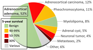
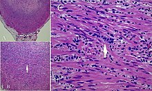
An adrenal tumor or adrenal mass is any benign or malignant neoplasms of the adrenal gland, several of which are notable for their tendency to overproduce endocrine hormones. Adrenal cancer is the presence of malignant adrenal tumors, and includes neuroblastoma, adrenocortical carcinoma and some adrenal pheochromocytomas. Most adrenal pheochromocytomas and all adrenocortical adenomas are benign tumors, which do not metastasize or invade nearby tissues, but may cause significant health problems by unbalancing hormones.

<span class="mw-page-title-main">Salivary gland tumour</span> Medical condition

Salivary gland tumours, also known as mucous gland adenomas or neoplasms, are tumours that form in the tissues of salivary glands. The salivary glands are classified as major or minor. The major salivary glands consist of the parotid, submandibular, and sublingual glands. The minor salivary glands consist of 800 to 1000 small mucus-secreting glands located throughout the lining of the oral cavity. Patients with these types of tumours may be asymptomatic.

Large cell lung carcinoma with rhabdoid phenotype (LCLC-RP) is a rare histological form of lung cancer, currently classified as a variant of large cell lung carcinoma (LCLC). In order for a LCLC to be subclassified as the rhabdoid phenotype variant, at least 10% of the malignant tumor cells must contain distinctive structures composed of tangled intermediate filaments that displace the cell nucleus outward toward the cell membrane. The whorled eosinophilic inclusions in LCLC-RP cells give it a microscopic resemblance to malignant cells found in rhabdomyosarcoma (RMS), a rare neoplasm arising from transformed skeletal muscle. Despite their microscopic similarities, LCLC-RP is not associated with rhabdomyosarcoma.

<span class="mw-page-title-main">Hyperthermic intraperitoneal chemotherapy</span>

Intraperitoneal hyperthermic chemoperfusion is a type of hyperthermia therapy used in combination with surgery in the treatment of advanced abdominal cancers. In this procedure, warmed anti-cancer medications are infused and circulated in the peritoneal cavity (abdomen) for a short period of time. The chemotherapeutic agents generally infused during IPHC are mitomycin-C and cisplatin.

Neuroendocrine adenoma middle ear (NAME) is a tumor which arises from a specific anatomic site: middle ear. NAME is a benign glandular neoplasm of middle ear showing histologic and immunohistochemical neuroendocrine and mucin-secreting differentiation.

Hyperthermic intrathoracic chemotherapy (HITOC) is part of a surgical strategy employed in the treatment of various pleural malignancies. The pleura in this situation could be considered to include the surface linings of the chest wall, lungs, mediastinum, and diaphragm. HITOC is the chest counterpart of HIPEC. Traditionally used in the treatment of malignant mesothelioma, a primary malignancy of the pleura, this modality has recently been evaluated in the treatment of secondary pleural malignancies.

<span class="mw-page-title-main">Pancreatic neuroendocrine tumor</span> Medical condition

Pancreatic neuroendocrine tumours, often referred to as "islet cell tumours", or "pancreatic endocrine tumours" are neuroendocrine neoplasms that arise from cells of the endocrine (hormonal) and nervous system within the pancreas.

References

  1. 1 2 3 4 MedStar Georgetown Cancer Institute
  2. Rare Diseases article
  3. Moffitt Cancer Center
  4. University of Chicago Medicine
  5. cancer.net
  6. MD Anderson
  7. "What is the Appendix?". News-Medical.net. 2016-10-18. Retrieved 2023-02-08.
  8. Park, Jin Hwa; Kang, Bo-kyeong; Lee, Hang Lak; Yoon, Jai Hoon; Lee, Kang Nyeong; Jun, Dae Won; Lee, Oh Young; Han, Dong Soo; Yoon, Byung Chul; Choi, Ho Soon (September 2020). "Correlation between Three-Dimensional Volume and Malignant Potential of Gastrointestinal Stromal Tumors (GISTs)". Journal of Clinical Medicine. 9 (9): 2763. doi: 10.3390/jcm9092763 . ISSN   2077-0383. PMC   7564070 . PMID   32858945.
  9. Rosai, Juan (2004) [1953]. "11. Gastrointestinal tract". Rosai and Ackerman's surgical pathology (9th ed.). Mosby. pp. 761–769.
  10. Sabiston Principles of Surgery (20th ed.). p. 1308.
  11. 1 2 Marmor, Schelomo; Portschy, Pamela R.; Tuttle, Todd M.; Virnig, Beth A. (6 January 2015). "The Rise in Appendiceal Cancer Incidence: 2000–2009". Journal of Gastrointestinal Surgery. 19 (4): 743–750. doi:10.1007/s11605-014-2726-7. PMID   25560182. S2CID   24206562.
  12. "Carcinoid Tumors: Symptoms, Diagnosis & Treatment". Cleveland Clinic. Retrieved 2023-02-08.
  13. Teixeira, Frederico José Ribeiro; Couto Netto, Sérgio Dias do; Akaishi, Eduardo Hiroshi; Utiyama, Edivaldo Massazo; Menegozzo, Carlos Augusto Metidieri; Rocha, Marcelo Cristiano (9 March 2017). "Acute appendicitis, inflammatory appendiceal mass and the risk of a hidden malignant tumor: a systematic review of the literature". World Journal of Emergency Surgery. 12 (1): 12. doi: 10.1186/s13017-017-0122-9 . PMC   5343298 . PMID   28286544.
  14. Hajjar, Roy; Dubé, Pierre; Mitchell, Andrew; Sidéris, Lucas (2019). "Combined Mucinous and Neuroendocrine Tumours of the Appendix Managed with Surgical Cytoreduction and Oxaliplatin-based Hyperthermic Intraperitoneal Chemotherapy". Cureus. 11 (1): e3894. doi: 10.7759/cureus.3894 . ISSN   2168-8184. PMC   6424473 . PMID   30911451.
  15. Michael Feely, Raul S. Gonzalez. "Appendix - Other tumors - Mucinous neoplasms (LAMN and HAMN)". Topic Completed: 1 October 2017. Revised: 11 December 2019
  16. Griniatsos, J; Michail, O (2010). "Appendiceal neuroendocrine tumors: recent insights and clinical implications". World Journal of Gastrointestinal Oncology. 2 (4): 192–196. doi: 10.4251/wjgo.v2.i4.192 . PMC   2999180 . PMID   21160597.
  17. M. Townsend, Courtney (2012) [1969]. "51. The appendix". Sabiston (18th ed.). Elsevier. p. 1289.
  18. McCusker, M. E.; Coté, T. R.; Clegg, L. X.; Sobin, L. H. (2002). "Primary malignant neoplasms of the appendix". Cancer. 94 (12): 3307–3312. doi: 10.1002/cncr.10589 . PMID   12115365. S2CID   40814989.
  19. Bailey and Love's Short Practice of Surgery (27th ed.). p. 1315.
  20. Selim, Jocelyn (Fall 2009). "The Fairest of All". CR. Philadelphia: American Association for Cancer Research. 4 (4). Archived from the original on 19 April 2010. Retrieved 26 May 2023.
  21. "Stuart Scott Says, 'F U, Cancer!'". Men's Health. Retrieved 7 January 2015.
  22. Sandomir, Richard (4 January 2015). "Stuart Scott, ESPN's Voice of Exuberance, Dies at 49" . New York Times . Retrieved 26 May 2023.
  23. Nikolić, Aleksandar (30 January 2015). "Vlada Divljan poručio: Nisam životno ugrožen, osećam se vrlo dobro" [Vlada Divljan said: I am not in danger, I feel very well]. Blic (in Serbian). Retrieved 26 May 2023.
  24. Weiss, Haley (10 January 2024). "What to Know About Appendiceal Cancer". TIME. Retrieved 11 March 2024.

Further reading